The Ethidium Bromide-Agar Cartwheel Method: A Comprehensive Guide for Efflux Pump Activity Detection in Drug-Resistant Pathogens
This article provides a detailed, step-by-step guide to the Ethidium Bromide-Agar Cartwheel (EB-AC) method, a classic and accessible qualitative assay for detecting active efflux in bacterial pathogens.
The Ethidium Bromide-Agar Cartwheel Method: A Comprehensive Guide for Efflux Pump Activity Detection in Drug-Resistant Pathogens
Abstract
This article provides a detailed, step-by-step guide to the Ethidium Bromide-Agar Cartwheel (EB-AC) method, a classic and accessible qualitative assay for detecting active efflux in bacterial pathogens. We explore the foundational principles of efflux-mediated resistance, deliver a complete methodological protocol for the EB-AC assay, address common troubleshooting and optimization challenges, and validate its utility by comparing it to modern quantitative techniques like real-time fluorometry and PCR. Designed for researchers and drug development professionals, this guide synthesizes current best practices to enable reliable screening for efflux pump overexpression, a critical step in combating antimicrobial resistance.
Understanding Efflux Pumps and the Rationale Behind the EB-Agar Cartwheel Assay
The Growing Crisis of Antimicrobial Resistance and the Role of Efflux Pumps
Application Notes
Antimicrobial resistance (AMR) is a global health crisis, with efflux pumps being a primary mechanism of multidrug resistance (MDR) in bacteria. These membrane proteins actively extrude a wide range of antibiotics, reducing intracellular concentration and therapeutic efficacy. The Ethidium Bromide (EtBr)-agar Cartwheel method is a foundational, low-cost phenotypic assay for screening efflux pump activity in bacterial isolates, particularly relevant for research within resource-limited settings. This application note details its integration into a broader thesis investigating efflux-mediated resistance.
Key Principles: The assay exploits EtBr, a substrate for many broad-specificity efflux pumps (e.g., AcrAB-TolC in E. coli). Bacteria with active efflux prevent EtBr accumulation, exhibiting lower fluorescence under UV light. When an efflux pump inhibitor (EPI) like Carbonyl Cyanide m-Chlorophenylhydrazone (CCCP) is added, efflux is inhibited, leading to increased EtBr accumulation and fluorescence.
Quantitative Data Context: Recent surveillance data underscores the urgency of efflux research. The following table summarizes key AMR statistics and efflux prevalence:
Table 1: Global AMR Burden and Prevalence of Efflux-Mediated Resistance
| Metric | Value | Key Pathogens Involved | Notes |
|---|---|---|---|
| Annual AMR-attributable deaths (2019) | ~4.95 million | E. coli, S. aureus, K. pneumoniae, S. pneumoniae | (Source: Antimicrobial Resistance Collaborators, 2022) |
| Isolates exhibiting MDR phenotypes | 20-60% in clinical settings | P. aeruginosa, A. baumannii | Varies significantly by region and hospital. |
| Isolates where efflux contributes to resistance | 30-80% among MDR strains | Enterobacteriaceae, Neisseria spp. | Major contributor to fluoroquinolone, tetracycline, β-lactam resistance. |
| Reduction in MIC with EPI addition | 4 to 64-fold | Campylobacter spp., Salmonella spp. | Demonstrates the functional contribution of efflux. |
Table 2: Common Efflux Pump Systems and Substrates
| Efflux Pump System | Organism | Antibiotic Substrates |
|---|---|---|
| AcrAB-TolC | Escherichia coli, Salmonella enterica | Fluoroquinolones, β-lactams, tetracyclines, chloramphenicol, EtBr |
| MexAB-OprM | Pseudomonas aeruginosa | β-lactams, fluoroquinolones, tetracycline, chloramphenicol |
| AdeABC | Acinetobacter baumannii | Aminoglycosides, tetracyclines, fluoroquinolones |
| MepA | Staphylococcus aureus | Fluoroquinolones, biocides, EtBr |
Experimental Protocols
Protocol 1: Ethidium Bromide-Agar Cartwheel Method for Efflux Phenotype Screening
Objective: To phenotypically identify bacterial strains with active efflux pump activity against EtBr.
Thesis Context: This protocol serves as the initial high-throughput screening step within the thesis workflow, categorizing clinical isolates as "Efflux Positive" or "Efflux Negative" for downstream genetic and functional analyses.
Materials:
- Research Reagent Solutions:
- Mueller-Hinton Agar (MHA): Standardized growth medium for antimicrobial susceptibility testing.
- Ethidium Bromide Stock Solution (1 mg/mL): Fluorescent efflux pump substrate. CAUTION: Mutagen. Use PPE.
- Carbonyl Cyanide m-Chlorophenylhydrazone (CCCP) Stock Solution (100 mM in DMSO): Protonophore that disrupts proton motive force, inhibiting many RND-type efflux pumps.
- Sterile Phosphate Buffered Saline (PBS): For bacterial suspension standardization.
- 0.5 McFarland Standard: To prepare standardized bacterial inoculum (~1.5 x 10^8 CFU/mL).
Procedure:
- Prepare two sets of MHA plates: a. Plate A (Efflux Activity): MHA supplemented with a sub-inhibitory concentration of EtBr (e.g., 0.5 µg/mL). Optimize concentration for species. b. Plate B (Efflux Inhibition): MHA supplemented with the same EtBr concentration and an EPI (e.g., CCCP at 20 µM).
- Prepare bacterial suspensions of test isolates and a control strain (e.g., E. coli K-12 as negative, a known overproducer as positive) in PBS, adjusted to 0.5 McFarland.
- Using a sterile swab, spot 2-3 µL of each suspension onto both Plate A and Plate B in a "cartwheel" pattern. Allow spots to dry.
- Incubate plates aerobically at 37°C for 16-20 hours.
- After incubation, observe plates under UV light (302 nm). Caution: Wear UV-protective eyewear.
- Interpretation:
- Efflux Positive Strain: On Plate A (EtBr only), weak or no fluorescence. On Plate B (EtBr+EPI), bright fluorescence.
- Efflux Negative Strain: Bright fluorescence on both Plate A and Plate B.
- A positive result indicates active extrusion of EtBr, likely mediated by constitutive efflux pump expression.
Protocol 2: Ethidium Bromide Accumulation Assay (Fluorometric)
Objective: To quantitatively measure real-time EtBr accumulation in bacterial cells in the presence and absence of an EPI.
Thesis Context: This quantitative protocol follows the cartwheel screen to provide kinetic data on efflux activity, validating phenotypic results and allowing comparison of efflux rates between isolates.
Materials:
- Research Reagent Solutions:
- HEPES Buffer (50 mM, pH 7.0): Maintains stable pH during fluorescence measurement.
- Glucose Solution (20% w/v): Energy source to support active efflux.
- Ethidium Bromide Working Solution (10 µg/mL in HEPES): Substrate for the assay.
- CCCP Working Solution (100 µM in HEPES): Efflux pump inhibitor control.
- Bacterial Cell Suspension: Mid-log phase cells, washed and resuspended in HEPES buffer with glucose (0.4% final).
Procedure:
- In a quartz cuvette or black 96-well plate, mix 1.9 mL of bacterial suspension with 100 µL of EtBr working solution (Final EtBr: 0.5 µg/mL).
- Place the cuvette in a spectrofluorometer (excitation: 530 nm, emission: 600 nm, slits: 5 nm). Record baseline fluorescence (F0) for 1-2 minutes.
- At time t=0, rapidly add 20 µL of glucose solution (final 0.4%) to energize the cells. Continuously record fluorescence (Ft) for 10-15 minutes. The initial rapid increase represents passive uptake; the subsequent plateau or decrease indicates active efflux.
- At the plateau phase (e.g., t=10 min), add 20 µL of CCCP working solution (final 1 µM) and continue recording for an additional 5-10 minutes. A sharp increase in fluorescence confirms efflux inhibition.
- Data Analysis: Calculate normalized fluorescence: (Ft - F0) / F0. Plot against time. The rate of fluorescence increase after CCCP addition is inversely proportional to the baseline efflux activity.
Diagrams
Title: Thesis Experimental Workflow for Efflux Research
Title: RND Efflux Pump Mechanism and EPI Action
Efflux pumps are integral membrane proteins that actively transport toxic substrates, including a wide range of antimicrobial agents, out of bacterial cells. This activity is a major mechanism of intrinsic and acquired multidrug resistance (MDR). Within the broader thesis investigating the Ethidium Bromide (EtBr)-agar Cartwheel method for rapid phenotypic detection of efflux activity, this application note details the core principles, quantitative data, and protocols underpinning efflux pump research. The EtBr-agar Cartwheel method serves as a foundational, cost-effective screening tool to identify strains with enhanced efflux capacity, guiding subsequent molecular and biochemical analyses.
Core Quantitative Data on Major Efflux Pump Families
Table 1: Major Bacterial Efflux Pump Families and Their Characteristics
| Pump Family (Representative) | Typical Gram Classification | Energy Coupling | Substrate Profile (Examples) | Clinical Relevance |
|---|---|---|---|---|
| RND (AcrAB-TolC in E. coli) | Negative | Proton Motive Force (H+) | Broad: β-lactams, fluoroquinolones, tetracyclines, macrolides, dyes, detergents | Primary MDR determinant in Gram-negatives; often chromosomally encoded. |
| MFS (NorA in S. aureus) | Positive | Proton Motive Force (H+) | Narrower: Fluoroquinolones, dyes (EtBr), disinfectants | Contributes to resistance in Gram-positives; can be plasmid-encoded. |
| MATE (NorM in V. parahaemolyticus) | Primarily Negative | Sodium Ion Gradient (Na+) or H+ | Fluoroquinolones, aminoglycosides, dyes | Important in various pathogens; often drug-specific. |
| SMR (EmrE in E. coli) | Negative | Proton Motive Force (H+) | Small, lipophilic cations, disinfectants, dyes | Smaller pumps; can confer low-level resistance to biocides. |
| ABC (LmrA in L. lactis) | Positive | ATP Hydrolysis | Very broad: Lipophilic drugs, dyes, peptides | Less common in bacteria for drug resistance; primary in eukaryotes. |
Table 2: Quantitative Impact of Efflux on Minimum Inhibitory Concentrations (MICs)
| Antimicrobial Agent | MIC in Wild-type E. coli (µg/mL) | MIC in ΔacrB Mutant (µg/mL) | Fold Increase (Wild-type/Mutant) |
|---|---|---|---|
| Chloramphenicol | 4 - 8 | 0.5 - 1 | 8x |
| Tetracycline | 2 - 4 | 0.25 - 0.5 | 8x |
| Ciprofloxacin | 0.03 - 0.06 | 0.004 - 0.008 | ~8x |
| Erythromycin | 64 - 128 | 4 - 8 | 16x |
| Ethidium Bromide | 32 - 64 | 2 - 4 | 16x |
Detailed Protocols
Protocol A: Ethidium Bromide-Agar Cartwheel Method for Phenotypic Efflux Screening
Principle: Bacteria with active efflux pumps expel the DNA-intercalating dye EtBr, reducing intracellular accumulation and fluorescence. In the presence of an efflux pump inhibitor (EPI) like CCCP, dye retention increases. Materials: See "The Scientist's Toolkit" below. Procedure:
- Prepare two sets of LB-agar plates: one containing a sub-inhibitory concentration of EtBr (e.g., 0.5 µg/mL) and another containing the same EtBr concentration plus a sub-inhibitory concentration of an EPI (e.g., 25 µM CCCP).
- Using a sterile loop, pick 3-5 colonies of the test bacterium and streak them radially from the center to the edge of each plate in a "cartwheel" pattern (4-6 streaks per plate).
- Inoculate a known efflux-deficient mutant (e.g., ΔacrB) and its wild-type parent as controls on both plates.
- Incubate plates at 37°C for 16-24 hours.
- Visualize plates under a UV transilluminator (302 nm). Record fluorescence intensity and pattern. Interpretation: Strains with active efflux will show significantly less fluorescence (darker streaks) on the EtBr-only plate compared to the EtBr+EPI plate. Strains lacking efflux will be fluorescent on both plates. This visual assay provides a rapid, qualitative assessment of efflux capability.
Protocol B: Real-Time Fluorometric Assay of Efflux Activity Using EtBr
Principle: Measures the kinetics of EtBr accumulation and extrusion in a cell suspension, providing quantitative data on pump activity. Procedure:
- Cell Preparation: Grow test and control strains to mid-log phase (OD600 ~0.5). Harvest cells by centrifugation, wash twice, and resuspend in assay buffer (e.g., 50 mM phosphate buffer, pH 7.0, with 5 mM MgCl2) to an OD600 of 0.2.
- Baseline Measurement: Aliquot 2 mL of cell suspension into a quartz cuvette. Place in a spectrofluorometer with thermostatic control (37°C). Set excitation to 530 nm and emission to 600 nm. Record baseline fluorescence for 60 seconds.
- Energy Poisoning & Accumulation: Add CCCP (final conc. 50 µM) to dissipate the proton motive force and inhibit active efflux. Immediately add EtBr (final conc. 2 µg/mL). Monitor fluorescence increase until it plateaus (2-5 minutes). This represents maximum intracellular dye accumulation (F_max).
- Efflux Induction: Add a metabolizable energy source (e.g., 0.4% glucose) to re-energize the cells and reactivate efflux pumps. Observe the rapid decrease in fluorescence as EtBr is expelled.
- Data Analysis: Calculate the initial rate of efflux (fluorescence units/second) after glucose addition. Compare rates between strains or between conditions (e.g., with/without an EPI).
Diagrams of Key Concepts and Workflows
The Scientist's Toolkit: Research Reagent Solutions
Table 3: Essential Materials for Efflux Pump Research (Focus on EtBr Methods)
| Item | Function & Application in Research | Example/Note |
|---|---|---|
| Ethidium Bromide (EtBr) | Fluorescent substrate dye for efflux pumps; used in Cartwheel and fluorometric assays to visualize and quantify pump activity. | Caution: Mutagen. Use appropriate PPE and waste disposal. Stock: 10 mg/mL in H2O. |
| Carbonyl Cyanide m-chlorophenyl hydrazone (CCCP) | Protonophore; dissipates the proton motive force (PMF), inhibiting secondary transporter pumps (RND, MFS, etc.). Standard efflux pump inhibitor (EPI) for controls. | Working conc.: 25-50 µM. Light sensitive. Stock: 10 mM in DMSO or EtOH. |
| Phenylalanine-Arginine β-naphthylamide (PAβN) | Broad-spectrum EPI; competitively inhibits RND pumps. Used to assess pump-specific contribution to resistance in MIC assays. | Often used at 20-50 µg/mL in combination with antibiotics. |
| Glucose | Metabolizable energy source. Used in fluorometric assays to re-energize cells after CCCP treatment, triggering active efflux. | 0.4% (w/v) final concentration is typical. |
| LB Agar & Broth | Standard growth medium for cultivating bacterial strains for phenotypic assays. | For Cartwheel method, agar is supplemented with EtBr ± EPI. |
| Spectrofluorometer & Cuvettes | Instrumentation for real-time, quantitative measurement of fluorescence changes in cell suspensions (Protocol B). | Requires temperature control and appropriate wavelength settings (Ex: 530nm, Em: 600nm for EtBr). |
| UV Transilluminator | Essential for visualizing fluorescence of bacterial streaks on EtBr-agar Cartwheel plates. | Caution: UV light. Use protective face shield. 302 nm is optimal for EtBr. |
| Microbial Strains: Wild-type, Δefflux mutant, Overexpression strain | Critical controls. Mutant confirms pump-specific effect. Overexpression strain serves as a positive control. | e.g., E. coli K-12 (wild-type) vs. E. coli K-12 ΔacrB. |
Ethidium bromide (EtBr) is a phenanthridine fluorescent dye widely employed as a universal substrate for studying bacterial efflux pump activity. Its utility stems from its physicochemical properties, which allow it to be recognized and extruded by a broad range of efflux pump systems across Gram-positive and Gram-negative bacteria.
Key Properties Facilitating Universal Efflux:
- Cationic Nature: At physiological pH, EtBr is positively charged, making it a substrate for many MultiDrug Resistance (MDR) efflux pumps that often expel cationic, amphiphilic compounds.
- Planar, Intercalating Structure: Its planar aromatic ring structure allows it to intercalate into DNA, providing a simple fluorescent readout for intracellular accumulation. This same structure is a common pharmacophore recognized by efflux pumps.
- Fluorescence Enhancement Upon Binding: EtBr exhibits a significant increase in fluorescence quantum yield upon binding to nucleic acids, enabling sensitive detection of intracellular vs. extruded dye.
- Broad Recognition: It is a documented substrate for major efflux pump families, including RND (e.g., AcrAB-TolC in E. coli), MFS (e.g., NorA in S. aureus), MATE, and SMR.
Quantitative Data on Ethidium Bromide Efflux
Table 1: Key Physicochemical and Efflux Parameters of Ethidium Bromide
| Parameter | Value / Description | Experimental Context |
|---|---|---|
| Molecular Weight | 394.3 g/mol | - |
| Charge at pH 7 | +1 (monocation) | - |
| Primary Excitation/Emission | ~518 nm / ~605 nm | In complex with nucleic acids. |
| Common Working Concentration | 0.5 - 2.0 µg/mL | Agar cartwheel, fluorescence assays. |
| Efflux Pump Families | RND, MFS, MATE, SMR | Demonstrated across bacterial species. |
| Typical MIC Reduction with EPI* | 4 to 16-fold | e.g., with CCCP (50 µM) or PAβN (20-40 mg/L). |
| Standard Positive Control Inhibitor | Carbonyl Cyanide m-chlorophenyl hydrazone (CCCP) 50-100 µM | Protonophore uncoupler. |
*EPI: Efflux Pump Inhibitor
Core Mechanisms of Efflux Recognition and Transport
EtBr serves as a model substrate to elucidate the functional operation of MDR efflux pumps. The general mechanism involves recognition by a broad-substrate-binding pocket, energy-dependent expulsion, and resultant phenotypic resistance.
Table 2: Efflux Pump Families that Transport EtBr
| Efflux Family | Example Pump (Organism) | Energy Source | Role in EtBr Resistance |
|---|---|---|---|
| RND | AcrAB-TolC (E. coli) | Proton Motive Force | Primary high-level resistance in Gram-negatives. |
| MFS | NorA (S. aureus) | Proton Motive Force | Major contributor in Gram-positives like S. aureus. |
| MATE | NorM (V. cholerae) | Na+ or H+ Gradient | Secondary efflux, often Na+-coupled. |
| SMR | EmrE (E. coli) | Proton Motive Force | Transports specific cationic dyes like EtBr. |
The Ethidium Bromide-Agar Cartwheel Method: Protocol
This protocol is central to the thesis context, providing a high-throughput, phenotypic screen for efflux pump activity in bacterial isolates.
A. Materials and Reagent Solutions
Table 3: Research Reagent Toolkit for EtBr-Agar Cartwheel Assay
| Item / Reagent | Function / Description | Typical Preparation/Concentration |
|---|---|---|
| Ethidium Bromide Stock | Efflux pump substrate & fluorescent indicator. | 10 mg/mL in sterile water. Store dark at 4°C. |
| Mueller-Hinton Agar (MHA) | Growth medium for the assay. | Prepare per manufacturer instructions. |
| Efflux Pump Inhibitor (EPI) | Positive control inhibitor to confirm efflux-mediated resistance. | e.g., CCCP (50 µM final) or PAβN (20-40 mg/L final). |
| Bacterial Test Strains | Isolates with known (control) and unknown efflux activity. | Overnight cultures adjusted to 0.5 McFarland standard. |
| Sterile Petri Dishes | Platform for agar-based assay. | Standard 90-100 mm diameter. |
| Carbenicillin or Chloramphenicol | Control antibiotic for standard susceptibility. | Used as a disk control (optional). |
| UV Transilluminator (312 nm) | Visualization of EtBr fluorescence. | Critical for reading results. |
B. Detailed Protocol
Step 1: Preparation of EtBr-Agar Plates.
- Prepare and autoclave Mueller-Hinton Agar.
- Cool the agar to approximately 50°C in a water bath.
- Aseptically add Ethidium Bromide stock solution to achieve a final concentration of 1.0 µg/mL. Swirl gently to mix thoroughly, avoiding bubbles.
- Pour approximately 25 mL of the EtBr-supplemented agar into each sterile Petri dish. Let solidify at room temperature, then store plates protected from light at 4°C for up to 1 week.
Step 2: Inoculation (Cartwheel Method).
- Dilute fresh overnight bacterial cultures in sterile saline or broth to a 0.5 McFarland standard (~1.5 x 10^8 CFU/mL).
- Using a sterile swab, lawn-inoculate the entire surface of an EtBr-agar plate.
- Allow the inoculum to dry for 5-10 minutes.
- Using sterile forceps, place a filter paper disk impregnated with an Efflux Pump Inhibitor (e.g., 50 µL of 1 mM CCCP) at the exact center of the plate.
- Streak the test strains in a "cartwheel" pattern from the edge of the central disk to the periphery of the plate. Typically, 6-8 strains can be tested per plate.
- Incubate the plate aerobically at 37°C for 16-20 hours.
Step 3: Visualization and Interpretation.
- After incubation, observe plates under short-wavelength UV light (312 nm) in a darkroom or using a UV transilluminator.
- Positive Efflux Activity: Bacterial growth streaks will show diminished red-orange fluorescence compared to the brightly fluorescent agar. The area around the central EPI disk will exhibit a zone of increased fluorescence (halo) where efflux is inhibited, allowing intracellular EtBr accumulation.
- Negative Efflux Activity: Bacterial growth will be brightly fluorescent, indicating no active efflux. No fluorescent halo around the EPI disk is expected.
Complementary Quantitative Protocols
Protocol: Real-Time Fluorometric Efflux Assay This kinetic assay measures active EtBr extrusion in real-time.
- Cell Preparation: Grow bacteria to mid-log phase. Harvest, wash twice in efflux assay buffer (e.g., PBS with 5 mM glucose, pH 7.0). Resuspend to an OD600 of ~0.4.
- Loading with EtBr: Add EtBr to cell suspension to a final concentration of 2 µg/mL. Incubate at 37°C with shaking for 30-60 minutes to allow accumulation.
- Energy Poison Control: For a negative control, pre-treat an aliquot of loaded cells with 100 µM CCCP for 10 minutes.
- Efflux Measurement: Pellet loaded cells, wash rapidly once with warm buffer to remove external dye, and resuspend in pre-warmed buffer. Immediately transfer to a fluorometer cuvette maintained at 37°C with continuous stirring.
- Data Acquisition: Monitor fluorescence (Ex: 518 nm, Em: 605 nm) for 60-120 seconds to establish a baseline efflux rate. Then, add 20 mM glucose (energy source) to energize efflux and record the rapid decrease in fluorescence as EtBr is extruded.
- Analysis: The initial rate of fluorescence decrease after glucose addition is proportional to efflux pump activity. Compare rates with and without EPIs.
Protocol: Minimum Inhibitory Concentration (MIC) Modulation Assay Determines the contribution of efflux to antimicrobial resistance.
- Prepare two-fold serial dilutions of the antibiotic of interest (e.g., ciprofloxacin) in Mueller-Hinton Broth in a microtiter plate.
- To one set of dilutions, add a sub-inhibitory concentration of an EPI (e.g., 25 mg/L PAβN or 50 µM CCCP).
- Inoculate each well with ~5 x 10^5 CFU/mL of the test bacterium.
- Incubate at 37°C for 18-24 hours.
- Determine the MIC with and without the EPI. A ≥4-fold reduction in MIC in the presence of the EPI is indicative of significant efflux-mediated resistance.
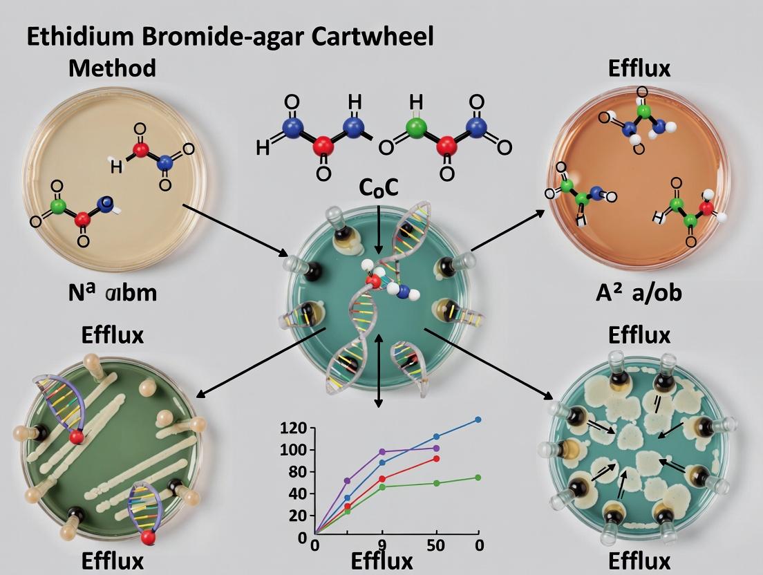
The Ethidium Bromide (EtBr)-agar Cartwheel method emerged from a critical need in antimicrobial resistance (AMR) research for accessible, high-throughput phenotypic assays to detect active efflux pump systems in bacterial pathogens. Prior to its adoption, efflux activity assessment often relied on complex, equipment-intensive methods like real-time fluorometry or PCR-based gene expression analysis, which were not feasible for many clinical or field laboratories. The Cartwheel method, developed as a derivative of the classical agar dilution assay, provided a simple, visual, and semi-quantitative solution. Its integration into a broader thesis on efflux research lies in its role as a foundational screening tool, enabling the rapid identification of efflux-positive strains and the preliminary evaluation of efflux pump inhibitors (EPIs) before committing to more resource-intensive molecular analyses.
Application Notes
The primary application is the detection and semi-quantitative comparison of Ethidium Bromide efflux capability in bacterial isolates, particularly Staphylococcus aureus, Escherichia coli, and Pseudomonas aeruginosa. The method is instrumental in:
- Phenotypic Screening: Rapidly classifying clinical isolates as efflux-positive or efflux-negative based on fluorescence under UV light.
- EPI Screening: Pre-screening potential efflux pump inhibitors by observing enhanced fluorescence retention in the presence of the inhibitor.
- Epidemiological Studies: Surveying the prevalence of efflux-mediated resistance in bacterial populations.
Key Advantages and Limitations
| Advantage | Limitation |
|---|---|
| Low cost and technical requirement | Semi-quantitative; does not provide kinetic parameters |
| High-throughput capability (multiple strains per plate) | Subjective visual endpoint interpretation |
| Visual, intuitive results | Specific to EtBr as a substrate; may not detect all efflux systems |
| Compatible with EPI assessment | Agar concentration and EtBr batch can affect results |
Quantitative Data Summary from Representative Studies Table 1: Typical Minimum Inhibitory Concentration (MIC) of EtBr for Common Bacterial Species with/without Efflux Pump Inhibitors (EPIs).
| Bacterial Species | Efflux Status | EtBr MIC (µg/mL) Range | EtBr MIC + EPI (e.g., CCCP 50µM) | Fold Reduction |
|---|---|---|---|---|
| S. aureus | Wild-type (Basal efflux) | 0.5 - 1.5 | 0.25 - 0.5 | 2-3 |
| S. aureus | Overexpressor (e.g., NorA) | 8 - 32 | 1 - 4 | 8-16 |
| E. coli | Wild-type | 10 - 20 | 2.5 - 5 | 4-8 |
| P. aeruginosa | Wild-type | 40 - 80 | 10 - 20 | 4-8 |
Table 2: Interpretation of Cartwheel Method Results Based on Fluorescence Intensity.
| Fluorescence under UV (366 nm) | Visual Description | Interpretation |
|---|---|---|
| Bright pink/red | Intense, clear fluorescence | Efflux-negative or EPI-active (EtBr retained) |
| Faint pink or orange | Dim, weak fluorescence | Moderate efflux activity |
| No fluorescence | Only background agar color | Strong efflux activity (EtBr fully expelled) |
Experimental Protocols
Protocol 1: Standard Agar Cartwheel Method for Efflux Phenotype Screening
- Objective: To determine the baseline efflux activity of bacterial isolates using EtBr-agar plates.
- Materials: See "The Scientist's Toolkit" below.
- Procedure:
- Prepare Mueller-Hinton Agar (MHA) supplemented with EtBr at a sub-inhibitory concentration (typically 0.5 - 1.0 µg/mL, optimized per species). Pour into Petri dishes.
- Suspend 2-3 fresh colonies of test and control strains in sterile saline to a 0.5 McFarland standard (~1.5 x 10⁸ CFU/mL).
- Using a sterile swab, create a radial "spoke" pattern from the center of the plate. Each spoke is inoculated with a different bacterial suspension. The center is left uninoculated.
- Incubate plates aerobically at 35°C for 18-24 hours.
- Observe plates under UV light (366 nm) in a dark room. Record the fluorescence intensity along each inoculated spoke.
- Interpretation: Compare fluorescence to control strains. Strains with bright fluorescence are considered efflux-negative (or impaired); strains with little to no fluorescence are efflux-positive.
Protocol 2: Cartwheel Method for EPI Potency Assessment
- Objective: To evaluate the ability of a compound to inhibit efflux activity.
- Procedure:
- Prepare two sets of EtBr-agar plates: one without EPI (control) and one supplemented with a sub-inhibitory concentration of the test EPI (e.g., 20-50 µg/mL for CCCP or PaβN).
- Inoculate both plates with the efflux-positive test strain and a known efflux-negative control using the cartwheel pattern.
- Incubate and examine under UV light as in Protocol 1.
- Interpretation: A marked increase in fluorescence intensity on the EPI-containing plate compared to the control plate indicates successful inhibition of the efflux pump.
Mandatory Visualization
Title: Agar Cartwheel Method Workflow & Interpretation
Title: EtBr Intracellular Fate & Efflux Mechanism
The Scientist's Toolkit Table 3: Essential Research Reagents and Materials for the Agar Cartwheel Method.
| Item | Function/Brief Explanation |
|---|---|
| Ethidium Bromide (EtBr) Stock Solution (e.g., 10 mg/mL in H₂O) | Fluorescent efflux pump substrate. Handle as a mutagen with appropriate PPE. |
| Mueller-Hinton Agar (MHA) | Standardized, low-inhibitor medium for antimicrobial susceptibility testing. |
| Carbonyl Cyanide m-chlorophenyl hydrazone (CCCP) | A protonophore used as a positive control EPI to collapse proton motive force. |
| Phenylalanine-arginine β-naphthylamide (PaβN) | Broad-spectrum, competitive EPI for RND pumps in Gram-negative bacteria. |
| Bacterial Control Strains | Known efflux-positive (e.g., S. aureus SA-1199B) and efflux-negative strains. |
| UV Lamp (366 nm) | Light source for exciting EtBr retained within bacterial cells. |
| 0.5 McFarland Standard | Turbidity standard for preparing standardized bacterial inocula. |
| Sterile Cotton Swabs | For inoculating agar plates in the distinct cartwheel/spoke pattern. |
Application Notes
Within the thesis on the Ethidium Bromide (EtBr)-agar Cartwheel method, this principle serves as a foundational assay for phenotypic detection of efflux pump activity in bacteria, particularly multidrug-resistant strains. The method leverages the natural fluorescence of EtBr, a substrate for many broad-specificity efflux pumps like those in the RND family. Active efflux prevents intracellular accumulation, resulting in a quantifiable loss of fluorescence on an agar medium. The assay is a critical, low-cost tool for initial screening and mechanistic studies in antibiotic resistance and efflux pump inhibitor (EPI) discovery.
Key Quantitative Data from Current Literature:
Table 1: Representative Efflux Phenotype Results Using EtBr-Agar Cartwheel Method
| Bacterial Strain (Phenotype) | Agar EtBr Concentration (µg/mL) | Fluorescence Under UV (365 nm) | Inferred Efflux Activity | Reference Context |
|---|---|---|---|---|
| E. coli K-12 (Wild-type) | 0.5 | Moderate | Baseline | Control strain |
| E. coli ΔacrB (Efflux-deficient) | 0.5 | High (Bright) | Negligible | Isogenic mutant control |
| K. pneumoniae MDR (Clinical isolate) | 1.0 | Low/None | High | Confirmed by EPI addition |
| P. aeruginosa PAO1 (Wild-type) | 1.0 - 2.0 | Low/None | Very High | Intrinsic resistance |
| S. aureus NorA overexpressor | 0.5 | Low | High (NorA-mediated) | Confirmed with reserpine |
| A. baumannii MDR (Clinical isolate) | 1.0 | Variable (Weak) | Moderate/High | Correlates with MIC data |
Table 2: Effect of Efflux Pump Inhibitors (EPIs) on Fluorescence Quenching
| EPI Added to Agar | Target Pump/Class | Result on Fluorescence in MDR Strain | Interpretation |
|---|---|---|---|
| Phenylalanine-arginine β-naphthylamide (PAβN) | RND family (e.g., AcrAB-TolC) | Restored (Bright) | Inhibition of major efflux system |
| Carbonyl cyanide m-chlorophenyl hydrazone (CCCP) | Proton Motive Force | Restored (Bright) | Depletes energy for active transport |
| Reserpine | MFS family (e.g., NorA) | Strain-specific Restoration | Confirms specific pump involvement |
| (1-(1-Naphthylmethyl)-piperazine (NMP) | RND family | Partial Restoration | Competitive inhibition observed |
Experimental Protocols
Protocol 1: Standard EtBr-Agar Cartwheel Method for Efflux Phenotyping
Objective: To screen bacterial isolates for baseline ethidium bromide efflux activity.
Materials (Research Reagent Solutions Toolkit):
Table 3: Essential Materials and Reagents
| Item | Function/Description |
|---|---|
| Mueller-Hinton Agar (MHA) | Standard medium for antimicrobial susceptibility testing. |
| Ethidium Bromide Stock (10 mg/mL) | Fluorescent efflux pump substrate. Handle as a mutagen. |
| Bacterial Overnight Broth Cultures | Test and control strains, adjusted to 0.5 McFarland standard. |
| Sterile Cotton Swabs or Inoculation Loops | For lawn culture preparation. |
| UV Transilluminator (365 nm) | To visualize EtBr fluorescence; must have appropriate UV shielding. |
| Efflux Pump Inhibitors (e.g., PAβN) | Optional, for confirmation assays. |
| Positive Control Strain (e.g., E. coli ΔacrB) | Efflux-deficient, expected bright fluorescence. |
| Negative Control Strain (e.g., P. aeruginosa PAO1) | High intrinsic efflux, expected fluorescence quenching. |
Procedure:
- Prepare molten Mueller-Hinton Agar and cool to ~50°C.
- Supplement with EtBr to a final concentration of 0.5 µg/mL (or as optimized, typically 0.5-2.0 µg/mL). Mix gently to avoid bubbles.
- Pour approximately 20-25 mL into sterile Petri dishes. Allow to solidify completely.
- Using a sterile swab, prepare a confluent lawn of each bacterial test strain on the EtBr-agar plate. A "cartwheel" pattern can be used by dividing the plate into sectors for multiple strains.
- Incubate plates at 37°C for 16-18 hours. Do not over-incubate, as this can lead to false-positive fluorescence from cell death and EtBr uptake.
- Observe plates under a UV transilluminator at 365 nm in a darkroom. Wear appropriate personal protective equipment for UV light.
- Interpretation: Strains with active efflux pumps will show little to no fluorescence (quenching). Strains deficient in efflux will display bright pink-orange fluorescence.
Protocol 2: Confirmatory Assay with Efflux Pump Inhibitors (EPIs)
Objective: To confirm that reduced fluorescence is due to active efflux.
Procedure:
- Prepare two sets of EtBr-agar plates as in Protocol 1.
- To one set, add an EPI (e.g., PAβN at 20-50 µg/mL, or CCCP at 10-20 µM) to the molten agar along with the EtBr.
- Inoculate paired plates (with and without EPI) with the test and control strains.
- Incubate and visualize as in Protocol 1.
- Interpretation: Restoration of fluorescence in the presence of the EPI on the test strain confirms that the initial quenching was due to inhibited efflux activity.
Diagrams
Title: EtBr-Agar Cartwheel Assay Workflow & Principle
Title: RND Efflux Pump Mechanism & EtBr Transport
Step-by-Step Protocol: Performing the EB-Agar Cartwheel Assay in Your Lab
Ethidium Bromide (EB)-supplemented agar plates are a cornerstone reagent in the Ethidium Bromide-agar Cartwheel method for studying bacterial efflux pump activity. This protocol details the preparation of these plates, which are used to screen and characterize efflux proficiency, particularly in pathogens like Campylobacter and Escherichia coli. The plates allow for semi-quantitative assessment of efflux by observing bacterial growth inhibition in the presence of EB, a classic efflux pump substrate.
Research Reagent Solutions & Essential Materials
| Item | Specification/Concentration | Function & Rationale |
|---|---|---|
| Agar | Bacteriological Grade (e.g., 1.5% w/v) | Provides solid support matrix for bacterial growth and diffusion of reagents. |
| Growth Medium | Mueller-Hinton Broth (MHB) or Agar (MHA) | Standard, nutrient-rich medium for non-fastidious organisms. Provides consistent growth conditions. |
| Ethidium Bromide (EB) Stock Solution | 10 mg/mL in sterile water | Fluorescent DNA intercalator used as a substrate for many RND-type efflux pumps. Aliquot and store protected from light at 4°C. |
| Test Antibiotics/Inhibitors | e.g., CCCP (100 mM in DMSO), PAβN (20 mg/mL in water) | Efflux pump inhibitors (EPIs) used as positive controls to confirm efflux-mediated resistance. |
| Phosphate Buffered Saline (PBS) | 1X, pH 7.4 | For bacterial suspension standardization. |
| Sterile Petri Dishes | 90 x 15 mm | Plate format for the cartwheel inoculation method. |
| McFarland Standard | 0.5 McFarland (~1.5 x 10^8 CFU/mL) | Reference for standardizing bacterial inoculum density. |
Protocol: Preparation of EB-Supplemented Agar Plates
Materials and Reagent Preparation
Safety Note: Ethidium bromide is a mutagen. Wear appropriate PPE (lab coat, gloves, safety goggles). Dispose of waste according to institutional guidelines for hazardous chemicals.
- Prepare Base Agar: Suspend 38 g of Mueller-Hinton Agar powder in 1 L of deionized water. Autoclave at 121°C for 15 minutes. Allow to cool in a water bath to approximately 50-55°C (handle-able but not solidifying).
- Prepare EB Working Solution: Thaw the 10 mg/mL stock solution. Perform serial dilution in sterile water to prepare a 1 mg/mL intermediate working solution.
- Supplement Agar: To the cooled, molten MHA, add the required volume of the 1 mg/mL EB working solution to achieve the final desired concentration (see Table 1). Swirl gently to mix thoroughly, avoiding bubbles.
- Pour Plates: Under a fume hood or with appropriate containment, pour approximately 20-25 mL of the EB-supplemented agar into each sterile Petri dish. Let plates solidify on a level surface at room temperature.
- Dry and Store: Leave plates slightly ajar under a laminar flow hood for 20-30 minutes to condense and dry the surface. Store plates in sealed bags, protected from light, at 4°C for up to 2 weeks.
Quantitative Data: EB Concentration Ranges
Table 1: EB Concentrations for Efflux Phenotyping in Different Bacteria
| Bacterial Species | Typical EB Test Range (µg/mL) | Common Screening Concentration (µg/mL) | Purpose & Interpretation |
|---|---|---|---|
| Campylobacter jejuni | 0.5 - 2.0 | 1.0 | Standard for CmeABC efflux pump activity. Growth indicates efflux proficiency. |
| Escherichia coli | 0.5 - 2.5 | 1.0 - 2.0 | Screening for AcrAB-TolC system activity. |
| Salmonella enterica | 0.5 - 2.0 | 1.0 | Assessment of AcrAB-TolC homolog activity. |
| Pseudomonas aeruginosa | 1.0 - 5.0 | 2.5 - 5.0 | Higher concentrations often needed due to intrinsic resistance. |
Protocol: Cartwheel Method for Efflux Screening
- Bacterial Preparation: Grow test and control strains to mid-log phase. Adjust turbidity of bacterial suspensions in PBS to 0.5 McFarland standard.
- Inoculation: Using a sterile swab, dip into the standardized suspension and streak from the center of the EB-supplemented agar plate outward in a straight line. Repeat to create 6-8 "spokes" (different strains/controls) on a single plate, forming a "cartwheel."
- Controls: Always include:
- An efflux-proficient positive control strain (e.g., wild-type C. jejuni 81-176).
- An efflux-deficient negative control strain (e.g., an isogenic cmeB knockout mutant).
- A strain spot inoculated adjacent to a disk containing an EPI (e.g., CCCP) to observe enhanced inhibition.
- Incubation: Incubate plates aerobically (or under microaerobic conditions for Campylobacter) at 37°C for 18-48 hours.
- Analysis: Measure the length of bacterial growth from the center along each spoke. Shorter growth indicates greater inhibition by EB and suggests reduced efflux activity. Compare test strains to controls.
Visualizing the Workflow and Mechanism
Title: Workflow from EB Plate Prep to Cellular Efflux Mechanism
Title: Experimental Protocol & Phenotype Interpretation Flowchart
Application Notes
Within the broader thesis investigating the Ethidium Bromide (EtBr)-agar Cartwheel method for assessing efflux pump activity in bacteria, the standardization of initial steps is paramount. This protocol establishes the critical foundation: the preparation of a standardized bacterial inoculum and its precise application via the cartwheel streaking pattern. In efflux research, minor variations in inoculum density or streaking technique can lead to significant discrepancies in the observed efflux-mediated fluorescence halos, compromising data reproducibility. This standardized approach ensures that subsequent phenotypic observations of EtBr extrusion are attributable to differential efflux activity and not technical artifacts.
Protocol: Standardized Inoculum Preparation
Objective: To prepare a bacterial suspension of standardized optical density for consistent application on EtBr-agar plates.
Materials (Research Reagent Solutions):
- Cation-Adjusted Mueller Hinton Broth (CAMHB): Standard growth medium for antimicrobial and efflux studies.
- Sterile Physiological Saline (0.85% NaCl): Diluent for adjusting bacterial density without osmotic shock.
- Ethidium Bromide Stock Solution (1 mg/mL): Efflux substrate. Handle with extreme care (mutagen).
- McFarland Standard (0.5): Reference for turbidimetric standardization.
- Sterile Polyester Swabs or Inoculation Loops: For sampling and transferring culture.
Methodology:
- From a fresh overnight culture on non-selective agar, pick 3-5 isolated colonies.
- Inoculate colonies into 3-5 mL of CAMHB.
- Incubate at 37°C with shaking (200 rpm) until the culture reaches the mid-logarithmic phase (typically 3-5 hours, OD600 ~0.4-0.6).
- Adjust the turbidity of the bacterial suspension to match that of a 0.5 McFarland standard using sterile saline. This yields a suspension of approximately 1.5 x 10^8 CFU/mL.
Data Presentation: Inoculum Standardization Metrics
| Parameter | Target Value | Purpose | Acceptable Range |
|---|---|---|---|
| Growth Phase | Mid-Log (OD600 ~0.5) | Ensures consistent metabolic & efflux pump activity. | OD600 0.4 - 0.6 |
| McFarland Standard | 0.5 | Standardizes initial cell density for plating. | 0.45 - 0.55 |
| Approx. CFU/mL | 1.5 x 10^8 | Quantifies viable cells for reproducible inoculation. | 1.0 - 2.0 x 10^8 |
| Diluent | 0.85% NaCl | Maintains osmotic balance during adjustment. | N/A |
Protocol: Cartwheel Pattern Streaking
Objective: To streak the standardized inoculum in a defined cartwheel pattern onto EtBr-agar plates, ensuring even radial growth and facilitating the measurement of efflux halos.
Materials:
- EtBr-Agar Plates: Mueller Hinton Agar supplemented with a sub-inhibitory concentration of EtBr (e.g., 0.5 - 2.0 µg/mL, optimized per species).
- Standardized Bacterial Inoculum: Prepared as per previous protocol.
- Sterile Cotton Swabs: For plate inoculation.
Methodology:
- Dip a sterile cotton swab into the standardized inoculum. Remove excess fluid by gently rotating the swab against the inside wall of the tube.
- Inoculate the EtBr-agar plate by swabbing the entire surface in three directions (rotating the plate ~60° each time) to ensure confluent lawn growth at the center.
- Using a sterile 10 µL inoculation loop, initiate the cartwheel streak. Start at the center of the plate and draw the loop straight out to the edge in a single, continuous radial line.
- Repeat to create 6-8 evenly spaced radial lines (like spokes of a wheel), using a new sterile loop for each line if quantifying multiple strains on one plate.
- Allow the inoculum to absorb into the agar (5-10 minutes, lid ajar in a biosafety cabinet).
- Invert and incubate plates at 37°C for 16-20 hours.
Data Presentation: Cartwheel Streaking Specifications
| Component | Specification | Rationale |
|---|---|---|
| Central Inoculum Zone | Confluent lawn, ~3-4 cm diameter | Provides a consistent reservoir of cells for radial efflux. |
| Number of Radii | 6 or 8 | Optimizes plate space for replicates/controls. |
| Streak Tool | Standard 10 µL loop | Ensures consistent streak width and cell deposition. |
| Incubation Time | 16-20 hours | Standardizes growth for halo development. |
Visualization: Experimental Workflow
Title: Workflow for Inoculum Prep and Cartwheel Streaking
The Scientist's Toolkit: Essential Research Reagents & Materials
| Item | Function in EtBr Cartwheel Assay |
|---|---|
| Ethidium Bromide (EtBr) | Fluorescent efflux pump substrate. Its extrusion by active pumps creates a measurable fluorescence halo. |
| Cation-Adjusted Mueller Hinton Broth/Agar | Standardized, reproducible media for susceptibility and efflux testing. |
| 0.5 McFarland Standard | Essential reference for standardizing bacterial inoculum density prior to plating. |
| Sterile 0.85% NaCl Solution | Isotonic diluent for adjusting bacterial suspension turbidity without affecting cell viability. |
| 10 µL Inoculation Loops (Sterile) | Critical for creating consistent, defined radial streaks in the cartwheel pattern. |
| UV Transilluminator or Gel Doc System | Required for visualizing and photographing the fluorescence halos around streaks after incubation. |
1. Introduction Within the broader thesis on the Ethidium Bromide-agar Cartwheel (EtBr-AC) method for studying bacterial efflux pump activity, this protocol details the critical post-inoculation phases. Consistent and optimized incubation conditions are paramount for accurate visualization and quantification of efflux-mediated fluorescence patterns. This section provides Application Notes for standardizing temperature, atmosphere, and duration, alongside protocols for interpreting results at defined time points.
2. Incubation Conditions: Application Notes Optimal incubation ensures reproducible bacterial growth and efflux pump expression, directly influencing the clarity of the "cartwheel" halos. Deviations can lead to false negatives or overestimation of activity.
Table 1: Standardized Incubation Parameters for the EtBr-AC Method
| Parameter | Recommended Condition | Rationale & Impact |
|---|---|---|
| Temperature | 37°C ± 0.5°C | Standard physiological temperature for mesophilic pathogens (e.g., E. coli, S. aureus). Ensures optimal enzyme kinetics and pump assembly. |
| Atmosphere | Ambient Air | For routine study of constitutive efflux. For obligate anaerobes, use an anaerobic chamber or gas-pack system. |
| Duration | 16-18 hours (Overnight) | Standard for clear, measurable halo formation. Shorter times (<12h) may yield faint halos; longer times (>24h) can lead to EtBr diffusion artifacts and overgrowth. |
| Humidity | >90% RH (Sealed container with moist towel) | Prevents agar plate desiccation, which can restrict bacterial motility and alter diffusion gradients. |
| Agitation | None (Static incubation) | Essential for establishing a stable concentration gradient of EtBr from the agar into the cells, driving the efflux-dependent fluorescence pattern. |
3. Experimental Protocol: Post-Inoculation Incubation Title: Protocol for Incubation and Initial Inspection of EtBr-agar Cartwheel Plates. Materials: Inoculated EtBr-agar plates (from Protocol Part 1), incubator at 37°C, hygrometer, sealed plastic container, distilled water. Procedure:
- Place all inoculated plates right-side up in a single layer inside a large, sealable plastic container.
- Add a moistened (not dripping) paper towel or gauze pad to the container to maintain high humidity.
- Securely close the container lid.
- Place the container in a pre-warmed, calibrated incubator set at 37°C.
- Incubate statically for 16-18 hours (overnight).
- Critical: Avoid moving or disturbing the plates during incubation to prevent disruption of nascent fluorescence gradients.
4. Optimal Timing for Result Interpretation Interpretation timing is experiment-dependent. The following protocol outlines a multi-timepoint inspection strategy for precise characterization.
Table 2: Interpretation Timepoints and Key Observations
| Time Post-Inoculation | Primary Purpose | Expected Observation (Efflux Pump Positive Strain) | Measurement Action |
|---|---|---|---|
| 16-18 hours (Primary) | Standard endpoint analysis. | Distinct, fluorescent bacterial growth with a surrounding dark, non-fluorescent "halo" on the fluorescent agar background. | Photograph under UV (365 nm). Measure halo diameter (HD) and growth diameter (GD). |
| 20-24 hours (Extended) | Assessing efflux sustainability. | Halo remains distinct, though may slightly reduce in contrast as EtBr slowly diffuses into the depleted zone. | Compare HD/GD ratio to 18h data. A stable ratio indicates sustained efflux. |
| <12 hours (Pilot) | Determining earliest detection point. | Faint fluorescence in growth, with a nascent, thin dark halo. | Useful for kinetic studies of efflux induction. |
5. Protocol for Quantitative Interpretation at 18 Hours Title: Protocol for Quantifying Efflux Activity at the 18-Hour Timepoint. Materials: Incubated plates, UV transilluminator (365 nm), ruler or digital calipers, camera with UV filter, lab notebook. Procedure:
- Imaging: Carefully remove one plate from the incubator. Place it on the UV transilluminator in a darkened room. Capture a photograph with a consistent scale and exposure.
- Measurement: Using the image or the plate itself (under UV light), measure two diameters per bacterial spot:
- Growth Diameter (GD): The diameter of the fluorescent bacterial growth.
- Halo Diameter (HD): The outer diameter of the complete dark halo.
- Calculation: Calculate the Halo Ratio (HR) for each spot: HR = HD / GD.
- Interpretation: A higher HR indicates greater relative efflux activity, as more EtBr has been actively extruded from the cells into the surrounding agar. Compare HR values between test strains and controls (e.g., wild-type vs. efflux knockout mutant).
Diagram 1: Result Interpretation Workflow for EtBr-AC Method
Diagram 2: Research Reagent Solutions for EtBr-AC Assay
The Scientist's Toolkit: Essential Materials Table 3: Key Research Reagent Solutions for the EtBr-AC Protocol
| Reagent/Material | Function in the Assay | Critical Specifications/Notes |
|---|---|---|
| Ethidium Bromide Stock Solution | Fluorescent substrate for RND-family efflux pumps. Its extrusion creates the observable phenotype. | Prepare at 10 mg/mL in water. Store in dark at 4°C. Handle as a mutagen with appropriate PPE. Final conc. in agar typically 1-2 µg/mL. |
| Mueller-Hinton Agar (MHA) | Standardized growth medium supporting the growth of non-fastidious pathogens. | Autoclave and cool to ~55°C before adding sterile-filtered EtBr stock to avoid heat degradation. |
| Efflux Pump Inhibitor (EPI) Solutions | Negative control to confirm efflux-mediated fluorescence loss. | e.g., Carbonyl cyanide m-chlorophenyl hydrazone (CCCP, 50 µM) or Phenylalanine-arginine β-naphthylamide (PAβN, 40 mg/mL). Use in designated wells. |
| Sterile Phosphate Buffered Saline (PBS) or Saline (0.85% NaCl) | Diluent for standardizing bacterial inoculum. | Used to adjust McFarland standard for consistent spot inoculation density. |
| UV Transilluminator (365 nm) | Excitation source for visualizing EtBr fluorescence. | Essential for imaging. Always wear UV-protective goggles. Use a camera with a UV-blocking filter for documentation. |
Within the thesis on the Ethidium Bromide-agar Cartwheel method for efflux activity research, interpreting fluorescence patterns under UV light is the critical analytical endpoint. This protocol details the systematic analysis of fluorescence patterns to distinguish between bacterial strains with active efflux pumps and those without, a key determinant in assessing antibiotic resistance and screening efflux pump inhibitors (EPIs). The method leverages the principle that active efflux reduces intracellular Ethidium Bromide (EtBr) accumulation, resulting in diminished fluorescence compared to cells where efflux is compromised or absent.
The interpretation is based on comparing fluorescence intensities and patterns. The following table summarizes standard quantitative benchmarks derived from control strains.
Table 1: Fluorescence Pattern Interpretation Guide
| Observed Pattern Under UV (365 nm) | Relative Fluorescence Intensity | Interpretation | Implied Efflux Activity |
|---|---|---|---|
| Strong, uniform red-orange halo around colonies | High (+++) | Intracellular EtBr accumulation | Inactive (e.g., efflux-deficient mutant, or with potent EPI) |
| Faint pink or colorless halo around colonies | Low (+) | Reduced EtBr accumulation | Active (e.g., wild-type with functional efflux pumps) |
| No halo, only background agar fluorescence | Very Low/Negative (-) | Very low/no EtBr uptake or extreme efflux | Highly Active / Possible Permeability Issue |
| Concentric rings (variable intensity) | Variable | Possible mixed population or regulatory adaptation | Heterogeneous |
Table 2: Example Experimental Data from Control Strains
| Bacterial Strain / Condition | Mean Halo Diameter (mm) | Mean Fluorescence Score (0-3) | Efflux Phenotype Confirmation |
|---|---|---|---|
| E. coli AG100 (WT) | 5.2 ± 0.8 | 1.0 ± 0.3 | Active (Tetrahdroxychalcone) |
| E. coli AG100A (ΔacrB) | 15.7 ± 1.2 | 3.0 ± 0.1 | Inactive |
| E. coli AG100 + CCCP (50 µM) | 14.9 ± 1.5 | 2.8 ± 0.2 | Inhibited |
| P. aeruginosa PAO1 (WT) | 4.1 ± 0.5 | 1.2 ± 0.2 | Active (PAβN) |
| S. aureus RN4220 (WT) | 8.5 ± 1.0 | 2.1 ± 0.3 | Moderately Active |
Detailed Experimental Protocol: Cartwheel Method and Imaging
Protocol 1: Ethidium Bromide-Agar Cartwheel Assay
Purpose: To screen and compare efflux activity across multiple bacterial strains or conditions simultaneously.
Materials:
- Mueller-Hinton Agar (MHA) plates: Standard growth medium.
- Ethidium Bromide Stock Solution: 10 mg/mL in distilled water. CAUTION: Mutagen. Handle with appropriate PPE.
- Bacterial Cultures: Overnight cultures adjusted to 0.5 McFarland standard.
- Sterile swabs or inoculation loops.
- UV Transilluminator or UV Cabinet (365 nm): For visualization.
- Digital camera with orange filter: For documentation.
Procedure:
- Agar Preparation: Prepare MHA, autoclave, and cool to ~50°C. Add EtBr to a final concentration of 1.0 µg/mL from the stock solution. Mix thoroughly and pour into Petri dishes.
- Inoculation (Cartwheel Pattern): a. Divide the plate’s reverse side into 6-8 sectors. b. Using a sterile swab, streak a bacterial strain from the center of the plate outwards to the edge in a straight line within one sector. Each strain/condition occupies one sector. c. Repeat for all samples, creating a "cartwheel" pattern. d. Include known efflux-positive (e.g., E. coli AG100) and efflux-negative (e.g., E. coli AG100A) controls on each plate.
- Incubation: Incubate plates aerobically at 37°C for 18-24 hours.
- Visualization and Imaging: a. After incubation, view plates under UV light at 365 nm in a dark room/cabinet. b. SAFETY: Wear UV-protective goggles. c. Photograph the plates immediately using a digital camera mounted on a stand. Use an orange filter (e.g., Wratten #22) to enhance contrast and reduce UV scatter.
Protocol 2: Quantitative Analysis of Fluorescence Images
Purpose: To assign semi-quantitative scores or measure fluorescence area.
Software: ImageJ (Fiji) or equivalent.
Procedure:
- Convert the image to 8-bit grayscale.
- Set a consistent threshold to highlight fluorescent halos.
- Use the measurement tool to determine the area or integrated density of fluorescence for each sector.
- Normalize values against the negative control (AG100A, set to 100% accumulation) and positive control (AG100, set to baseline efflux activity).
The Scientist's Toolkit: Essential Research Reagent Solutions
Table 3: Key Reagents and Materials for EtBr-Agar Cartwheel Assays
| Item | Function / Purpose | Example / Specification |
|---|---|---|
| Ethidium Bromide (EtBr) | Fluorescent substrate for efflux pumps. Intercalates into DNA; its fluorescence increases upon binding. Accumulation inversely proportional to efflux activity. | 10 mg/mL aqueous stock solution. Store in dark at 4°C. |
| Efflux Pump Inhibitors (EPIs) | Positive control reagents to confirm efflux-mediated fluorescence reduction. They collapse proton motive force or compete for pump sites. | Carbonyl cyanide m-chlorophenyl hydrazone (CCCP, 50 µM), Phenylalanine-arginine β-naphthylamide (PAβN, 40 mg/L). |
| Defined Bacterial Strains | Essential controls for assay validation and comparison. | E. coli AG100 (WT, AcrAB-TolC+), E. coli AG100A (ΔacrB mutant). |
| Mueller-Hinton Agar (MHA) | Standardized, low-protein growth medium that minimizes nonspecific binding of EtBr. | Prepared according to CLSI guidelines. |
| UV Light Source (365 nm) | Excitation source for EtBr-DNA complex. Long-wave UV is safer and provides optimal excitation. | UV transilluminator or hand-held lamp, 365 nm emission. |
| Orange Filter (Wratten #22) | Placed on camera lens to block scattered UV/blue light and transmit EtBr's orange-red emission (~590 nm), improving image contrast. | Optical filter, absorbs below ~550 nm. |
Visualizing the Interpretative Workflow and Mechanism
Title: Efflux Assay Workflow & Mechanism
Title: Efflux Pump Inhibition vs. Activity Pathways
Within the broader thesis on the Ethidium Bromide (EtBr)-agar Cartwheel method, this protocol details its critical application in screening clinical bacterial isolates for active efflux pump systems. This phenotypic assay is a cornerstone for identifying multidrug-resistant (MDR) strains, particularly those resistant due to overexpression of efflux pumps, prior to genetic confirmation. It serves as a rapid, cost-effective frontline tool in both clinical microbiology and antimicrobial drug development pipelines.
Core Principles and Data Interpretation
The EtBr-agar Cartwheel method exploits the fluorescent properties of EtBr, a substrate for many broad-specificity efflux pumps (e.g., AcrAB-TolC in Enterobacteriaceae). Active efflux prevents intracellular accumulation, resulting in no or weak fluorescence under UV light. Strains with impaired or inhibited efflux accumulate EtBr and fluoresce brightly.
Table 1: Interpretation of EtBr-Agar Cartwheel Results
| Observation Under UV (365 nm) | Efflux Status | Implied Resistance Mechanism | Typical Next-Step Analysis |
|---|---|---|---|
| No fluorescence at all agar concentrations | Efflux-Positive (High-Level) | Likely constitutive overexpression of pumps | Genotyping (e.g., acrR, marR mutations); MIC profiling with/without EPIs. |
| Fluorescence only at higher EtBr concentrations (e.g., >0.5 µg/mL) | Efflux-Positive (Moderate-Level) | Baseline or inducible pump activity | Efflux inhibition assays; gene expression studies (qRT-PCR). |
| Bright fluorescence at all tested concentrations | Efflux-Negative | Resistance likely due to other mechanisms (e.g., enzymatic degradation, target modification). | PCR for specific resistance genes (e.g., ESBLs, carbapenemases). |
| Zone of fluorescence inhibition around a central EPI disk | Efflux Confirmation & EPI Screening | Active efflux susceptible to specific inhibitor. | Dose-response studies with the EPI; synergy testing with antibiotics. |
Table 2: Typical Quantitative Data from Screening E. coli Clinical Isolates
| Isolate Category (n=50) | Number Efflux-Positive (%) | Mean MIC Ciprofloxacin (µg/mL) | Mean MIC Cipro + CC₃₀* (µg/mL) | Common Co-Resistance Pattern |
|---|---|---|---|---|
| Susceptible Control | 0 (0%) | 0.03 | 0.03 | None |
| MDR, Non-ESBL | 18 (90%) | 4.8 | 0.2 | Tetracycline, Chloramphenicol |
| MDR, ESBL-Producing | 22 (44%) | 32.0 | 16.0 | Cephalosporins, Aminoglycosides |
| Carbapenem-Resistant | 10 (83%) | 64.0 | 4.0 | β-lactams, Fluoroquinolones |
*CC₃₀ = Carbonyl cyanide m-chlorophenylhydrazone (EPI) at 30 µM.
Detailed Experimental Protocols
Protocol 3.1: Primary Screening of Clinical Isolates Using EtBr-Agar Cartwheel
Objective: To phenotypically categorize clinical isolates as efflux-positive or efflux-negative.
Materials: See "The Scientist's Toolkit" below. Procedure:
- Agar Preparation: Prepare Mueller-Hinton Agar (MHA). For a standard assay, create plates with incremental EtBr concentrations: 0 mg/L, 0.25 mg/L, 0.5 mg/L, 1.0 mg/L, and 2.0 mg/L. Allow plates to solidify and dry.
- Inoculum Standardization: Adjust turbidity of fresh overnight broth cultures to 0.5 McFarland standard (~1.5 x 10⁸ CFU/mL).
- Inoculation: Using a sterile swab, create a dense central "hub" of inoculum (~5mm diameter) on each plate. From this hub, streak 4-6 "spokes" out to the plate edge. This cartwheel pattern allows multiple concentrations to be tested from a single inoculum hub per plate.
- Incubation: Incubate plates aerobically at 37°C for 18-24 hours.
- Visualization & Interpretation: Observe plates under UV light at 365 nm in a dark room. Score each spoke: lack of fluorescence along the growth streak indicates active efflux at that EtBr concentration. Use Table 1 for interpretation.
Protocol 3.2: Confirmatory Assay with Efflux Pump Inhibitors (EPIs)
Objective: To confirm efflux-mediated resistance and screen for EPI activity. Procedure:
- Prepare MHA plates containing a sub-inhibitory concentration of EtBr (e.g., 0.5 mg/L, determined from Protocol 3.1).
- Swab the entire surface with a standardized suspension of the efflux-positive isolate.
- Place a sterile blank filter paper disk at the center. Apply 20 µL of a known EPI solution (e.g., 10 mM CCCP, Phe-Arg-β-naphthylamide [PAβN], or novel compound) to the disk.
- Incubate at 37°C for 18-24h.
- Observe under UV light. A distinct zone of bright fluorescence around the EPI disk confirms that efflux inhibition allowed intracellular EtBr accumulation.
Visualizations
Title: Workflow for Screening Clinical Isolates with EtBr-Agar Cartwheel
Title: Mechanism of EtBr Fluorescence in Efflux +/- Cells
The Scientist's Toolkit: Research Reagent Solutions
Table 3: Essential Materials for EtBr-Agar Cartwheel Assays
| Item / Reagent | Function / Purpose | Key Considerations |
|---|---|---|
| Ethidium Bromide Stock Solution (e.g., 10 mg/mL in H₂O) | Fluorescent efflux pump substrate. Core of the assay. | CAUTION: Mutagen. Use PPE, liquid waste decontamination with bleach. |
| Mueller-Hinton Agar (MHA) | Standardized growth medium for antimicrobial susceptibility testing. | Ensures reproducible bacterial growth and drug diffusion. |
| Efflux Pump Inhibitors (EPIs) | CCCP, PAβN, MC-207,110. Used in confirmatory assays. | CCCP is a protonophore; PAβN competes for RND pumps. Solubility (DMSO) and cytotoxicity controls required. |
| UV Transilluminator / Lamp (365 nm) | Visualization of intracellular EtBr fluorescence. | Must be used in a dark room. Safety goggles for UV protection are mandatory. |
| McFarland Standard Turbidity Tubes | Standardizing bacterial inoculum density. | Critical for reproducible streak growth and fluorescence interpretation. |
| Positive Control Strain (e.g., E. coli ATCC 25922, efflux-negative) | Assay validation baseline (should fluoresce). | Confirms agar and EtBr are functional. |
| Negative Control Strain (e.g., E. coli KAM3/pMG306, acrB knockout, efflux-positive) | Assay validation baseline (should not fluoresce). | Confirms the assay detects efflux activity. |
| Sterile Blank Antimicrobial Disks | For EPI application in confirmatory disk assays. | Allows localized efflux inhibition. |
Solving Common Problems and Optimizing Your EB-Cartwheel Assay for Reliable Results
Within the broader thesis on the Ethidium Bromide-agar Cartwheel method for efflux activity research, a common technical hurdle is the observation of weak or absent fluorescence in bacterial samples post-incubation. This application note systematically addresses the two primary experimental factors—Ethidium Bromide (EB) concentration and agar depth—that critically influence fluorescence intensity and, consequently, the interpretation of efflux pump activity. Optimizing these parameters is essential for reliable, reproducible results in drug development research aimed at characterizing bacterial resistance mechanisms.
Key Factors and Quantitative Data
Ethidium Bromide Concentration
EB intercalates into bacterial DNA, and its fluorescence is quenched in the extracellular environment. Active efflux pumps reduce intracellular EB, leading to lower fluorescence. Insufficient EB leads to weak signal; excessive EB can mask efflux differences due to saturation or toxicity.
Table 1: Effect of Ethidium Bromide Concentration on Fluorescence Outcome
| EB Concentration (µg/mL) | Fluorescence Intensity | Interpretation & Recommendation |
|---|---|---|
| 0.5 | Very Weak/None | Below detection threshold. Avoid. |
| 1.0 | Weak | May suffice for strong hyper-expressers. Suboptimal. |
| 1.5 | Strong, Clear | Optimal for most Gram-positive bacteria (e.g., S. aureus). |
| 2.0 | Strong | Can mask moderate efflux activity. Use for screening weak pumps. |
| 2.5+ | Saturated/High Background | Diminished contrast, potential cell toxicity. Not recommended. |
Agar Depth (Thickness)
Agar depth directly affects dye diffusion, oxygen availability (for aerobic incubation), and the effective EB concentration reaching the cells. Uneven or incorrect depth leads to zone heterogeneity.
Table 2: Effect of Agar Depth on Assay Reproducibility
| Agar Depth (mm) | Diffusion Uniformity | Fluorescence Consistency | Recommendation |
|---|---|---|---|
| < 2 | Poor, rapid drying | Highly variable, high background | Unacceptable. |
| 3 - 4 | Excellent | Highly Consistent | Optimal range. |
| 5 - 6 | Good | Consistent | Acceptable. |
| > 6 | Slow, gradients may form | Central zones may be weaker | Suboptimal; avoid. |
Detailed Experimental Protocols
Protocol 1: Optimizing Ethidium Bromide Concentration
Objective: To determine the ideal EB concentration for a specific bacterial strain. Materials: See "Scientist's Toolkit" below. Procedure:
- Prepare a 10 mg/mL stock solution of EB in sterile distilled water. Filter sterilize (0.22 µm). CAUTION: EB is a mutagen. Wear appropriate PPE.
- Prepare Mueller-Hinton Agar (MHA) plates with varying final EB concentrations: 0.5, 1.0, 1.5, 2.0, and 2.5 µg/mL. For 20 mL agar: add 10 µL, 20 µL, 30 µL, 40 µL, and 50 µL of stock, respectively.
- Pour plates meticulously to a uniform depth of 4 mm (approx. 25 mL in a standard 90 mm Petri dish).
- Using a sterile swab, prepare a lawn of the target bacterium (e.g., S. aureus ATCC 25923) adjusted to 0.5 McFarland standard.
- Allow plates to dry, then aseptically place sterile blank paper disks (6 mm) in the center.
- Incubate plates at 35°C for 18-24 hours.
- Observe under a UV transilluminator (λ = 302 nm or 365 nm). Document fluorescence.
- Interpretation: The optimal concentration produces bright fluorescence in a control strain lacking efflux pumps (or treated with an efflux pump inhibitor like CCCP) and clearly dimmer zones in test strains with active efflux.
Protocol 2: Standardizing Agar Depth for Reproducibility
Objective: To achieve uniform agar thickness for consistent results. Materials: See "Scientist's Toolkit." Procedure:
- Calibrate the volume-to-depth relationship for your Petri dishes. For a 90 mm diameter dish, a 4 mm depth requires approximately 25 mL of molten agar.
- Use a sterile serological pipette to dispense a precise volume (e.g., 25.0 mL) of molten MHA (cooled to ~50°C) containing the optimal EB concentration (e.g., 1.5 µg/mL) into each plate.
- Immediately place the plate on a perfectly level surface. Gently swirl to ensure even distribution before solidification.
- Allow plates to solidify at room temperature for 30 minutes, then store sealed at 4°C for up to 1 week.
- Before use, bring plates to room temperature and check for surface condensation; dry in a laminar flow hood if necessary.
- Proceed with bacterial lawn preparation and incubation as in Protocol 1.
- Quality Control: Measure agar depth at multiple points with a digital caliper. Standard deviation should be < 0.3 mm.
Visualization of Workflow and Factors
Diagram Title: Troubleshooting Pathway for EB Cartwheel Assay Fluorescence
Diagram Title: EB Uptake, Efflux, and Fluorescence Signal Relationship
The Scientist's Toolkit: Essential Research Reagent Solutions
Table 3: Key Materials for the EB-Agar Cartwheel Assay
| Item | Specification/Example | Function in the Assay |
|---|---|---|
| Ethidium Bromide (EB) | Molecular biology grade, 10 mg/mL stock solution. | Fluorescent DNA intercalating dye; substrate for efflux pumps. |
| Mueller-Hinton Agar (MHA) | Prepared per CLSI guidelines. | Standardized growth medium ensuring reproducible bacterial growth and dye diffusion. |
| Efflux Pump Inhibitor (EPI) | Carbonyl cyanide m-chlorophenyl hydrazone (CCCP), 100 µM final concentration. | Positive control; dissipates proton motive force to inhibit active efflux, enhancing fluorescence. |
| Sterile Blank Disks | 6 mm diameter, paper or cellulose. | Placed centrally to create a concentration gradient for visual assessment of efflux activity. |
| UV Transilluminator | Wavelengths 302 nm or 365 nm. | Excites EB-DNA complexes for visualization and photography of fluorescence zones. |
| Digital Caliper | Precision ±0.01 mm. | Measures agar depth precisely to ensure uniformity across experimental plates. |
| Leveling Plate | Precision machined flat surface. | Ensures poured agar solidifies to a uniform depth, preventing thickness gradients. |
Application Notes
Within the research paradigm of the Ethidium Bromide-agar Cartwheel method for studying bacterial efflux pump activity, inconsistent zone of inhibition patterns are a major source of irreproducibility. This protocol addresses the two most critical variables: inoculum density and streaking technique. Proper standardization of these factors is essential for generating reliable, quantitative data on efflux activity, which is foundational for drug development targeting efflux-mediated resistance.
The core principle involves creating a concentration gradient of Ethidium Bromide (EtBr) in an agar plate, radially streaking test bacteria from the high-concentration center to the low-concentration periphery, and observing the fluorescence zone pattern. Inconsistent inoculum preparation leads to variable bacterial loads at the streak origin, altering the apparent efflux capacity. Similarly, non-uniform streaking pressure and path distort the gradient exposure, causing aberrant zone morphologies.
Table 1: Impact of Inoculum Density on Zone Patterns in EtBr Cartwheel Assay
| Inoculum Density (CFU/mL) | Optical Density (OD600) | Resulting Zone Pattern | Interpretation of Efflux Activity |
|---|---|---|---|
| 1.0 x 10^8 | ~0.1 (McFarland 0.5) | Clear, sharp fluorescence boundary, consistent width. | Reliable, quantitative measure. |
| 1.0 x 10^7 | ~0.01 | Faint, diffuse fluorescence, wider irregular zone. | Under-inoculation leads to false-positive efflux. |
| 1.0 x 10^9 | ~1.0 | No distinct zone, confluent fluorescence from origin. | Over-inoculation masks efflux, false negative. |
Table 2: Effect of Streaking Technique Variables on Zone Consistency
| Streaking Variable | Optimal Protocol | Common Error | Observed Artifact |
|---|---|---|---|
| Initial Spot Deposit Time | 1-2 minutes for absorption | Immediate streaking | Unequal starting biomass, skewed gradient. |
| Streak Pressure | Gentle, consistent, no agar gouging | Variable pressure | Wavy, discontinuous zone edges. |
| Streak Path Radii | Exactly 4, evenly spaced at 90° intervals | Overlapping or crowded paths | Zones merge, impossible to measure. |
| Streak Length | From center to ~3mm from plate edge | Stopping mid-plate | Incomplete gradient exposure. |
Experimental Protocols
Protocol 1: Standardized Inoculum Preparation for Cartwheel Assay
Objective: To prepare a bacterial suspension of precisely 1 x 10^8 CFU/mL for consistent efflux assay initiation.
Materials:
- Fresh overnight bacterial culture in appropriate broth.
- Sterile saline (0.85% NaCl) or phosphate-buffered saline (PBS).
- Spectrophotometer calibrated at 600 nm.
- Sterile culture tubes.
Procedure:
- Grow the test bacterial strain overnight (16-18 hours) under standard conditions.
- Vortex the culture thoroughly to ensure a homogeneous suspension.
- Dilute 100 µL of the overnight culture into 900 µL of sterile saline (1:10 dilution) in a spectrophotometer cuvette.
- Measure the OD600 against a saline blank.
- Calculate the required dilution using the formula: Volume of Culture (mL) = (Target OD / Measured OD) x Final Volume (mL). The target OD600 for a 1 x 10^8 CFU/mL suspension is typically 0.08 - 0.1 for most enteric bacteria (e.g., E. coli).
- Prepare the working inoculum in a sterile tube. Confirm density by serial dilution and plating if absolute accuracy is required for a new strain.
Protocol 2: The Four-Quadrant Radial Streaking Technique
Objective: To uniformly streak the standardized inoculum from the high-concentration EtBr center to the low-concentration periphery, creating four independent, measurable test gradients.
Materials:
- EtBr-agar Cartwheel plate (with EtBr gradient cast radially).
- Standardized inoculum (from Protocol 1).
- Sterile cotton swabs or 10 µL inoculating loop.
- Timer.
Procedure:
- Label the bottom of the Cartwheel plate with four sector identifiers (A, B, C, D).
- Using a sterile swab or loop, apply 10 µL of the standardized inoculum as a single, small spot directly onto the center of the agar plate where the EtBr concentration is highest.
- Allow the spot to absorb into the agar for exactly 2 minutes with the lid slightly ajar in a biosafety cabinet.
- Using a fresh sterile inoculating loop, begin streak A. Gently touch the loop to the absorbed spot and, in a single continuous motion, streak radially outward in a straight line to approximately 3mm from the plate's edge. Apply minimal, consistent pressure.
- Repeat Step 4 with a new sterile loop for streaks B, C, and D, creating four evenly spaced radial streaks (90° apart).
- Allow streaks to dry for 5 minutes.
- Incubate the plate under appropriate conditions (e.g., 37°C for 18-24 hours).
- Visualize under a UV transilluminator (302 nm) and document the fluorescence pattern. Measure the distance of fluorescence extrusion from the center for each quadrant.
Mandatory Visualization
Title: Inoculum Density Effects on EtBr Cartwheel Assay Results
Title: Standardized Radial Streaking Workflow for Cartwheel Assay
The Scientist's Toolkit: Research Reagent Solutions
Table 3: Essential Materials for the Ethidium Bromide Cartwheel Assay
| Item | Function in the Experiment | Critical Specification/Note |
|---|---|---|
| Ethidium Bromide Stock Solution | Efflux pump substrate. Fluoresces upon binding DNA, allowing visualization of cellular accumulation. | Typically 10 mg/mL in water. Handle as a mutagen with appropriate PPE. |
| Cation-Adjusted Mueller Hinton Agar (CAMHA) | Standardized growth medium for antimicrobial susceptibility testing, ensuring reproducible results. | Must be prepared and poured to a uniform depth (4 mm). |
| McFarland 0.5 Standard | Visual or densitometric reference for standardizing bacterial inoculum density. | Corresponds to ~1.5 x 10^8 CFU/mL. Use for calibrating spectrophotometer. |
| Sterile Saline (0.85% NaCl) | Diluent for adjusting bacterial inoculum to a precise optical density without promoting growth. | Must be sterile and particle-free for accurate OD measurement. |
| UV Transilluminator (302 nm) | Excitation source for visualizing EtBr fluorescence. Used to photograph and measure zones of efflux. | Must be used with appropriate UV-blocking face shield and cabinet. |
| Fresh Inoculating Loops (10 µL) | For applying and streaking the bacterial inoculum. Must be changed between streaks. | Disposable plastic loops prevent cross-contamination and ensure consistent volume. |
| Digital Spectrophotometer | For accurate measurement of bacterial culture optical density at 600 nm (OD600). | Must be calibrated regularly. Cuvettes must be clean and matched. |
The Ethidium Bromide (EtBr)-agar Cartwheel method is a foundational, semi-quantitative technique for detecting efflux pump activity in bacterial isolates, particularly mycobacteria. Within the broader thesis investigating this method, a critical limitation is its potential for false-positive results due to non-specific reductions in fluorescence. The primary thesis context is the optimization of this method's specificity through the controlled use of Efflux Pump Inhibitors (EPIs). Carbonyl cyanide m-chlorophenyl hydrazone (CCCP), a protonophore that dissipates the proton motive force (PMF), serves as a classic EPI to confirm that observed EtBr fluorescence accumulation is directly linked to active efflux. This application note details protocols and considerations for integrating CCCP and other EPIs to validate and refine efflux activity data obtained via the Cartwheel method.
Core Principles: How EPIs Validate Efflux Phenotypes
In the Cartwheel method, bacteria are embedded in agar containing sub-inhibitory concentrations of EtBr, a fluorescent efflux substrate. Active efflux pumps expel EtBr, resulting in lower bacterial fluorescence compared to a control. However, reduced fluorescence can also stem from impaired membrane integrity, altered metabolism, or non-specific binding. Inclusion of an EPI like CCCP in a parallel assay allows researchers to distinguish true efflux:
- With Active Efflux: CCCP inhibits the PMF-dependent pumps, leading to a significant increase in intracellular EtBr accumulation and fluorescence in the EPI-treated sample versus the untreated control.
- Without Active Efflux (e.g., Membrane Damage): Fluorescence remains unchanged or decreases further with CCCP, indicating an alternative mechanism for initial low fluorescence.
Table 1: Impact of CCCP on Ethidium Bromide Accumulation in Various Bacterial Strains
| Bacterial Species/Strain | Efflux Phenotype (Without EPI) | [CCCP] Used (µg/mL) | Fold-Increase in EtBr Fluorescence with CCCP* | Key Implication for Cartwheel Method |
|---|---|---|---|---|
| Mycobacterium smegmatis mc²155 | High (Low baseline fluorescence) | 20 | 3.5 - 4.2 | Confirms major role of PMF-driven efflux in EtBr extrusion. |
| Mycobacterium tuberculosis H37Ra | Moderate | 10 | 2.0 - 2.8 | Validates observed efflux is energy-dependent. |
| Staphylococcus aureus RN4220 | Positive (NorA overexpression) | 25 | 3.0 | Supports use of Cartwheel for Gram-positive efflux screening. |
| Escherichia coli K-12 | Low (High baseline fluorescence) | 50 | 1.1 - 1.3 | Indicates minimal PMF-dependent EtBr efflux; baseline fluorescence is due to other factors. |
*Values are generalized ranges from published fluorometric accumulation assays.
Table 2: Comparison of Common EPIs Used in Conjunction with EtBr Assays
| EPI Name (Abbrev.) | Primary Target/Mode of Action | Typical Working Concentration | Use in Cartwheel Method Validation | Key Consideration |
|---|---|---|---|---|
| CCCP | Protonophore; dissipates ΔpH & ΔΨ (entire PMF) | 10-50 µg/mL | Gold standard for confirming PMF-dependence. | Cytotoxic at higher doses; requires solvent control (e.g., DMSO). |
| Verapamil (VPL) | Inhibits RND pumps in mycobacteria; MFS pumps in S. aureus. | 100-200 µg/mL | Useful for identifying specific pump classes. | Less broad-spectrum than CCCP; species-specific efficacy. |
| Reserpine | Inhibits ABC and MFS transporters in Gram-positives. | 20-80 µg/mL | Validates efflux in Streptococci, Lactococci. | Poor activity against mycobacterial pumps. |
| Phenylalanine-arginine β-naphthylamide (PAβN) | Broad-spectrum RND pump inhibitor in Gram-negatives. | 20-40 mg/L | Not typically used for mycobacteria; for Gram-negative Cartwheel variants. | Limited permeability in mycobacteria. |
Detailed Experimental Protocols
Protocol 4.1: Modified EtBr-Agar Cartwheel Method with CCCP Control
Aim: To determine the PMF-dependence of observed efflux activity in bacterial isolates.
Materials: See "The Scientist's Toolkit" below.
Procedure:
- Prepare Agar Plates: a. Prepare two sets of Middlebrook 7H10 agar (or appropriate for target organism) with 0.5 µg/mL Ethidium Bromide. CAUTION: EtBr is a mutagen; use appropriate PPE. b. For the Test Set, add filter-sterilized CCCP from a 10 mg/mL DMSO stock to a final concentration of 20 µg/mL (e.g., 200 µL stock per 100 mL agar). Ensure agar is cooled to ~55°C before adding. c. For the Control Set, add an equal volume of sterile DMSO (e.g., 200 µL per 100 mL). d. Pour plates and allow to solidify.
Prepare Bacterial Inoculum: a. Grow test and control (e.g., known efflux-positive) strains to mid-log phase. b. Adjust turbidity to McFarland 1.0 standard. c. Perform a 10-fold serial dilution in sterile saline (10⁻¹, 10⁻², 10⁻³).
Inoculation (Cartwheel Pattern): a. Using a micropipette, place 10 µL aliquots of each bacterial dilution (undiluted to 10⁻³) onto the agar in a radial "cartwheel" pattern, with the highest concentration at the center. b. Inoculate both CCCP-containing and DMSO-control plates. c. Let spots dry completely.
Incubation: Incubate plates at optimal growth temperature (e.g., 37°C for mycobacteria) for the standard duration (e.g., 24h for rapid-growers, up to 7-10 days for M. tuberculosis).
Visualization & Analysis: a. Observe plates under a UV transilluminator (302 nm). Wear UV-protective eyewear. b. Scoring: Record the highest dilution at which fluorescence is visibly quenched (indicative of efflux activity). c. Interpretation: A higher fluorescence (i.e., quenching observed at a lower dilution) on the CCCP plate compared to the DMSO control plate confirms PMF-dependent efflux. The difference in dilution score quantifies the EPI effect.
Protocol 4.2: Quantitative Fluorometric EtBr Accumulation Assay (Broth-Based Validation)
Aim: To generate quantitative fluorescence data supporting Cartwheel method observations.
Procedure:
- Grow bacteria to mid-log phase. Wash and resuspend in assay buffer (e.g., PBS with 0.4% glucose) to an OD₆₀₀ of 0.4.
- Aliquot 200 µL of suspension into wells of a black 96-well microplate. Prepare triplicates for: Test + CCCP, Test + DMSO, Efflux+ Control + CCCP, Efflux+ Control + DMSO.
- Pre-incubate with CCCP (final conc. 20 µg/mL) or DMSO for 10 minutes.
- Rapidly add EtBr to a final concentration of 2 µg/mL. Immediately begin fluorescence measurement (excitation: 530 nm, emission: 600 nm) every 2 minutes for 30-60 minutes at growth temperature with shaking.
- Analysis: Plot Relative Fluorescence Units (RFU) vs. Time. Calculate the Accumulation Ratio as (RFUmax with CCCP) / (RFUmax with DMSO). A ratio >1.5 indicates significant PMF-dependent efflux.
Visualizations
Diagram 1: EtBr Cartwheel Method with CCCP Integration Workflow
Diagram 2: EPI Action on PMF-Dependent Efflux Pump
The Scientist's Toolkit: Key Research Reagent Solutions
Table 3: Essential Materials for EtBr Cartwheel Method with EPI Studies
| Reagent/Material | Function & Role in the Protocol | Critical Notes for Specificity |
|---|---|---|
| Ethidium Bromide (EtBr) | Fluorescent substrate dye for efflux pumps. Its accumulation inversely correlates with pump activity. | Use consistent, sub-inhibitory concentration (e.g., 0.5 µg/mL agar). Source purity affects background fluorescence. |
| Carbonyl Cyanide m-Chlorophenyl Hydrazone (CCCP) | Protonophore EPI; dissipates PMF to confirm energy-dependence of efflux. | Prepare fresh stock in DMSO. Optimize concentration to avoid bactericidal effects (start at 10-20 µg/mL). |
| Dimethyl Sulfoxide (DMSO), Molecular Biology Grade | Solvent control for CCCP and other EPI stocks. Ensures observed effects are due to EPI, not solvent. | Match DMSO concentration exactly in control plates. High concentrations can affect membrane fluidity. |
| Middlebrook 7H10 Agar | Standard medium for mycobacterial growth and efflux assays. Supports integration of EtBr and EPIs. | Ensure thorough mixing of additives. Batch variations can affect growth and fluorescence. |
| Verapamil Hydrochloride | Alternative EPI targeting specific pump families (e.g., RND in mycobacteria). Used for mechanistic studies. | Useful for differentiating between pump classes when used alongside CCCP. |
| UV Transilluminator (302 nm) | Essential for visualizing EtBr fluorescence quencing in the Cartwheel assay. | Calibrate exposure time for consistent scoring. Use photographic documentation for records. |
| Black 96-Well Microplates | For quantitative fluorometric accumulation assays to validate Cartwheel results. | Minimizes light scatter and crosstalk between wells for accurate RFU measurement. |
| Fluorescent Plate Reader | Enables kinetic measurement of EtBr accumulation in the presence/absence of EPIs. | Must have appropriate filters (Ex ~530 nm / Em ~600 nm) and temperature control. |
Adapting the Method for Fastidious Organisms and Anaerobic Cultures
Application Notes
The Ethidium Bromide (EtBr)-agar Cartwheel method is a foundational, semi-quantitative technique for detecting efflux pump activity in bacteria, visualized as fluorescence under UV light. Its adaptation for fastidious and anaerobic organisms is critical for comprehensive antimicrobial resistance (AMR) research, as these groups include significant pathogens (e.g., Helicobacter pylori, Prevotella spp., Clostridium difficile) with under-characterized efflux mechanisms. Integrating this method into a broader thesis on efflux research allows for comparative analysis of efflux prevalence across microbial physiologies, informing targeted drug development.
Key Challenges and Adaptations:
- Fastidious Organisms: Require enriched, specific media and often microaerophilic or capnophilic conditions. The standard Mueller-Hinton agar base must be replaced or supplemented.
- Anaerobic Organisms: Require complete oxygen exclusion during all phases: medium preparation, inoculation, incubation, and analysis. The protocol must be executed within an anaerobic workstation or using pre-reduced anaerobically sterilized (PRAS) media and anaerobic jars.
- Efflux Pump Substrate Specificity: EtBr is a broad-spectrum substrate for many pumps (e.g., RND family). However, for organisms with unique pumps, complementary assays with alternative substrates (e.g., Hoechst 33342 for ABC transporters) may be necessary.
Quantitative Data Summary: Table 1: Protocol Modifications for Different Microbial Groups
| Microbial Group | Example Species | Base Agar Medium | Incubation Conditions | Key Additive/Modification | Typical Result Interpretation (Zone of Fluorescence) |
|---|---|---|---|---|---|
| Standard Aerobes | E. coli ATCC 25922 | Mueller-Hinton Agar (MHA) | 35°C, Aerobic, 18-24h | CCCP (50 µM) as efflux pump inhibitor (EPI) control | No zone: No efflux. Outer ring only: Baseline efflux. Inner ring: Strong efflux. |
| Fastidious Aerobic | Haemophilus influenzae | Chocolate MHA or Brain Heart Infusion + 5% Fildes Enrichment | 35°C, 5% CO2, 18-24h | Supplement with NAD & hemin (XV factors) | Results interpreted as per standard, but compared to species-specific control strains. |
| Fastidious Microaerophile | Helicobacter pylori | Brucella Agar + 5-10% Sheep Blood | 37°C, Microaerophilic (5% O2, 10% CO2, 85% N2), 72-96h | Supplement with Vitamins, Skirrow's antibiotic mix may be omitted for some strains. | Longer incubation required. Fluorescence may be fainter; use positive control. |
| Anaerobes | Bacteroides fragilis | Brucella Agar + 5% Laked Sheep Blood, Vitamin K1, Heme | 37°C, Anaerobic (80% N2, 10% H2, 10% CO2), 48h | Media must be pre-reduced. Use anaerobic chamber for all steps. | Efflux activity can be pronounced; include EPI control with anaerobic-compatible inhibitor (e.g., NMP). |
Table 2: Efflux Pump Inhibitor (EPI) Stock Solutions for Cartwheel Method
| EPI Name | Target Pump Family | Stock Solution Concentration | Working Concentration in Agar | Solvent | Stability & Storage |
|---|---|---|---|---|---|
| Carbonyl Cyanide m-chlorophenyl hydrazone (CCCP) | Proton Motive Force (Uncoupler) | 10 mM | 50 µM | Ethanol or DMSO | -20°C, 6 months (light sensitive) |
| Phenylalanine-arginine β-naphthylamide (PAβN) | RND (Gram-negative) | 20 mg/mL | 20 µg/mL | Water | -20°C, 3 months |
| 1-(1-Naphthylmethyl)-piperazine (NMP) | RND (Gram-negative) | 100 mM | 100 µM | DMSO | -20°C, 12 months |
| Reserpine | MFS (Gram-positive) | 10 mM | 20 µM | DMSO | -20°C, 6 months (light sensitive) |
Experimental Protocols
Protocol 1: Standard EtBr-Agar Cartwheel Method for Aerobes
Objective: To detect baseline efflux pump activity in common aerobic bacteria. Materials: Mueller-Hinton Agar (MHA), Ethidium Bromide stock (1 mg/mL), EPI stock (e.g., CCCP), sterile petri dishes, UV transilluminator (312 nm). Procedure:
- Prepare MHA, autoclave and cool to 50°C.
- Prepare two agar preparations:
- Agar A: MHA + EtBr (final 0.5 µg/mL).
- Agar B: MHA + EtBr (0.5 µg/mL) + EPI (e.g., CCCP at 50 µM).
- Pour 25 mL of each agar into separate plates. Allow to solidify.
- Using a sterile swab, create a dense, confluent lawn of the test organism on each plate type.
- Immediately after inoculation, use a sterile cork borer or tip to punch a central well (diameter ~5 mm).
- Seal plates and incubate aerobically at 35°C for 18-24 hours.
- Observe under UV light (312 nm) in a darkroom. Document fluorescence patterns.
Protocol 2: Adapted Method for Fastidious Microaerophiles (e.g.,H. pylori)
Objective: To detect efflux activity in bacteria requiring complex media and reduced oxygen. Materials: Brucella agar, defibrinated sheep blood, EtBr stock, EPI stock, anaerobic jar or CO2 incubator, gas-generating sachets for microaerophilic conditions. Procedure:
- Prepare enriched Brucella agar, autoclave. Cool to 50°C and aseptically add 5-10% sterile sheep blood.
- Prepare Agar A (with EtBr) and Agar B (with EtBr+EPI) as in Protocol 1, using the enriched blood agar base.
- Pour plates and allow to solidify.
- Inoculate plates as per Cartwheel method (Steps 4-5 of Protocol 1).
- Place plates in an anaerobic jar with a microaerophilic gas-generating sachet. Evacuate and fill the jar according to manufacturer instructions to achieve ~5% O2, 10% CO2.
- Incubate at 37°C for 72-96 hours.
- Carefully transfer plates to a darkroom and examine under UV light immediately after removal from the jar to minimize oxygen exposure effects.
Protocol 3: Adapted Method for Strict Anaerobes (e.g.,B. fragilis)
Objective: To detect efflux activity under strict anaerobic conditions. Materials: Pre-reduced Brucella agar supplemented with hemin and vitamin K1, laked sheep blood, anaerobic chamber (glove box), anaerobic gas mix (N2/H2/CO2), PRAS media. Procedure:
- All steps are performed inside an anaerobic chamber.
- Bring pre-reduced, supplemented Brucella agar to 50°C within the chamber.
- Add sterile, pre-reduced solutions of EtBr and EPI to create Agar A and Agar B.
- Pour plates inside the chamber and allow to solidify.
- Inoculate plates with the test anaerobe using the standard Cartwheel lawn and well technique.
- Seal plates in anaerobic bags or keep within the chamber.
- Incubate plates at 37°C inside the anaerobic chamber for 48 hours.
- Visualize fluorescence inside the anaerobic chamber using a portable, battery-powered UV lamp. Document immediately.
Visualizations
Title: EtBr Cartwheel Method Core Principle
Title: Workflow for Adapting Method to Fastidious/Anaerobic Cultures
The Scientist's Toolkit
Table 3: Essential Research Reagent Solutions & Materials
| Item | Function in Adapted Cartwheel Method | Specification/Note |
|---|---|---|
| Ethidium Bromide (EtBr) Stock | Fluorescent efflux pump substrate. Accumulation indicates low efflux activity. | 1 mg/mL in water. Handle as mutagen with appropriate PPE. |
| Carbonyl Cyanide m-chlorophenyl hydrazone (CCCP) | Protonophore uncoupler; positive control EPI for many pumps by dissipating proton motive force. | 10 mM stock in DMSO. Light-sensitive, aliquot for single use. |
| Pre-Reduced, Anaerobically Sterilized (PRAS) Media | Supports anaerobic growth without oxidative damage. Essential for strict anaerobe protocol. | Purchase commercially (e.g., Brucella, Schaedler agar) or prepare via rigorous boiling/anaerobic dispensing. |
| Defibrinated or Laked Sheep Blood | Enrichment for fastidious organisms (provides X & V factors, hemin). Laked blood (lysed cells) releases nutrients more readily. | Use 5-10% v/v. Laked blood is preferred for Bacteroides and other anaerobes. |
| Vitamin K1 & Hemin Solutions | Essential growth cofactors for many fastidious and anaerobic bacteria (e.g., Prevotella, Porphyromonas). | Filter sterilize. Typical final concentrations: 0.5 µg/mL (Vitamin K1), 5 µg/mL (Hemin). |
| Microaerophilic & Anaerobic Gas Generating Systems | Creates the requisite low-oxygen atmosphere for incubation of fastidious/anaerobic cultures. | Use sealed jars with chemical sachets (e.g., CampyGen, AnaeroGen) or maintain in an anaerobic chamber. |
| Portable UV Lamp (312 nm) | For visualizing EtBr fluorescence inside an anaerobic chamber or workstation. | Battery-powered, handheld. Ensure wavelength is appropriate for EtBr excitation. |
| Selective Antibiotic Mix (e.g., Skirrow's) | For isolation of specific fastidious pathogens (e.g., Campylobacter) from mixed cultures prior to assay. | May be omitted for pure strain efflux testing to avoid confounding EPI effects from antibiotics. |
Within the broader thesis on the Ethidium Bromide (EtBr)-agar Cartwheel method for efflux activity research, consistent documentation is paramount. The cartwheel method, a qualitative and semi-quantitative assay, relies on visualizing EtBr efflux from bacterial cells plated on EtBr-agar under UV light. Standardized imaging and scoring are critical for generating reproducible, comparable data on efflux pump activity, especially in screening for efflux pump inhibitors (EPIs) in drug development.
Application Notes: Core Principles
UV Imaging Best Practices
- Equipment Calibration: Regularly calibrate UV transilluminator (typically 302 nm or 365 nm) with a radiometer to ensure consistent output. Document the wavelength, make, model, and calibration date.
- Camera & Settings: Use a dedicated CCD camera with fixed settings (exposure time, aperture, ISO, white balance). A standardized color scale bar should be included in every image.
- Environmental Control: Perform imaging in a darkroom. Document ambient conditions. Always include a sample identifier and scale ruler in the frame.
- Safety: Wear appropriate PPE (UV-blocking face shield, lab coat, gloves) when working with UV light and EtBr.
Standardized Scoring System
A semi-quantitative scoring system translates visual fluorescence patterns into comparable data. The following system is adapted for cartwheel assays:
Table 1: Standardized Scoring System for EtBr-Agar Cartwheel Assay
| Score | Fluorescence Phenotype | Interpretation of Efflux Activity |
|---|---|---|
| 0 | No bacterial growth observed. | Toxic; cannot assess. |
| 1 | Dull red fluorescence across entire colony. | Negligible/No efflux. |
| 2 | Bright red center with dull red periphery. | Weak efflux activity. |
| 3 | Dark center (quenched) with bright red outer ring. | Moderate efflux activity. |
| 4 | Entire colony is dark/black (fluorescence fully quenched). | Strong efflux activity. |
| +EPI | Score decreases (e.g., from 4 to 2) in presence of putative EPI. | Confirmation of EPI activity. |
Detailed Experimental Protocols
Protocol: EtBr-Agar Cartwheel Assay
Objective: To assess baseline efflux activity and screen for EPIs using a standardized cartwheel method.
Materials: See "Scientist's Toolkit" below. Procedure:
- Agar Preparation: Prepare Mueller-Hinton Agar (MHA). Autoclave and cool to ~55°C. Add Ethidium Bromide stock solution to a final concentration of 1.0 µg/mL. Pour 20-25 mL per plate. Let solidify.
- Bacterial Preparation: Grow test organism to 0.5 McFarland standard in appropriate broth (e.g., cation-adjusted Mueller-Hinton Broth).
- Inoculation: Using a sterile swab, create a confluent lawn of bacteria on the EtBr-agar plate. Let surface dry for 5-10 mins.
- Disk Application: Aseptically place blank antimicrobial disks (6 mm) onto the lawn. For EPI screening, apply 20 µL of the putative EPI solution (at sub-inhibitory concentration) to one disk. Apply 20 µL of solvent control to another disk.
- Incubation: Incubate plates right-side-up at 35±2°C for 16-20 hours.
- Imaging & Scoring:
- Place plate under UV transilluminator (302 nm) in darkroom.
- Capture image using standardized camera settings.
- Score fluorescence pattern in the zone surrounding each disk using the 0-4 scale in Table 1. Perform scoring in triplicate by independent, blinded researchers.
Protocol: Quantitative Analysis of UV Images
Objective: To extract semi-quantitative fluorescence intensity data from standardized UV images. Software: ImageJ/FIJI (open source). Procedure:
- Import standardized image (TIFF format preferred).
- Convert image to 8-bit grayscale.
- Using the "Oval" selection tool, define a consistent area adjacent to the test disk.
- Measure the mean gray value (MGV) for the selected area (Analyze > Measure). A lower MGV indicates less retained EtBr (higher efflux).
- Repeat for control disks and background agar.
- Calculate Normalized Fluorescence Intensity (NFI):
NFI = (MGV_sample - MGV_background) / (MGV_solvent_control - MGV_background).
Table 2: Example Quantitative Data from Cartwheel Assay
| Strain/Condition | Visual Score (Avg.) | Mean Gray Value (SD) | Normalized Fluorescence Intensity |
|---|---|---|---|
| Wild-type + Solvent | 4 | 45.2 (±3.1) | 1.00 |
| Wild-type + EPI A | 2 | 118.7 (±5.4) | 2.63 |
| Efflux Mutant + Solvent | 1 | 165.3 (±4.8) | 3.66 |
Diagrams
UV Imaging and Scoring Workflow for Cartwheel Assay
EtBr Influx and Efflux Pathway with EPI Action
The Scientist's Toolkit
Table 3: Essential Research Reagent Solutions & Materials
| Item | Function/Brief Explanation |
|---|---|
| Ethidium Bromide Stock Solution (1 mg/mL) | Fluorescent substrate for efflux pumps. Intercalates DNA, fluoresces under UV. |
| Mueller-Hinton Agar (MHA) | Standardized, low-inhibitor medium for antimicrobial susceptibility testing. |
| Blank Antimicrobial Disks (6 mm) | Sterile cellulose disks for application of test compounds (EPIs). |
| Putative EPI Solutions | Compounds suspected of inhibiting efflux pumps (e.g., PAβN, CCCP analogs). |
| UV Transilluminator (302 nm) | Light source to excite EtBr fluorescence. Preferred over 365 nm for EtBr. |
| CCD Camera with UV Filter | For capturing consistent, digital images of fluorescence patterns. |
| ImageJ/FIJI Software | Open-source image analysis software for quantifying fluorescence intensity. |
| Personal Protective Equipment (PPE) | UV-blocking face shield, nitrile gloves, lab coat for safe handling of EtBr and UV. |
Beyond the Cartwheel: Validating Results and Comparing to Modern Efflux Assays
1.0 Introduction & Thesis Context Within the broader thesis on the Ethidium Bromide-Agar Cartwheel (EB-AC) method as a phenotypic screen for efflux pump activity, a critical validation step is the correlation of its results with quantitative antimicrobial susceptibility data. This application note details the protocol for correlating EB-AC positivity/negativity with Minimum Inhibitory Concentration (MIC) values obtained via reference broth microdilution (BMD) methods. This correlation is essential for confirming that observed efflux activity directly contributes to reduced drug susceptibility, thereby bridging a simple phenotypic assay with standardized MIC metrics used in clinical and pharmaceutical research.
2.0 Research Toolkit: Essential Reagents & Materials
| Item | Function in Experiment |
|---|---|
| Cation-adjusted Mueller Hinton Broth (CAMHB) | Standardized growth medium for BMD, ensuring reproducible cation concentrations for accurate MIC determination. |
| Ethidium Bromide (EtBr) Stock Solution (1 mg/mL) | Efflux pump substrate and fluorescent dye used in the EB-AC assay to identify efflux-active strains. |
| 96-Well U-Bottom Microdilution Trays | Plates used for preparing serial dilutions of antimicrobial agents in the BMD assay. |
| Test Antimicrobial Agents | The drug(s) of interest (e.g., fluoroquinolones, tetracyclines) whose MIC will be correlated with efflux status. |
| Bacterial Isolates (Characterized) | Includes known efflux-positive (EB-AC+) and efflux-negative (EB-AC-) control strains, plus test isolates. |
| Plate Reader (Spectrophotometer) | For measuring optical density (OD) to determine bacterial growth endpoints in BMD. |
| UV Transilluminator | For visualizing EtBr fluorescence in EB-AC plates to assign efflux status. |
| Statistical Analysis Software | For performing statistical tests (e.g., Mann-Whitney U test) on MIC distributions between EB-AC+ and EB-AC- groups. |
3.0 Integrated Experimental Workflow Protocol
3.1 Step 1: Ethidium Bromide-Agar Cartwheel (EB-AC) Assay
- Protocol: Prepare Mueller Hinton Agar (MHA) plates containing a sub-inhibitory concentration of EtBr (typically 0.5 - 1.0 µg/mL). Streak test and control bacterial isolates radially from the center to the edge of the plate in a "cartwheel" pattern. Incubate at 35°C for 18-24 hours. Visualize under UV light (302 nm).
- Interpretation: Efflux-positive (EB-AC+) strains exhibit bright orange fluorescence at the streak site due to intracellular EtBr accumulation. Efflux-negative (EB-AC-) strains show little to no fluorescence.
3.2 Step 2: Broth Microdilution (BMD) MIC Determination
- Protocol: Following CLSI guidelines (M07), prepare serial two-fold dilutions of the target antimicrobial agent in CAMHB in a 96-well microdilution tray. Inoculate each well with a standardized bacterial suspension adjusted to ~5 x 10⁵ CFU/mL. Include growth control and sterility control wells. Incubate at 35°C for 16-20 hours.
- Interpretation: The MIC is the lowest concentration of antimicrobial that completely inhibits visible growth, as read visually or spectrophotometrically.
3.3 Step 3: Data Correlation & Statistical Analysis
- Protocol: Tabulate the EB-AC result (positive or negative) and the corresponding BMD MIC value for each isolate. Group isolates by their efflux status.
- Analysis: Compare the distribution of MICs between EB-AC+ and EB-AC- groups using non-parametric statistics (e.g., Mann-Whitney U test). Calculate the median MIC and MIC range for each group.
4.0 Data Presentation & Correlation Analysis Table 1: Example Correlation Data for *Pseudomonas aeruginosa Isolates and Ciprofloxacin*
| Isolate ID | EB-AC Phenotype | BMD MIC (µg/mL) Ciprofloxacin | Fold-Change vs. Median EB-AC- |
|---|---|---|---|
| PA01 (Control) | Negative | 0.25 | 1x |
| PA04 | Negative | 0.5 | 2x |
| PA07 | Negative | 0.25 | 1x |
| Median (EB-AC- Group) | Negative | 0.25 | -- |
| PA02 (Control) | Positive | 4 | 16x |
| PA03 | Positive | 8 | 32x |
| PA05 | Positive | 2 | 8x |
| Median (EB-AC+ Group) | Positive | 4 | -- |
Statistical Note: In this example dataset, the median MIC for EB-AC+ isolates is 16-fold higher than for EB-AC- isolates (p < 0.05, Mann-Whitney U Test).
5.0 Visualized Workflows and Pathways
Title: EB-AC and BMD Correlation Workflow
Title: Efflux-Mediated Resistance Mechanism
Within the thesis exploring the Ethidium Bromide-agar Cartwheel (EtBr-AC) method as a primary, semi-quantitative screen for bacterial efflux pump activity, this document details the critical validation protocol. The EtBr-AC method provides a visual, zone-based readout of efflux proficiency. The EtBr Accumulation-Flux Method serves as the essential real-time, quantitative follow-up assay. It validates cartwheel results, quantifies efflux pump inhibition by potential drugs, and generates precise kinetic data, bridging initial screening with detailed mechanistic studies.
Application Notes
- Primary Purpose: To measure the real-time kinetics of Ethidium Bromide (EtBr) accumulation and efflux in bacterial cells, providing a direct, quantitative measure of efflux pump activity.
- Key Applications: 1) Validation of phenotypes observed in the EtBr-agar Cartwheel assay. 2) High-throughput screening for Efflux Pump Inhibitors (EPIs). 3) Characterization of efflux-deficient mutants. 4) Comparison of efflux activity across bacterial strains or growth conditions.
- Quantitative Output: The assay generates two key parameters: Accumulation Rate (RFU/min) and Efflux Velocity (RFU/min). The difference in efflux velocity in the presence and absence of an EPI defines the level of inhibition.
- Advantages: Real-time, kinetic data; amenable to multi-well plate formats; highly sensitive to modulation by inhibitors.
- Considerations: Requires a fluorescence plate reader with temperature control; data normalization to cell density (OD600) is critical; specificity for efflux must be controlled using energy poisons (e.g., CCCP) or mutant strains.
Detailed Experimental Protocol
Protocol 1: Real-Time EtBr Accumulation and Efflux Assay
Objective: To quantify the baseline efflux activity of a bacterial strain and assess the potency of an Efflux Pump Inhibitor (EPI).
Materials & Reagents:
- Bacterial culture in mid-log phase (OD600 ~0.4-0.6)
- Appropriate broth medium (e.g., Mueller-Hinton Broth, Cation-Adjusted)
- Ethidium Bromide stock solution (e.g., 10 mg/mL in water)
- Efflux Pump Inhibitor (EPI) stock solution (e.g., 20 mM in DMSO)
- Carbonyl Cyanide m-Chlorophenyl Hydrazone (CCCP) stock solution (e.g., 50 mM in DMSO) - energy poison control
- Phosphate Buffered Saline (PBS) or HEPES buffer (pH 7.0-7.4)
- 96-well black-walled, clear-bottom microplate
- Fluorescence microplate reader capable of kinetic reads (Ex/Em: ~530/590 nm for EtBr) with temperature control (37°C)
Procedure:
Sample Preparation:
- Harvest bacterial cells by centrifugation (e.g., 3000 x g, 10 min). Wash twice in assay buffer (PBS/HEPES). Resuspend to a standardized OD600 of 0.5 in warm assay buffer.
- For EPI testing: Pre-incubate aliquots of cell suspension with the desired final concentration of EPI (e.g., 20-100 µM) or equivalent volume of vehicle control (DMSO) for 10 minutes at 37°C.
- For negative control: Pre-incubate an aliquot with CCCP (final conc. 50-100 µM) for 10 minutes to abolish active efflux.
Plate Setup & Kinetic Read:
- Dispense 180 µL of each cell suspension into designated wells of the microplate. Include a well with buffer only as background.
- Place the plate in the pre-warmed (37°C) plate reader and initiate a baseline kinetic cycle (measure fluorescence every 60 sec for 2-3 cycles).
- Accumulation Phase: Pause the reader, rapidly add 20 µL of EtBr (pre-warmed) to each well to achieve a final concentration (typically 0.5-2 µg/mL). Resume kinetic measurement immediately. Measure fluorescence every 60 seconds for 15-20 minutes. This phase shows intracellular EtBr accumulation as fluorescence increases.
- Efflux Phase: Pause the reader again. Add 20 µL of glucose (or another energy source) in buffer to a final concentration of 0.2-0.4% w/v to re-energize the cells. Resume kinetic measurement for an additional 20-30 minutes. A rapid decrease in fluorescence indicates active efflux.
Data Analysis:
- Subtract the background fluorescence (buffer-only well) from all readings.
- Normalize fluorescence readings to the cell density (OD600) of the suspension.
- Plot normalized Relative Fluorescence Units (RFU) vs. Time.
- Calculate the Initial Accumulation Rate (linear slope of the first 5-10 min of accumulation phase).
- Calculate the Initial Efflux Velocity (linear slope of the first 5-10 min after glucose addition). Compare velocities between EPI-treated and untreated cells.
Key Research Reagent Solutions & Materials
| Item | Function/Explanation |
|---|---|
| Ethidium Bromide (EtBr) | Fluorescent substrate for many Multi-Drug Resistance (MDR) efflux pumps (e.g., AcrAB-TolC in E. coli). Its accumulation is inversely proportional to efflux activity. |
| Carbonyl Cyanide m-chlorophenyl hydrazone (CCCP) | Protonophore that dissipates the proton motive force (PMF). Serves as a negative control by inhibiting PMF-dependent efflux pumps, confirming the assay measures active transport. |
| HEPES Buffer (pH 7.4) | Provides a stable, physiologically relevant pH environment during the assay, maintaining pump activity. |
| Glucose Solution (20% w/v) | Energy source added during the efflux phase to replenish cellular ATP/PMF, driving maximal efflux pump activity. |
| Efflux Pump Inhibitor (EPI) Stocks | Compounds like PAβN (Phe-Arg β-naphthylamide), a broad-spectrum EPI, used to demonstrate assay sensitivity and quantify inhibitor potency. |
| Black-walled, Clear-bottom 96-well Plate | Minimizes cross-well optical interference (fluorescence) while allowing for optional OD600 measurement from the bottom. |
Table 1: Representative Data from EtBr Accumulation-Flux Assay on E. coli ATCC 25922
| Condition | Final [EPI] | Accumulation Rate (RFU/min/OD) | Efflux Velocity (RFU/min/OD) | % Efflux Inhibition* |
|---|---|---|---|---|
| No Inhibitor (Control) | - | 12.5 ± 1.2 | -22.3 ± 2.1 | 0% |
| + PAβN | 50 µM | 35.6 ± 2.8 | -5.4 ± 1.5 | 75.8% |
| + CCCP (Control) | 100 µM | 48.2 ± 3.1 | -0.8 ± 0.3 | 96.4% |
| Efflux-deficient Mutant | - | 52.7 ± 4.0 | -1.2 ± 0.4 | 94.6% |
*% Efflux Inhibition = [1 - (Velocity+EPI / VelocityControl)] x 100. Data presented as mean ± SD from n=3 replicates.
Table 2: Assay Validation Metrics
| Parameter | Typical Value/Outcome | Notes |
|---|---|---|
| Z'-Factor (for EPI screening) | 0.5 - 0.8 | Indates a robust assay suitable for screening (CCC vs. EPI-treated cells). |
| Signal-to-Background Ratio | 5:1 to 15:1 | Ratio of fluorescence in CCCP-treated (max accumulation) vs. buffer-only wells. |
| Key Kinetic Time Points | Accumulation: 15-20 min; Efflux: 20-30 min | Standard duration for clear signal differentiation. |
Visualizations
Title: EtBr Accumulation-Flux Assay Workflow
Title: Efflux Mechanism & Assay Readout
Application Notes
Within a thesis investigating the Ethidium Bromide (EtBr)-agar Cartwheel method for screening efflux pump activity in bacterial isolates, phenotypic positivity requires genetic confirmation. This protocol details the quantitative Reverse Transcription Polymerase Chain Reaction (qRT-PCR) methodology to link observed efflux activity (reduced EtBr accumulation) to the overexpression of specific efflux pump genes. The workflow ensures that cartwheel assay results are robustly validated at the molecular level.
Core Protocol: RNA Extraction, cDNA Synthesis, and qRT-PCR for Efflux Genes
I. Sample Preparation
- Bacterial Strains: EtBr-agar Cartwheel positive isolates (test) and a defined efflux-negative or wild-type control strain.
- Growth Conditions: Grow cultures to mid-log phase (OD600 ~0.5) under standard conditions. Induce with sub-inhibitory concentrations of relevant antibiotics/efflux substrates if required by the experimental thesis design.
- Stress Control: Include a sample treated with a known efflux pump inducer (e.g., CCCP for Enterobacteriaceae) as a positive control for gene overexpression.
II. Total RNA Extraction (Hot Phenol-Chloroform Method)
- Pellet 2 mL of bacterial culture by centrifugation (12,000 x g, 1 min, 4°C).
- Resuspend pellet in 200 µL of TE buffer (pH 8.0) containing 1 mg/mL lysozyme. Incubate for 5 min at room temperature.
- Add 1 mL of TRIzol reagent. Vortex thoroughly.
- Add 200 µL of chloroform, shake vigorously for 15 sec, and incubate for 3 min at room temperature.
- Centrifuge at 12,000 x g for 15 min at 4°C. Transfer the upper aqueous phase to a new tube.
- Precipitate RNA with 500 µL of isopropanol. Incubate at -20°C for 1 hour.
- Centrifuge at 12,000 x g for 15 min at 4°C. Wash pellet with 1 mL of 75% ethanol.
- Air-dry pellet and resuspend in 30-50 µL of RNase-free water.
- Treat with DNase I (RNase-free) according to manufacturer's instructions.
- Quantify RNA using a spectrophotometer (A260/A280 ratio ~2.0 is acceptable). Verify integrity via 1% agarose gel electrophoresis.
III. cDNA Synthesis Use 1 µg of total RNA per reaction with a reverse transcription kit.
- Combine RNA template, random hexamer/primer, dNTP mix, and RNase-free water.
- Incubate at 65°C for 5 min, then place on ice.
- Add 5X reaction buffer, RNase inhibitor, and reverse transcriptase.
- Run in a thermal cycler: 25°C for 10 min (priming), 50°C for 30-60 min (extension), 85°C for 5 min (inactivation). Store cDNA at -20°C.
IV. Quantitative PCR (qPCR)
- Reaction Mix (20 µL total): 10 µL of 2X SYBR Green Master Mix, 1 µL each of forward and reverse primer (10 µM), 2 µL of cDNA template (diluted 1:10), 6 µL of nuclease-free water.
- Cycling Conditions (Standard SYBR Green): 95°C for 3 min; 40 cycles of 95°C for 15 sec, 60°C (primer-specific Tm) for 30 sec, 72°C for 30 sec; followed by a melt curve analysis.
- Primer Design: Target key efflux genes relevant to the studied organism and EtBr efflux (e.g., acrB, mexB, adeB, qacA). Include at least two stable reference genes (e.g., rpoB, gyrB, 16S rRNA).
- Data Analysis: Calculate relative gene expression using the 2-ΔΔCt method. Normalize target gene Ct values to the geometric mean of reference genes in each sample, and compare to the control strain.
Data Presentation
Table 1: qRT-PCR Validation of EtBr Cartwheel-Positive Isolates
| Isolate ID | Phenotype (EtBr Cartwheel) | Target Gene | ΔCt (vs. Control) | Fold Change (2-ΔΔCt) | Genetic Confirmation (Y/N) |
|---|---|---|---|---|---|
| Control | Negative | acrB | 0.0 | 1.0 | - |
| ISO-01 | Positive | acrB | -3.2 | 9.2 | Y |
| ISO-02 | Positive | acrB | -1.8 | 3.5 | Y |
| ISO-03 | Positive | acrB | 0.5 | 0.7 | N |
| ISO-01 | Positive | tolC | -2.9 | 7.5 | Y |
| ISO-02 | Positive | tolC | -1.5 | 2.8 | Y |
Table 2: Essential qRT-PCR Reagents and Controls
| Component | Function | Example/Note |
|---|---|---|
| TRIzol / QIAzol | Monophasic solution for simultaneous cell lysis and RNA stabilization. | Maintains RNA integrity. |
| DNase I (RNase-free) | Degrades genomic DNA contamination in RNA preps. | Critical for accurate Ct values. |
| Reverse Transcriptase | Synthesizes complementary DNA (cDNA) from RNA template. | Use enzymes with high efficiency. |
| SYBR Green Master Mix | Contains hot-start Taq polymerase, dNTPs, buffer, and DNA-binding dye. | Enables real-time detection of amplicons. |
| Validated Primer Pairs | Target-specific oligonucleotides for efflux and reference genes. | Must be optimized for efficiency (90-110%). |
| Nuclease-free Water | Solvent free of RNases and DNases. | Prevents sample degradation. |
| Positive Control cDNA | cDNA from a strain with known efflux gene overexpression. | Validates the qPCR assay setup. |
| No-Template Control (NTC) | Reaction mix with water instead of cDNA. | Detects primer-dimer or contamination. |
| No-Reverse Transcriptase Control | RNA sample processed without reverse transcriptase. | Confirms absence of gDNA contamination. |
Visualization
Workflow: Phenotype to Genotype Validation
qPCR Reaction Setup and Cycling
The Ethidium Bromide-Agar Cartwheel (EB-AC) method is a phenotypic, low-cost assay for detecting efflux pump activity in bacterial isolates, particularly relevant for Gram-negative pathogens. Its primary output is the visualization of fluorescence under UV light, where decreased fluorescence indicates active efflux of the ethidium bromide (EtBr) substrate. Integrating this standalone technique into a comprehensive Antimicrobial Resistance (AMR) workflow enhances its diagnostic power and research utility, transforming a simple positive/negative result into a node in a decision tree for mechanistic investigation and therapeutic strategy.
Key Application Notes:
- Triage Tool: EB-AC serves as an initial, high-throughput screen to identify isolates with constitutive or inducible efflux activity from a large collection (e.g., surveillance isolates, pre- and post-treatment pairs).
- Phenotypic Confirmation: It provides phenotypic validation for genotypic data (e.g., detection of mex, ade, acr genes via PCR/NGS), confirming the functional expression of these efflux pump genes.
- Modulator Screening: The EB-AC platform is ideal for preliminary screening of Efflux Pump Inhibitors (EPIs). Incorporating EPIs into the agar can restore fluorescence, visually identifying compounds that block efflux.
- Correlative Diagnostics: Results should be correlated with minimum inhibitory concentration (MIC) profiles to establish the contribution of efflux to resistance phenotypes against specific antibiotic classes (e.g., fluoroquinolones, tetracyclines, β-lactams).
Table 1: Correlation Between EB-AC Results and MIC Increases in Pseudomonas aeruginosa
| EB-AC Phenotype | Avg. EtBr MIC (μg/mL) | Avg. Ciprofloxacin MIC (μg/mL) | Avg. Meropenem MIC (μg/mL) | Predicted Pump Expression |
|---|---|---|---|---|
| Strong Efflux (No Fluorescence) | 256 | 4.8 | 3.2 | High (e.g., MexAB-OprM) |
| Weak Efflux (Dim Fluorescence) | 32 | 1.2 | 1.0 | Moderate/Low |
| No Efflux (Bright Fluorescence) | 4 | 0.5 | 0.8 | Basal |
Table 2: Impact of EPI (PAβN at 20 μg/mL) on EB-AC Results and MIC Fold-Changes
| Initial EB-AC Phenotype | EB-AC Result with EPI | Fold Reduction in Ciprofloxacin MIC with EPI | Fold Reduction in EtBr MIC with EPI |
|---|---|---|---|
| Strong Efflux | Fluorescence Restored | 4-8x | 16-32x |
| Weak Efflux | Fluorescence Enhanced | 2-4x | 4-8x |
| No Efflux | No Change | <2x | <2x |
Detailed Experimental Protocols
Protocol 1: Standard EB-AC Assay for Efflux Phenotype Screening
- Objective: To determine the baseline efflux activity of a bacterial isolate.
- Materials: See Scientist's Toolkit.
- Procedure:
- Prepare EtBr-agar: Add Ethidium Bromide to molten Mueller-Hinton Agar (MHA) for a final concentration of 1.0 μg/mL. Pour plates.
- Standardize bacterial inoculum: Adjust a fresh broth culture to 0.5 McFarland standard (~1.5 x 10^8 CFU/mL).
- Spot inoculation: Using a sterile loop, dip into the standardized suspension and stab inoculate onto the center of the EtBr-agar plate. Repeat for all test isolates.
- Incubation: Incubate plates aerobically at 35°C ± 2°C for 16-20 hours.
- Visualization: Observe plates under UV light (302 nm) in a dark room. Document fluorescence.
- Interpretation: No fluorescence = Positive for active efflux. Bright pink fluorescence = Negative for active efflux. Dim fluorescence = Intermediate/weak efflux activity.
Protocol 2: EB-AC EPI Modulation Assay
- Objective: To confirm efflux-mediated resistance and screen for EPI activity.
- Procedure:
- Prepare two sets of EtBr-agar plates (1.0 μg/mL EtBr). To one set, add a candidate EPI (e.g., PAβN at 20 μg/mL, CCCP at 25 μM) to the molten agar before pouring.
- Inoculate test isolates (with known strong efflux phenotype) and appropriate controls onto both EPI-containing and EPI-free plates as in Protocol 1.
- Incubate and visualize as in Protocol 1.
- Interpretation: Restoration of fluorescence on EPI plate confirms efflux is responsible for the EtBr phenotype and indicates EPI efficacy. Compare fluorescence intensity between plates.
Visualizations
Diagram 1: EB-AC Integrated AMR Diagnostic Workflow
Diagram 2: RND Efflux Pump Mechanism & EPI Action
The Scientist's Toolkit
Table 3: Essential Reagents & Materials for EB-AC Integration Workflow
| Item | Function/Description |
|---|---|
| Ethidium Bromide (EtBr) Stock Solution (1 mg/mL) | Fluorescent efflux pump substrate. CAUTION: Mutagenic; handle with appropriate PPE. |
| Cation-Adjusted Mueller Hinton Agar (CAMHA) | Standardized medium for antimicrobial susceptibility and efflux assays. |
| Efflux Pump Inhibitors (EPIs): Phenylalanine-Argine β-Naphthylamide (PAβN), Carbonyl Cyanide m-Chlorophenyl Hydrazone (CCCP) | Chemical modulators used to confirm efflux activity; inhibit pump function or uncouple proton motive force. |
| Antibiotic MIC Strips/E-Test or Microbroth Dilution Panels | For determining precise MICs to correlate with EB-AC phenotype. |
| UV Transilluminator (302 nm) or UV Cabinet | Essential for visualizing EtBr fluorescence emitted by bacterial biomass. |
| Bacterial Strains: Wild-type, Efflux Knockout Mutant, Efflux Overexpression Strain | Critical positive and negative controls for validating EB-AC assay performance. |
| DNA Extraction & PCR Kit for Efflux Pump Genes (mexR, mexB, acrB, adeB, etc.) | For genotypic confirmation of efflux pump presence/absence. |
| Spectrophotometer (for McFarland standardization) | Ensures consistent and comparable inoculum density across tests. |
Conclusion
The Ethidium Bromide-Agar Cartwheel method remains a valuable, cost-effective first-line phenotypic tool for detecting efflux pump activity, particularly in resource-limited settings or for high-throughput initial screening. Its strength lies in its visual simplicity and direct link to functional efflux. However, as detailed, its qualitative nature necessitates complementary validation with quantitative fluorometric assays and genetic analysis for definitive mechanistic studies. Future directions involve standardizing the method with digital image analysis for semi-quantification and adapting it for screening novel efflux pump inhibitors (EPIs), directly contributing to the pipeline for adjuvant therapies aimed at restoring antibiotic efficacy against multidrug-resistant pathogens.