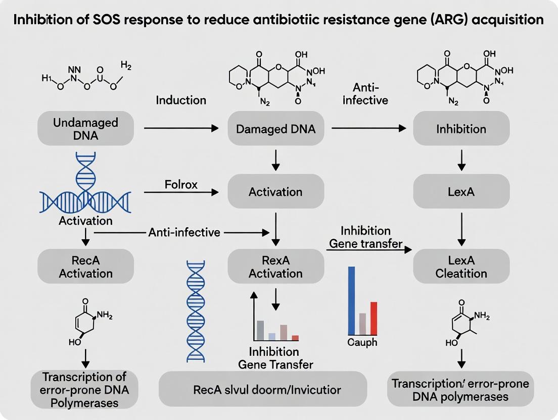
Targeting the SOS Response: A Novel Strategy to Curb Antibiotic Resistance Gene Acquisition in Bacteria
This article provides a comprehensive analysis of the SOS response as a critical bacterial pathway facilitating horizontal acquisition of antibiotic resistance genes (ARGs).
Targeting the SOS Response: A Novel Strategy to Curb Antibiotic Resistance Gene Acquisition in Bacteria
Abstract
This article provides a comprehensive analysis of the SOS response as a critical bacterial pathway facilitating horizontal acquisition of antibiotic resistance genes (ARGs). It explores the foundational molecular mechanisms linking SOS induction to ARG uptake, reviews current methodologies for SOS inhibition using small molecules and genetic tools, addresses common challenges in experimental design and compound efficacy, and validates this approach through comparative analysis with traditional antibiotics and other resistance mitigation strategies. Tailored for researchers and drug development professionals, the review synthesizes recent advances to inform the development of next-generation antimicrobial adjuvants.
The SOS-ARG Nexus: Understanding the Molecular Link Between Bacterial Stress and Resistance Spread
Troubleshooting Guide: SOS Response Inhibition Experiments
Issue 1: Poor SOS Response Induction in Control Cultures
- Problem: Expected upregulation of SOS genes (e.g., recA, umuDC, sulA) is not observed after DNA damage treatment.
- Solution: Verify DNA-damaging agent concentration and exposure time. For Mitomycin C, a typical range is 0.5-2 µg/mL for 30-60 minutes. Check strain integrity; ensure it is not a LexA non-cleavable mutant. Use a positive control like a recA::gfp reporter strain and quantify fluorescence.
- Preventative Step: Perform a kill curve assay for each new batch of DNA-damaging agent to confirm activity.
Issue 2: High Background Cytotoxicity from SOS Inhibitor Compounds
- Problem: Test compounds intended to inhibit the SOS response cause significant bacterial cell death even without DNA damage, confounding results.
- Solution: Titrate compound concentration. Establish a sub-inhibitory concentration (MIC90) for viability assays. Use a viability stain (e.g., propidium iodide) alongside SOS reporter assays to differentiate SOS inhibition from general toxicity.
- Alternative Approach: Switch to a genetically encoded inhibitor (e.g., LexA repressor mutant) as a control to distinguish pharmacological from genetic effects.
Issue 3: Variable Horizontal Gene Transfer (HGT) Assay Results
- Problem: Measurements of plasmid conjugation or transduction efficiency are inconsistent when SOS is inhibited.
- Solution: Standardize donor and recipient cell densities to precise optical density (OD600). For conjugation, typical ratios are 1 donor:10 recipient. Ensure consistent membrane contact time on filters. Include a known SOS-inducing antibiotic (e.g., trimethoprim) in the recipient counterselection to prevent donor overgrowth.
- Protocol Refinement: Perform assays in biological triplicate with internal plasmid copy number controls (qPCR for oriT sequence).
Issue 4: Inefficient LexA Cleavage Assay In Vitro
- Problem: LexA protein is not cleaved by activated RecA (RecA*) in reconstituted biochemical assays.
- Solution: Ensure RecA is properly activated with ATPγS (a non-hydrolyzable ATP analog) and single-stranded DNA (ssDNA) cofactor. A typical reaction uses a 1:5 molar ratio of LexA:RecA. Verify buffer conditions: 20-50 mM Tris-HCl (pH 7.5), 10 mM MgCl₂, 1 mM DTT. Run negative controls without ssDNA or ATPγS.
Frequently Asked Questions (FAQs)
Q1: What are the most reliable transcriptional reporters for quantifying SOS induction in real-time? A: The most common and reliable reporters are based on promoters for sulA (cell division inhibition) or umuDC (error-prone repair) fused to gfp, luciferase, or lacZ. The recA promoter is also used but has a more complex regulation. For drug discovery, a P_sulA-gfp reporter in E. coli provides a strong, dose-dependent signal.
Q2: Which bacterial strains are most relevant for studying SOS inhibition in the context of antibiotic resistance evolution? A: Clinical isolates of Escherichia coli, Klebsiella pneumoniae, and Pseudomonas aeruginosa are highly relevant. Standard lab strains (e.g., E. coli MG1655) are used for foundational mechanism studies. Consider using strains with functional error-prone polymerases (Pol IV, Pol V) for assays related to mutagenesis-induced resistance.
Q3: Beyond RecA-LexA, what other targets are being explored for SOS inhibition? A: Current research targets include:
- Error-Prone Polymerases (Pol V UmuD'₂C): Direct inhibitors to block mutagenic bypass.
- RecA Filament Disruption: Molecules that prevent RecA nucleation on ssDNA or its stabilization.
- LexA Repressor Stabilization: Compounds that prevent LexA cleavage.
- SOS-Regulated Toxin-Antitoxin Systems: Exploiting SOS-induced cellular stress.
Q4: How do I distinguish between general bacterial growth inhibition and specific SOS pathway inhibition? A: Employ a two-tier assay. First, measure growth (OD600) and viability (CFU) with the inhibitor alone. Second, measure SOS gene induction (via reporter) in the presence of a DNA-damaging agent +/- inhibitor. A specific SOS inhibitor will show minimal effect on growth but a significant reduction in the reporter signal post-damage.
Table 1: Common SOS-Inducing Agents and Experimental Parameters
| Inducing Agent | Typical Working Concentration | Exposure Time | Primary DNA Lesion | Key Readout |
|---|---|---|---|---|
| Mitomycin C | 0.5 - 2 µg/mL | 30 - 60 min | Interstrand Crosslink | P_sulA-gfp fluorescence |
| Ciprofloxacin | 10 - 100 ng/mL | 60 - 120 min | Double-Strand Break | P_recA-lacZ activity |
| UV Radiation | 20 - 50 J/m² | N/A (pulse) | Pyrimidine Dimer | Western blot for LexA cleavage |
| Trimethoprim | 10 - 20 µg/mL | 90 - 180 min | Replication Fork Stall | RT-qPCR for umuD |
Table 2: Efficacy of Representative SOS Inhibitors in Reducing HGT
| Inhibitor Class | Example Compound | Target | Reduction in Conjugation (%)* | Reduction in Transduction (%)* |
|---|---|---|---|---|
| RecA Cofactor Competitor | suramin | RecA-ssDNA binding | 60-75% | 20-30% |
| LexA Stabilizer | Peptide aptamers | LexA cleavage site | 40-60% | 10-20% |
| Pol V Inhibitor | TZ39 series | UmuD' interaction | 15-25% | 70-85% |
| Data is model-dependent and approximate. Values represent ranges observed in *E. coli models with specific plasmids/phages.* |
Key Experimental Protocols
Protocol 1: Measuring SOS Inhibition Using a P_sulA-gfp Reporter Assay
- Grow reporter strain to mid-log phase (OD600 ~0.3-0.4) in appropriate medium.
- Aliquot culture into a microplate. Add a sub-inhibitory concentration of the test SOS inhibitor compound. Incubate 15 min.
- Add Mitomycin C to a final concentration of 1 µg/mL. Include controls: no treatment, Mitomycin C only, inhibitor only.
- Monitor GFP fluorescence (ex/em ~485/520 nm) and OD600 in a plate reader for 3-5 hours.
- Calculate the normalized fluorescence (Fluorescence/OD600) and plot over time. The area under the curve (AUC) for the inhibitor + Mitomycin C sample, compared to Mitomycin C alone, quantifies inhibition.
Protocol 2: Conjugation Assay to Assess SOS-Dependent HGT Inhibition
- Prepare donor strain (carrying mobilizable plasmid with antibiotic resistance) and recipient strain (with a different chromosomal resistance) by growing to OD600 ~0.6.
- Mix donor and recipient at a 1:10 ratio in a final volume of 1 mL. Pellet and resuspend in 100 µL of fresh LB.
- Spot mixture onto a 0.22 µm filter placed on non-selective LB agar. Include test SOS inhibitor in the agar.
- Incubate for 2 hours to allow conjugation.
- Resuspend filter in liquid medium, serially dilute, and plate on agar containing antibiotics that select for transconjugants (recipient marker + plasmid marker). Plate controls for donor and recipient viability.
- Calculate conjugation frequency = (Number of transconjugant CFU) / (Number of recipient CFU).
Pathway and Workflow Visualizations
Title: SOS Response Pathway and Link to Antibiotic Resistance
Title: Experimental Workflow for SOS Inhibition Screening
The Scientist's Toolkit: Key Research Reagent Solutions
| Item | Function in SOS Research | Example/Notes |
|---|---|---|
| Mitomycin C | Classic, potent inducer of DNA crosslinks and the SOS response. | Use at low µg/mL range. Handle as a toxic mutagen. |
| Ciprofloxacin | Fluoroquinolone antibiotic that causes DSBs via topoisomerase inhibition; clinically relevant inducer. | Useful for studying SOS in antibiotic treatment contexts. |
| LexA Non-Cleavable Mutant Strain | Genetic control where SOS cannot be induced (e.g., lexA3). | Essential for confirming on-target effects of pharmacological inhibitors. |
| RecA-GFP Fusion Protein | Allows visualization of RecA filament formation in live cells. | Critical for inhibitors targeting RecA nucleation/polymerization. |
| UmuD' Antibody | Detects the cleaved, active form of UmuD (part of Pol V). | Key for assessing error-prone polymerase activation. |
| Mobilizable Plasmid with oriT | Plasmid containing an origin of transfer for conjugation assays. | Required for measuring SOS-dependent horizontal gene transfer. |
| ATPγS | Non-hydrolyzable ATP analog used to activate RecA in vitro. | Stabilizes the RecA* nucleoprotein filament for biochemical assays. |
| SOS Response Reporter Plasmids | Plasmids with SOS promoter (PsulA, PumuDC) driving fluorescent/luminescent reporters. | High-throughput screening for inhibitor compounds. |
Technical Support Center
Troubleshooting Guides & FAQs
Q1: During conjugation inhibition assays, my donor and recipient cells are not forming stable mating pairs, leading to low transfer frequency. What could be wrong? A1: This is often due to suboptimal culture conditions or issues with the pilus. Ensure both strains are in the late exponential growth phase (OD600 ~0.6-0.8). For E. coli, perform mating on solid, pre-warmed LB agar (not in liquid broth) at 37°C for 60-90 minutes. Check the donor strain's pilus functionality using a pilus-specific phage (e.g., M13 for F-pili). If using an SOS response inhibitor (e.g., RecA inhibitor), verify it does not impair bacterial growth at the concentration used, as this can indirectly reduce conjugation.
Q2: In transformation experiments with environmental DNA containing ARGs, my negative controls are showing colonies on selective plates. How do I eliminate this contamination? A2: Contamination in transformation controls typically indicates nuclease degradation of selective antibiotics or carryover of antibiotic resistance. First, prepare fresh antibiotic stocks and plates. Treat your competent cells (e.g., with DNase I) prior to transformation to degrade any contaminating DNA. Implement a "no-DNA" control where you also plate the competent cells without antibiotic selection to check for inherent resistance. For assays focusing on SOS inhibition, ensure your inhibitor compound is dissolved in a vehicle (e.g., DMSO) that does not induce stress or DNA damage responses.
Q3: When measuring transduction rates of ARGs by bacteriophages, I'm getting inconsistent plaque counts between replicates. How can I standardize my phage titer? A3: Inconsistent plaques usually stem from uneven bacterial lawn or variable phage adsorption. Always use a soft agar overlay method with a fresh, mid-log phase recipient culture. For adsorption, include 5mM CaCl₂ or MgCl₂ in your medium to facilitate phage binding. Standardize the phage infection time (e.g., 20 minutes adsorption at 37°C without shaking) before plating. If your research involves SOS inhibitors, note that some may directly impact phage lytic/lysogenic cycles; include a phage-only control with the inhibitor to assess its direct effect on phage viability.
Q4: My assay to measure the impact of an SOS inhibitor on ARG acquisition via all three HGT pathways shows no effect. How can I validate that the SOS response is actually being inhibited? A4: You must include a positive control for SOS inhibition. Co-transform your experimental strain with a reporter plasmid (e.g., pUA66-sulA-gfp) that expresses GFP under control of an SOS-inducible promoter. Treat with a known SOS inducer (e.g., 0.5 µg/mL mitomycin C) with and without your inhibitor. Measure fluorescence (Ex488/Em520) or perform qPCR on key SOS genes (recA, lexA, umuC). A lack of reporter induction confirms SOS inhibition. Ensure your inhibitor is present throughout the HGT assay.
Table 1: Typical Frequencies of Horizontal Gene Transfer Under Standard Lab Conditions
| HGT Mechanism | Donor System | Recipient Strain | Approx. Transfer Frequency (Events/Cell) | Key Influencing Factors |
|---|---|---|---|---|
| Conjugation | E. coli (F⁺ plasmid) | E. coli F⁻ | 10⁻¹ to 10⁻³ | Mating time, temperature, surface, pilus type |
| Transformation | Purified plasmid DNA | B. subtilis competent cells | 10⁻⁵ to 10⁻⁷ | DNA concentration/purity, heat-shock parameters, cell competence state |
| Transduction | P1 phage (lysate) | E. coli | 10⁻⁶ to 10⁻⁸ | Phage MOI, adsorption time, presence of divalent cations |
Table 2: Impact of SOS Response Inhibitors on HGT Frequency of a Model ARG (blaTEM-1)
| SOS Inhibitor (10µM) | Conjugation Frequency (% of Control) | Transformation Efficiency (% of Control) | Transduction Frequency (% of Control) | Reference Strain(s) |
|---|---|---|---|---|
| Control (DMSO) | 100 ± 12 | 100 ± 8 | 100 ± 15 | E. coli MG1655 |
| RecA Inhibitor (e.g., Bisaniline) | 35 ± 7 | 95 ± 10 | 210 ± 25 | E. coli MG1655 |
| LexA Stabilizer | 60 ± 9 | 102 ± 9 | 180 ± 20 | E. coli MG1655 |
| Mechanism Note | Inhibits relaxosome/pilus synthesis? | Minimal effect expected | Potential increase due to phage shift to lytic cycle |
Experimental Protocols
Protocol 1: Standardized Solid-Surface Conjugation Assay with SOS Inhibition Objective: Quantify plasmid-mediated ARG transfer in the presence of an SOS response inhibitor.
- Culture: Grow donor (carrying conjugative plasmid with ARG) and recipient (chromosomal counter-selectable marker, e.g., rpsL for streptomycin resistance) to late exponential phase in LB with appropriate antibiotics.
- Inhibitor Pre-treatment: Add SOS inhibitor (or vehicle control) to both cultures 60 minutes prior to mating.
- Mating: Mix 100 µL of donor and recipient cells. Pellet, resuspend in 50 µL LB, and spot onto pre-warmed, non-selective LB agar plate. Incubate at 37°C for 90 minutes.
- Harvest & Plate: Resuspend mating spot in 1 mL saline, serially dilute, and plate on selective media: a) Donor count (antibiotic for plasmid), b) Recipient count (antibiotic for chromosomal marker), c) Transconjugant count (antibiotics for both plasmid and recipient marker).
- Calculate: Transfer Frequency = (Transconjugant CFU/mL) / (Recipient CFU/mL).
Protocol 2: Natural Transformation Assay with Environmental DNA (eDNA) Extract Objective: Measure acquisition of ARGs from fragmented eDNA by competent bacteria.
- eDNA Extraction: Extract DNA from environmental sample (e.g., wastewater biofilm) using a kit optimized for metagenomic DNA. Fragment by sonication to ~10-20 kb.
- Competent Cell Preparation: Grow recipient strain (e.g., Acinetobacter baylyi ADP1) to OD600 ~0.3. Induce competence by shifting to low-nutrient medium. Treat with SOS inhibitor for 30 minutes.
- Transformation: Incubate 100 ng of eDNA with 200 µL competent cells for 30 minutes at 30°C. Include a no-DNA control and a positive control (known plasmid DNA).
- Selection & Analysis: Plate on selective antibiotic plates. Confirm ARG acquisition via colony PCR on random colonies.
Protocol 3: Phage Transduction Frequency Assay Objective: Determine the rate of ARG transfer via generalized transduction.
- Phage Lysate Prep: Propagate phage (e.g., P1vir) on a donor strain carrying the ARG. Filter sterilize (0.22 µm).
- Titer Determination: Use double-layer agar method to determine phage titer (PFU/mL) on the recipient strain.
- Transduction: Mix recipient cells (OD600=0.5) with phage lysate at an MOI of 0.1. Add CaCl₂ to 5mM. Adsorb for 20 min at 37°C. Add phage antiserum or dilute 10-fold to stop adsorption.
- Selection: Pellet cells, resuspend in saline, and plate on selective media for transductants (antibiotic for ARG). Plate on non-selective media for recipient count.
- Calculate: Transduction Frequency = (Transductant CFU/mL) / (Recipient CFU/mL).
Visualizations
Diagram 1: SOS Response and HGT Interplay
Title: SOS Pathway Promotes Horizontal Gene Transfer
Diagram 2: HGT Inhibition Experiment Workflow
Title: Workflow for Testing SOS Inhibitors on HGT
The Scientist's Toolkit: Research Reagent Solutions
Table 3: Essential Reagents for HGT & SOS Inhibition Research
| Item | Function & Application | Example Product/Catalog |
|---|---|---|
| RecA Inhibitor | Small molecule inhibitor (e.g., Bisaniline) used to directly block RecA nucleoprotein filament formation, preventing SOS induction. | Researcher-synthesized; See PMID: 28392552 |
| Mitomycin C | DNA crosslinking agent; standard positive control for robust induction of the SOS response in Gram-negative and Gram-positive bacteria. | Sigma-Aldrich, M4287 |
| SOS Reporter Plasmid | Plasmid with fluorescent protein (GFP/mCherry) under control of an SOS promoter (sulA, recA, umuDC). Quantifies SOS activity via fluorescence. | Addgene, pUA66 derivatives |
| DNase I, RNase-free | Critical for destroying contaminating free DNA in transformation and transduction control reactions to prevent false positives. | Thermo Fisher, EN0521 |
| Phage P1vir Lysate | Generalized transducing phage for E. coli; essential for standardizing transduction assays and moving ARGs between strains. | ATCC, 25404-B1 |
| Competent Cell Prep Kit | High-efficiency chemically competent cells for transformation assays, ensuring reproducibility in DNA uptake experiments. | Zymo Research, T3001 |
| Synth. Oligopeptide (PI) | Pilus Inhibitor peptide that blocks conjugation by preventing F-pilus assembly; used as a conjugation-specific control. | Custom synthesis (e.g., sequence: NTVGYL... ) |
| qPCR Mix for SOS Genes | SYBR Green master mix with primer sets for recA, lexA, umuC, and a housekeeping gene to quantify SOS induction at transcriptional level. | Bio-Rad, 1725271 |
How SOS Induction Promotes Integrons, Mobile Genetic Elements, and Competence
Troubleshooting & FAQs for SOS Response and ARG Acquisition Experiments
Q1: In my reporter assay (e.g., sulA-gfp), I observe high baseline fluorescence even without an inducing agent. What could be causing this? A: High baseline SOS induction is common. Troubleshoot in this order:
- Check Strain Genotype: Ensure your strain lacks mutations in key SOS repressors (e.g., lexA) or repair genes that cause constitutive SOS expression.
- Assess Media & Growth Conditions: Certain media components or physical stress (e.g., temperature shifts, poor aeration) can induce SOS. Use fresh, pre-warmed media and optimize growth conditions.
- Verify Inducer Purity: Contaminants in your stock solutions may act as indirect inducing agents. Prepare fresh stocks of antibiotics (e.g., ciprofloxacin) or use a known positive control like mitomycin C.
- Rule Out Plasmid Copy Number Effects: If using a plasmid-borne reporter, high copy number can lead to leaky expression. Consider integrating the reporter into the chromosome or using a low-copy vector.
Q2: When testing a putative SOS inhibitor, I see reduced reporter signal, but bacterial growth is also severely inhibited. How do I distinguish specific SOS inhibition from general toxicity? A: This is a critical specificity control. Perform a parallel experiment:
- Measure the minimum inhibitory concentration (MIC) of your compound.
- Conduct the reporter assay at a sub-inhibitory concentration (e.g., 1/4 or 1/2 MIC).
- If SOS inhibition persists without growth defect, it suggests specific activity. Also, test the compound in a ΔrecA strain; a specific SOS inhibitor should have no additional effect in this background, as the SOS pathway is already genetically disabled.
Q3: My conjugation or transformation assay shows high variability when co-treating with DNA-damaging agents and potential inhibitors. How can I improve reproducibility? A: Variability often stems from timing and cell state.
- Synchronize Cultures: Use fresh colonies to inoculate pre-cultures and ensure all experimental cultures are in the same growth phase (mid-log phase is standard).
- Standardize Induction Timing: Pre-treat the donor/recipient cells with the SOS-inducing agent for a defined period (e.g., 30-60 min) before adding the inhibitor or initiating mating/competence. This mimics the physiological sequence of induction then potential inhibition.
- Include Essential Controls: Always include (a) no-treatment, (b) inducer-only, and (c) inhibitor-only controls in every experiment to baseline your measurements.
Q4: I am quantifying integron cassette shuffling via PCR, but my results are inconsistent. What are the key parameters to optimize? A: Cassette excision/reintegration is a low-frequency event.
- Maximize Input DNA: Use a high-quality plasmid or chromosomal prep from a large number of cells (~10^9 CFU).
- PCR Protocol: Use a polymerase with high processivity and fidelity. Increase cycle number (35-40 cycles) and use a touchdown PCR program to improve specificity for low-abundance products.
- Validation: Sequence all PCR products to confirm they represent genuine cassette excision events and not non-specific amplification.
Experimental Protocols
Protocol 1: Quantifying SOS Induction with a Fluorescent Reporter Assay
- Objective: Measure SOS response kinetics upon treatment with DNA-damaging agents and/or inhibitors.
- Materials: Bacterial strain with chromosomally integrated P[sulA]-gfp or similar reporter; 96-well black-walled, clear-bottom plate; plate reader with temperature control and fluorescence capabilities.
- Steps:
- Grow overnight culture in appropriate medium.
- Dilute 1:100 in fresh medium and grow to mid-log phase (OD600 ~0.3-0.5).
- Dispense 180 µL of culture per well.
- Add 20 µL of treatment solutions: vehicle control, SOS inducer (e.g., 0.2 µg/mL mitomycin C), test inhibitor, or inducer + inhibitor.
- Immediately load plate into reader. Measure OD600 and GFP fluorescence (Ex: 485 nm, Em: 520 nm) every 10-15 minutes for 6-8 hours, with orbital shaking before each read.
- Normalize fluorescence to OD600 for each time point. Calculate fold induction relative to the vehicle control at the peak response time.
Protocol 2: Measuring SOS-Mediated Horizontal Gene Transfer (Conjugation)
- Objective: Assess the effect of SOS induction/inhibition on plasmid conjugation frequency.
- Materials: Donor strain (carrying mobilizable plasmid, e.g., RP4), recipient strain (plasmid-free, antibiotic marker distinct from donor), conjugation filters (0.22 µm pore size).
- Steps:
- Grow donor and recipient cultures separately to mid-log phase.
- (Optional Induction/Inhibition): Pre-treat donor culture with SOS inducer and/or inhibitor for 30-60 minutes.
- Mix donor and recipient at a defined ratio (e.g., 1 donor:10 recipient) in a small volume. Pass the mixture through a sterile membrane filter.
- Place the filter, bacteria-side-up, on a non-selective agar plate. Incubate for conjugation (e.g., 37°C for 1-2 hours).
- Resuspend cells from the filter in liquid medium. Plate serial dilutions on selective agar that counts only transconjugants (recipient marker + plasmid marker).
- Plate dilutions on selective agar for donor and recipient counts to determine input numbers.
- Calculate conjugation frequency = (Number of Transconjugants) / (Number of Recipient cells).
Data Presentation
Table 1: Impact of SOS-Inducing Agents on Mobile Genetic Element Transfer Frequencies
| Inducing Agent (Concentration) | Conjugation Frequency (Δlog10) | Natural Transformation Efficiency (Δlog10) | Integron Cassette Excision (Fold Change) | Key Experimental Organism |
|---|---|---|---|---|
| Mitomycin C (0.2 µg/mL) | +2.1 ± 0.3 | +1.8 ± 0.4 | 15.5 ± 3.2 | E. coli, V. cholerae |
| Ciprofloxacin (0.05x MIC) | +3.0 ± 0.5 | +2.5 ± 0.3 | 22.1 ± 4.7 | P. aeruginosa, S. pneumoniae |
| UV Irradiation (20 J/m²) | +1.5 ± 0.4 | +1.2 ± 0.3 | 8.7 ± 2.1 | A. baylyi, E. coli |
| None (Control) | 0.0 ± 0.1 | 0.0 ± 0.1 | 1.0 ± 0.3 | Various |
Table 2: Efficacy of Putative SOS Inhibitors in Reducing ARG Acquisition
| Inhibitor (Class) | Target | SOS Reporter Inhibition (IC50, µM) | Reduction in Conjugation (at 10 µM) | Reduction in Competence (at 10 µM) | Cytotoxicity (Mammalian Cells CC50, µM) |
|---|---|---|---|---|---|
| Zant | RecA | 0.8 ± 0.2 | >99% | 98% | >100 |
| Acetovanillone | LexA | 25.0 ± 5.0 | 75% ± 10% | 65% ± 8% | >200 |
| Aminoglycoside | RecA* | N/D | 40% ± 15% | N/D | >100 |
*Indirect effect via membrane disruption. N/D: Not Determined.
Visualizations
The Scientist's Toolkit: Key Research Reagent Solutions
| Reagent / Material | Primary Function in SOS/ARG Research | Example Product / Note |
|---|---|---|
| Mitomycin C | Classic DNA crosslinker; standard, potent SOS-inducing positive control. | Sigma-Aldrich, M0503. Light-sensitive, prepare fresh. |
| Ciprofloxacin | Fluoroquinolone antibiotic; induces SOS via DNA gyrase inhibition, clinically relevant. | Sigma-Aldrich, 17850. Use at sub-MIC for induction studies. |
| SOS Reporter Strain | Provides quantitative, real-time readout of SOS response. | E. coli MG1655 P[sulA]-gfp (available from Addgene). |
| RecA Inhibitor (Zant) | Small molecule that blocks RecA nucleoprotein filament formation; gold-standard experimental inhibitor. | Tocris Bioscience, 5974. Use DMSO vehicle control. |
| Mobilizable Plasmid (e.g., RP4) | Contains origin of transfer (oriT); used to measure conjugation frequency under SOS conditions. | Standard in HGT studies. Often carries an ARG for selection. |
| Chromosomal Integron Model | Engineered strain with a defined integron platform to study cassette excision/integration dynamics. | e.g., E. coli with pSW integron platform. |
| ΔrecA / ΔlexA Strains | Genetic controls to confirm SOS-specific effects of inducers or inhibitors. | Essential for validating mechanism. |
| Cell-Free Transcription-Translation (TX-TL) System | Reconstitutes SOS pathway components to test inhibitors without cell permeability issues. | PURExpress (NEB) or homemade E. coli extract. |
Technical Support Center
FAQs & Troubleshooting
Q1: In my E. coli ΔrecA strain, I am not observing the expected suppression of ARG acquisition from environmental DNA, even with my SOS inhibitor. What could be wrong? A1: The SOS response is RecA-dependent, but ARG acquisition can occur via RecA-independent pathways. Verify your strain genotype. Check for alternative integron or phage-mediated pathways. Quantify extracellular DNA concentration; high levels can overwhelm suppression. Run a positive control with a known RecA-dependent DNA damaging agent (e.g., mitomycin C) to confirm your inhibitor's efficacy.
Q2: My SOS response inhibitor (e.g., peptide mimic, small molecule) shows high efficacy in vitro but fails in my murine polymicrobial infection model. How can I troubleshoot this? A2: This often indicates pharmacokinetic (PK) challenges. Check:
- Stability: Is the compound degraded in vivo? Use HPLC/MS to measure plasma/tissue concentrations over time.
- Penetration: Does it reach the infection site (e.g., abscess)? Perform bioimaging or homogenize tissue for compound quantification.
- Microbiota Effect: The inhibitor may affect commensals, altering model dynamics. Use 16s rRNA sequencing on control groups.
Q3: When measuring genetic permissiveness via qPCR for integrase or transposase expression, my control samples (no DNA damage) show high background signal. How do I reduce noise? A3: High background suggests basal SOS activation.
- Troubleshoot Culture Conditions: Ensure medium is fresh and not contaminated with trace antibiotics or H₂O₂. Use thorough washing steps before assay.
- Check Strain Integrity: Even lab strains can acquire mutations that increase basal recA or lexA expression. Re-streak from a validated frozen stock.
- Optimize Primers: Ensure they do not amplify genomic regions of paralogous genes. Run a melt curve and gel electrophoresis to confirm single amplicon.
Q4: I am using a PₛᵤₗA-gfp reporter to visualize SOS activation in single cells, but fluorescence is weak and inconsistent after DNA damage induction. A4:
- Confirm Induction: Run a plate reader assay with the same culture to confirm the inducing agent (e.g., ciprofloxacin) is active at your standard concentration.
- Reporter Issues: Check for plasmid loss (include antibiotic). The gfp variant may have poor folding/fluorescence in your host; consider a different variant (e.g., sfGFP).
- Microscopy Settings: Ensure exposure time and gain are set appropriately. Use a positive control strain with a constitutive GFP.
Key Experimental Protocols
Protocol 1: Quantifying ARG Acquisition Frequency via Conjugation or Transformation
- Objective: Measure the frequency of antibiotic resistance gene (ARG) transfer under SOS-inhibited vs. control conditions.
- Method:
- Grow donor (carrying plasmid-borne ARG) and recipient (chromosomally marked, e.g., Rifᴿ) strains to mid-log phase.
- Pre-treat recipient with SOS inhibitor or vehicle control for 30 min. Induce DNA damage in donor with sub-inhibitory ciprofloxacin (0.1x MIC) for 1 hr to activate SOS.
- Mix donor and recipient at a 1:10 ratio on a filter placed on agar. Incubate 2 hrs.
- Resuspend cells, perform serial dilutions, and plate on selective media containing antibiotics for donor count, recipient count, and transconjugant count (selecting for both donor and recipient markers).
- Calculation: Acquisition frequency = (Transconjugant CFU/mL) / (Recipient CFU/mL).
Protocol 2: Assessing Genetic Permissiveness via RT-qPCR of Mobile Genetic Element (MGE) Genes
- Objective: Quantify expression of integrases (intI1) or transposases as a proxy for genetic permissiveness.
- Method:
- Treat bacterial culture with DNA-damaging agent ± SOS inhibitor. Include untreated and inhibitor-only controls.
- At timepoints (e.g., 30, 60, 120 min), collect cells and immediately stabilize RNA (e.g., RNAprotect).
- Extract total RNA, treat with DNase I, and synthesize cDNA using random hexamers.
- Perform qPCR using primers for intI1, tnpA, and housekeeping genes (rpoB, gyrB).
- Analyze using the 2^(-ΔΔCt) method, normalizing to housekeeping genes and the untreated control.
Data Presentation
Table 1: Efficacy of SOS Inhibitor Candidates on ARG Acquisition Frequency
| Inhibitor (Class) | Target | E. coli ARG Acquisition (Conjugation) Frequency (Log Reduction vs. Control) | P. aeruginosa Biofilm-Associated Plasmid Uptake (% Reduction) | Cytotoxicity (Mammalian Cell IC₅₀, μM) |
|---|---|---|---|---|
| Zanthonamide #9 (Natural Product) | RecA-ssDNA filament | -2.7 ± 0.3 | 68% ± 12% | >250 |
| Pep. Mimic A2 (Peptide) | LexA autoproteolysis | -1.8 ± 0.4 | 42% ± 9% | 155 ± 22 |
| Small Molecule X17 (Synthetic) | RecA ATPase | -3.2 ± 0.2* | 81% ± 7%* | 98 ± 15 |
Table Footnote: * p<0.01 vs. other inhibitors in same column. Data from minimum n=3 experiments.
Table 2: Impact of DNA Damage Agents on MGE Gene Expression
| DNA Damage Agent | Concentration | intI1 Expression Fold-Change (ΔΔCt) | tnpA Expression Fold-Change (ΔΔCt) | SOS Reporter Activation (RFU) |
|---|---|---|---|---|
| None (Control) | - | 1.0 ± 0.2 | 1.0 ± 0.3 | 100 ± 15 |
| Ciprofloxacin | 0.05 μg/mL | 12.5 ± 2.1 | 8.7 ± 1.5 | 1850 ± 210 |
| Mitomycin C | 0.5 μg/mL | 22.3 ± 3.8 | 15.2 ± 2.9 | 3200 ± 405 |
| UV Radiation | 10 J/m² | 5.6 ± 1.2 | 4.1 ± 0.8 | 920 ± 130 |
The Scientist's Toolkit
| Research Reagent / Material | Function in SOS/ARG Research |
|---|---|
| SOS Reporter Plasmids (e.g., PₛᵤₗA-gfp/lux) | Visualize or quantify SOS activation in real-time at population or single-cell level. |
| Sub-inhibitory Antibiotics (e.g., Ciprofloxacin, Trimethoprim) | Induce the SOS response via specific DNA damage pathways without killing the entire culture. |
| ΔrecA / ΔlexA Mutant Strains | Essential genetic controls to confirm the SOS-dependence of observed ARG acquisition phenotypes. |
| RecA ATPase Activity Assay Kit (e.g., colorimetric) | Screen and characterize potential SOS inhibitor compounds in vitro. |
| Exogenous DNA (e.g., ARG-bearing plasmid, genomic DNA) | Used as substrate in transformation assays to measure genetic permissiveness directly. |
| SOS Inhibitor Candidates (e.g., Zanthonamide #9, Peptide Mimics) | Pharmacological tools to dissect the SOS-ARG link and potential therapeutic leads. |
Visualizations
Diagram 1: SOS Pathway from DNA Damage to Genetic Permeability
Diagram 2: Key Experiment Workflow for SOS-ARG Research
Evidence Linking SOS Suppression to Reduced Plasmid and Phage-mediated ARG Transfer
Technical Support Center
Welcome to the SOS Suppression Research Technical Hub This center provides troubleshooting guidance and FAQs for experiments investigating the inhibition of the SOS response to curb the acquisition of antibiotic resistance genes (ARGs) via plasmids and bacteriophages.
Frequently Asked Questions (FAQs) & Troubleshooting
Q1: Our SOS response inhibitor (e.g., LexA stabilizer, RecA inhibitor) is not showing a reduction in plasmid conjugation efficiency in our E. coli model. What could be wrong? A: This is a common issue. Please check the following:
- Inhibitor Stability & Concentration: Verify the compound's stability in your growth medium and ensure you are using a concentration that fully suppresses the SOS response without causing general toxicity. Perform a dose-response curve with a known SOS-inducing agent (e.g., mitomycin C) and a reporter gene (e.g., sulA::gfpmut3) to confirm effective suppression.
- Conjugation Conditions: Ensure the mating conditions (time, temperature, cell density ratio of donor and recipient) are optimal for the control (untreated) group. High stress from overcrowding or antibiotic carryover can independently induce SOS.
- Plasmid Type: Confirm your plasmid utilizes an SOS-dependent conjugation mechanism. Some plasmids (e.g., F-type) are less SOS-dependent than others (e.g., some IncI and IncN plasmids). Check the literature for your specific plasmid's regulation.
Q2: During transduction assays, we observe high variance in phage titer between replicates when using SOS-suppressed cells. How can we improve consistency? A: High variance often stems from the lysogeny decision.
- Lytic vs. Lysogenic Phage: Use well-characterized lytic phages (e.g., T4) for consistent infection titers. If studying temperate phages, the frequency of lysogeny increases under SOS suppression, which can drastically alter plaque counts. Monitor lysogen formation explicitly.
- Cell Physiology: Synchronize the growth phase of your bacterial host. The SOS suppression state can sensitively affect receptor expression. Use mid-log phase cells (OD600 ~0.4-0.6) harvested and processed identically for all replicates.
- Inhibitor Pre-treatment: Pre-incubate cells with the SOS inhibitor for at least 30 minutes prior to phage addition to ensure the pathway is fully suppressed.
Q3: How do we distinguish between reduced ARG transfer due to SOS inhibition versus general growth impairment or toxicity from our compound? A: Critical controls are required.
- Growth Curves: Run parallel growth curves in rich media for treated and untreated cells. An OD600 measurement at the end of the conjugation/transduction assay is insufficient.
- Viability Counts: Plate for colony-forming units (CFUs) of donor, recipient, and transconjugant/transductant cells on appropriate selective media to calculate transfer frequency normalized to the recipient count (e.g., transconjugants per recipient).
- SOS-Specific Reporter: Include a positive control using a non-toxic SOS inducer (e.g., sub-inhibitory trimethoprim) with and without your inhibitor to confirm SOS is being modulated independently of growth.
Q4: What are the best methods to quantify SOS activity in real-time during these transfer experiments? A: Fluorescent transcriptional reporters are the gold standard.
- Protocol: Integrate a reporter construct (e.g., PsulA-gfp, PrecA-mCherry) into the chromosome of your model strain. During the co-culture or infection, sample aliquots at intervals.
- Measurement: Measure fluorescence (e.g., GFP: Ex/Em 488/510 nm) via microplate reader or flow cytometry. Normalize fluorescence to OD600. This provides direct, quantitative evidence of SOS suppression correlating with transfer events.
Q5: Our qPCR data for ARG copy number in transconjugants is inconsistent. Any tips? A: This relates to DNA extraction and normalization.
- Normalization Gene: Always normalize the ARG gene to a single-copy chromosomal housekeeping gene (e.g., rpoB, gyrB) from the transconjugant/transductant. This controls for variation in DNA extraction efficiency and cell number.
- Purification: Use a high-fidelity DNA purification kit designed for plasmid DNA to ensure both chromosomal and plasmid DNA are recovered efficiently. Verify DNA quality with a 260/280 ratio (~1.8).
Table 1: Impact of SOS Suppression on Horizontal Gene Transfer Frequencies
| Transfer Mechanism | Experimental Model | SOS Condition | Average Transfer Frequency | Reduction vs. Control | Key Reference Compound |
|---|---|---|---|---|---|
| Plasmid Conjugation | E. coli (IncN plasmid) | Induced (Mitomycin C) | 2.5 x 10⁻² | (Baseline) | - |
| E. coli (IncN plasmid) | Suppressed (Zadarivir) | 4.1 x 10⁻⁴ | ~60-fold | RecA inhibitor | |
| Phage Transduction | Salmonella / P22 phage | Induced (Ciprofloxacin) | 8.7 x 10⁻⁵ | (Baseline) | - |
| Salmonella / P22 phage | Suppressed (LexA peptide) | 1.2 x 10⁻⁶ | ~70-fold | LexA stabilizer | |
| Natural Transformation | A. baylyi (competent) | Induced (DNA damage) | 5.0 x 10⁻³ | (Baseline) | - |
| A. baylyi (competent) | Suppressed (RecA inhibitor) | 3.0 x 10⁻⁴ | ~16-fold | - |
Table 2: Efficacy of Different SOS Inhibitor Classes
| Inhibitor Class | Example Compound | Primary Target | Typical Working Conc. | Pros | Cons |
|---|---|---|---|---|---|
| RecA Inhibitors | Zadarivir | RecA nucleoprotein filament | 10-50 µM | Blocks all SOS functions | Can be bacteriostatic at high doses |
| LexA Stabilizers | Peptide Mimetics | LexA repressor cleavage | Varies (µM-mM) | Highly specific | Poor cell permeability often requires expression vectors |
| Small Molecule SOS Blockers | SPI-1 | Unknown (upstream) | 20 µM | Novel mechanism | Mechanism may be indirect |
Experimental Protocols
Protocol 1: Standard Filter Mating Assay with SOS Modulation Purpose: To quantify plasmid conjugation frequency under SOS-suppressed conditions. Materials: Donor strain (plasmid with selectable ARG, e.g., Ampᴿ), Recipient strain (chromosomal counterselection, e.g., Strᴿ), SOS inhibitor, LB broth, 0.22µm membrane filters, sterile forceps. Steps:
- Grow donor and recipient cultures separately to mid-log phase (OD600 ~0.5).
- Pre-treatment: Add SOS inhibitor or vehicle control to both cultures. Incubate with shaking for 30 min.
- Mating: Mix donor and recipient at a 1:10 ratio on a membrane filter placed on a non-selective LB agar plate. Incubate for 90 min at 37°C.
- Harvesting: Resuspend cells from the filter in sterile saline. Perform serial dilutions.
- Plating: Plate appropriate dilutions on agar selective for: i) donor (Amp), ii) recipient (Str), and iii) transconjugants (Amp + Str).
- Calculation: Incubate plates and count colonies. Transfer Frequency = (Transconjugant CFU/mL) / (Recipient CFU/mL).
Protocol 2: Phage Transduction Assay with SOS Suppression Purpose: To measure the effect of SOS suppression on ARG transfer via generalized transduction. Materials: Donor strain (lysogen or ARG donor), Recipient strain, purified phage stock, SOS inhibitor, CaCl₂/MgSO₄ solution, soft agar. Steps:
- Prepare Recipient: Grow recipient strain to mid-log phase. Pre-treat with SOS inhibitor for 30 min.
- Phage Infection: Mix 100 µL of treated recipient cells with a known MOI (~0.1-1) of phage particles and 1mM CaCl₂. Incubate for 15 min at 37°C without shaking to allow adsorption.
- Outgrowth & Selection: Add the mixture to 3 mL soft agar, pour onto selective agar plates (selecting for the transduced ARG). Alternatively, add antibiotic directly after adsorption, incubate with broth for expression, then plate.
- Titer Control: Perform parallel plaque assays on non-selective plates to determine total infectious phage particles.
- Calculation: Incubate plates. Transduction Frequency = (Transductant CFU) / (Total Plaque-Forming Units added).
Visualizations
Title: SOS Pathway and Inhibitor Action on HGT Promotion
Title: SOS Suppression Conjugation Experiment Workflow
The Scientist's Toolkit: Research Reagent Solutions
Table 3: Essential Reagents for SOS & HGT Inhibition Studies
| Reagent/Material | Function/Application | Example Product/Strain |
|---|---|---|
| RecA Inhibitors | Directly binds RecA to prevent filament formation and LexA cleavage. | Zadarivir (research compound), Nicotinamide (weaker analog). |
| LexA Stabilizer Peptides | Mimics LexA's cleavage site, competing for RecA interaction. | Custom synthetic peptide (sequence: VWQCSM). |
| SOS Reporter Strain | Provides real-time, quantitative readout of SOS activity via fluorescence. | E. coli MG1655 PsulA-gfpmut3 (chromosomal). |
| Model Conjugative Plasmid | Well-characterized plasmid with SOS-regulated transfer machinery. | IncN plasmid R46 or IncI1 plasmid R64. |
| Generalized Transducing Phage | Efficiently packages and transfers random host DNA fragments. | P1vir (for E. coli), P22 (for Salmonella). |
| SOS-Inducing Antibiotic | Positive control for inducing the SOS response at sub-inhibitory levels. | Trimethoprim, Ciprofloxacin, Mitomycin C. |
| Flow Cytometer / Plate Reader | Essential for quantifying SOS reporter fluorescence and normalizing to cell density. | BD Accuri C6, BioTek Synergy H1. |
Inhibiting SOS to Block Resistance: Experimental Strategies and Drug Discovery Pathways
High-Throughput Screening Assays for SOS Response Inhibitors
Technical Support Center
Troubleshooting Guides & FAQs
Q1: Our high-throughput luminescence-based reporter assay (e.g., sulA::lux) shows consistently high background luminescence in negative controls, obscuring signal detection. What could be the cause? A1: High background is frequently caused by contaminating antibiotics or DNA-damaging agents. Ensure your growth media, buffers, and compound libraries are free from trace antibiotics (e.g., quinolones) or chemicals that induce DNA damage. Check for bacterial contamination in your reporter strain stock. Perform a dose-response with a known inducer like Mitomycin C to confirm the dynamic range of your assay. If the issue persists, consider using a reporter with a tighter promoter (e.g., recA::GFP) and ensure adequate washing steps in a microplate format.
Q2: During the fluorescence-based (GFP) reporter assay for lexA repression, we observe poor Z'-factor (<0.5), indicating low assay robustness for HTS. How can we improve it? A2: A low Z'-factor often stems from high well-to-well variability. Key optimizations include:
- Cell Density: Use a precise, low-variance method for inoculating assay plates (e.g., using a multichannel pipette with calibrated tips or a liquid dispenser). Maintain OD600 within a narrow range (e.g., 0.05 ± 0.005).
- Incubation Conditions: Ensure consistent temperature and humidity in your plate incubator/shaker to prevent edge-effect evaporation.
- Data Normalization: Implement dual-normalization: first, to cell density (using a stain like resazurin), then to internal controls on every plate (Positive Control: 1µg/mL Mitomycin C; Negative Control: DMSO vehicle).
- Strain Stability: Use a reporter strain with the construct chromosomally integrated to avoid plasmid loss-related variability.
Q3: In the counter-screening assay for cytotoxicity (e.g., resazurin reduction), our potential SOS inhibitors show reduced fluorescence, suggesting toxicity. How do we distinguish general toxicity from specific SOS inhibition? A3: This is a critical step in the thesis context to identify specific inhibitors that reduce ARG acquisition without killing the bacterium, minimizing selective pressure. Perform parallel assays:
- Specificity Assay: Test compounds in a reporter strain where the lexA box is mutated, making expression constitutive. A true SOS inhibitor will not reduce signal here.
- Time-Course: SOS inhibition should precede growth inhibition. Measure GFP (reporter) and OD600 simultaneously every 30 minutes.
- Check Non-SOS DNA Damage: Use a
ΔrecAstrain. If the compound is toxic here, its mechanism is independent of the SOS pathway. - Alternative Viability Assays: Confirm with a CFU count assay, as some compounds may interfere with resazurin chemistry.
Q4: The β-galactosidase complementation assay (e.g., LexA-F[1], LexA-F[2]) yields inconsistent results upon the addition of candidate inhibitors. What are potential sources of error? A4: This protein-fragment complementation assay is sensitive to conditions affecting protein folding and interaction.
- Compound Interference: Some compounds may denature proteins or inhibit β-galactosidase enzyme activity directly. Run an enzyme activity control with purified β-galactosidase and your compounds.
- Auto-induction: Ensure your growth medium (e.g., LB) does not contain lactose or other inducing sugars. Use a defined medium like M9 with glycerol.
- Lysis Efficiency: Standardize the lysis protocol (time, temperature, and concentration of lysis agents like polymyxin B or chloroform/SDS).
Q5: When performing the essential secondary assay—monitoring RecA filament formation via FRET—we get weak FRET signal changes even with strong inducers. What should we check? A5: FRET assay optimization is technically demanding.
- Protein Labeling: Verify the dye-to-protein ratio for both donor (e.g., Cy3) and acceptor (e.g., Cy5) labeled RecA/ssDNA. Suboptimal ratios severely impact signal.
- ssDNA Co-factor: Ensure the presence of a sufficient concentration of the correct co-factor (e.g., GT-rich oligonucleotide) for RecA nucleation.
- Instrument Calibration: Check for photobleaching and ensure your plate reader's filters are optimal for your FRET pair. Use control proteins with known FRET behavior.
Experimental Protocols
Protocol 1: HTS-Compatible Luminescent Reporter Assay for SOS Induction Objective: Identify compounds that inhibit Mitomycin C-induced SOS response.
- Day 1: Inoculate reporter strain (e.g., E. coli MG1655 sulA::luxCDABE) in LB (no antibiotic) and grow overnight at 37°C, 220 rpm.
- Day 2: a. Dilute culture 1:100 in fresh, pre-warmed LB and grow to mid-log phase (OD600 ~0.5). b. Dilute cells in assay buffer (LB + 0.1% DMSO) to OD600 0.05. c. Using an automated liquid handler, dispense 45 µL of cell suspension into each well of a white, clear-bottom 384-well plate. d. Pin-transfer 100 nL of compound from library (final typical concentration 10 µM) or manually add 5 µL of control solutions (Positive Control: 1µg/mL Mitomycin C; Negative Control: 0.5% DMSO). e. Seal plate, incubate at 37°C with shaking for 90 minutes. f. Measure luminescence on a plate reader (integration time: 500 ms).
Protocol 2: Flow Cytometry-Based GFP Reporter Assay for Hit Validation Objective: Quantitatively validate hits from HTS at single-cell resolution.
- Prepare cells and treat in a 96-well deep-well block as in Protocol 1, but with a final volume of 1 mL.
- After 2 hours of induction with sub-inhibitory Mitomycin C (200 ng/mL) ± inhibitor, add 50 µL of culture to 150 µL of PBS in a U-bottom 96-well plate.
- Analyze immediately on a flow cytometer equipped with a 488 nm laser and 530/30 nm filter.
- Collect at least 10,000 events per sample. Gate on forward/side scatter to exclude debris.
- Data Analysis: Calculate the geometric mean fluorescence intensity (gMFI) of the GFP-positive population. Normalize to the Mitomycin C-only control (100% induction) and DMSO control (0% induction).
Protocol 3: Counter-Screen for Cytotoxicity via Resazurin Reduction Objective: Discard compounds that inhibit SOS response via general bactericidal/bacteriostatic effects.
- Following the GFP assay, add resazurin sodium salt to the remaining culture to a final concentration of 25 µM.
- Incubate the plate at 37°C for 30-60 minutes (protected from light).
- Measure fluorescence (Ex: 560 nm, Em: 590 nm).
- Calculate % viability relative to DMSO-treated cells. Compounds showing <80% viability at the screening concentration should be flagged for dose-response analysis.
Table 1: Performance Metrics of Common SOS Reporter Assays in HTS
| Assay Type | Readout | Approx. Z'-factor | Throughput (wells/day) | Cost per Well | Key Interference |
|---|---|---|---|---|---|
| Luminescence (Promoter::lux) | Light Emission | 0.6 - 0.8 | 10,000+ | Low | Chemical quenchers, background ATP |
| Fluorescence (Promoter::GFP) | Fluorescence Intensity | 0.5 - 0.7 | 5,000 - 8,000 | Medium | Autofluorescent compounds |
| β-Galactosidase Complementation | Absorbance (420 nm) | 0.4 - 0.6 | 3,000 - 5,000 | Medium-High | Enzyme inhibitors, lysis variability |
| FRET (RecA filamentation) | FRET Ratio | 0.3 - 0.5 | 1,000 - 2,000 | High | Compound fluorescence, protein stability |
Table 2: Example Hit Characterization Data from a Thesis Study
| Compound ID | Primary Screen (% Inhibition) | IC50 (SOS Inhibition) | CC50 (Cytotoxicity) | Selectivity Index (CC50/IC50) | ΔrecA Toxicity (Y/N) |
Effect on Conjugative ARG Transfer* |
|---|---|---|---|---|---|---|
| SOSI-001 | 95% | 1.2 µM | >50 µM | >41 | N | 75% Reduction |
| SOSI-002 | 88% | 5.5 µM | 12 µM | 2.2 | Y | 30% Reduction |
| SOSI-003 | 78% | 8.1 µM | >100 µM | >12 | N | 65% Reduction |
| Mitomycin C | N/A (Inducer) | N/A | 0.05 µM | N/A | N/A | 300% Increase |
Measured in an *E. coli conjugation model with a plasmid encoding β-lactamase.
Diagrams
Diagram 1: SOS Response Pathway and Inhibitor Targets
Diagram 2: HTS Workflow for SOS Inhibitor Discovery
The Scientist's Toolkit: Research Reagent Solutions
Table 3: Essential Materials for SOS Response HTS Assays
| Item | Function & Rationale | Example Product/Catalog # |
|---|---|---|
| Reporter Strain | Genetically engineered bacteria where an SOS-responsive promoter drives a reporter gene (lux, gfp). Essential for primary screening. | E. coli DPD1715 (recA::luxCDABE) or in-house sulA::GFP construct. |
| Inducing Agent (Positive Control) | DNA-damaging agent to fully induce the SOS response for assay validation and normalization. | Mitomycin C (Sigma, M4287) or Ciprofloxacin (Sigma, 17850). |
| HTS-Compatible LexA Protease Assay Kit | For secondary biochemical confirmation of inhibitors targeting LexA cleavage. | Not commercially ubiquitous; often in-house FRET or fluorescence polarization assays using purified LexA and RecA*ssDNA. |
| Resazurin Sodium Salt | Cell-permeant dye used for viability counter-screening. Reduction to fluorescent resorufin indicates metabolic activity. | AlamarBlue Cell Viability Reagent (Thermo Fisher, DAL1025). |
| 384-Well, White, Solid-Bottom Microplates | Optimal for luminescence assays, minimizing cross-talk and maximizing signal capture. | Corning 3570 or Greiner 781074. |
| Fluorophore-Labeled RecA & ssDNA | Key reagents for the FRET-based RecA filamentation assay (critical secondary assay). | Purified RecA labeled with Cy3/Cy5 (e.g., Cytiva) + complementary GT-rich oligonucleotide. |
ΔrecA Isogenic Strain |
Essential control strain to determine if compound toxicity is SOS-dependent or general. | Construct via lambda-red recombination or obtain from KEIO collection. |
| Conjugation-Proficient Strain Pair | For final thesis-context assay: donor (with ARG plasmid) and recipient (SOS reporter) to measure impact of inhibitors on horizontal gene transfer. | E. coli HB101 (RP4 plasmid) & MG1655 rifR recipient. |
Technical Support Center
Troubleshooting Guide: SOS Inhibition Experiments
Q1: My assay shows no reduction in LexA cleavage despite adding a putative RecA inhibitor. What could be wrong? A: This could indicate an issue with inhibitor specificity, stability, or assay conditions.
- Check 1: Inhibitor Stability. Many RecA inhibitors are nucleoside analogs or small molecules susceptible to degradation. Prepare fresh stock solutions in recommended solvent (e.g., DMSO) and confirm storage conditions (-20°C or -80°C, under nitrogen if recommended).
- Check 2: Assay Buffer Conditions. RecA's nucleoprotein filament formation and co-protease activity are Mg²⁺-dependent. Ensure your reaction buffer contains the optimal 5-10 mM MgCl₂. Also, verify the pH is maintained at 7.5-8.0.
- Check 3: Positive Control Failure. Include a known RecA ATPase inhibitor (e.g., ATPγS, a non-hydrolyzable ATP analog) as a control. If this also fails, your LexA cleavage assay protocol may need optimization.
Q2: I am using an upstream signal inhibitor (e.g., ROS scavenger, DNA damage preventer), but bacterial viability is drastically reduced, confounding my antibiotic resistance acquisition results. How do I proceed? A: This is a common pitfall when targeting upstream pathways critical for general metabolism.
- Solution 1: Dose-Response Titration. Perform a thorough titration to find a sub-inhibitory concentration (sub-MIC) that dampens the SOS signal without affecting growth. Use the table below as a starting guide for common agents.
| Upstream Inhibitor | Typical Working Range | Critical Consideration |
|---|---|---|
| Thiourea (ROS Scavenger) | 10-50 mM | Can be toxic at >50 mM; monitor OD600 closely. |
| Curcumin (ROS/RecA modulator) | 20-100 µM | Poor aqueous solubility; use DMSO carrier <0.5% v/v. |
| Ciprofloxacin (Control, induces SOS) | 0.01-0.1 µg/mL | Use sub-MIC to induce SOS without killing. |
- Solution 2: Use a Time-Delayed Addition. Add the upstream inhibitor after bacteria have entered log-phase growth, but before the primary SOS-inducing agent (e.g., antibiotic).
Q3: My qPCR data for SOS gene expression (e.g., recA, lexA, umuDC) is inconsistent when testing indirect inhibitors. What are key validation steps? A: Inconsistency often stems from inadequate SOS induction normalization or RNA quality.
- Step 1: Validate Induction Level. Always include a no-inhibitor, SOS-induced control (e.g., + 0.05 µg/mL Ciprofloxacin). The fold-change in recA or umuDC expression should be significant (typically 5-50 fold) over the uninduced baseline.
- Step 2: Normalize to Multiple Housekeeping Genes. Use at least two stable genes (e.g., rpoD, gapA) for normalization, as some treatments may affect common single references like 16S rRNA.
- Step 3: Correlate with Functional Phenotype. Support qPCR data with a functional SOS reporter assay (e.g., Chromosomal sfiA::lacZ fusion). See protocol below.
Frequently Asked Questions (FAQs)
Q: What is the most definitive assay to confirm a direct vs. indirect mode of action for an SOS inhibitor? A: A biochemical LexA cleavage assay using purified components is definitive for direct RecA targeting.
- Incubate purified RecA protein (1 µM) with single-stranded DNA (ssDNA, e.g., dT30, 3 µM) and ATP (1 mM) in buffer (25 mM Tris-HCl pH 7.5, 10 mM MgCl₂, 1 mM DTT) for 10 min at 37°C to form the active RecA* filament.
- Add purified LexA protein (1 µM) and the inhibitor. Include DMSO-only and no-RecA controls.
- Stop the reaction after 60 min with SDS-PAGE loading buffer.
- Analyze by SDS-PAGE (15% gel) stained with Coomassie Blue. Direct RecA inhibitors will prevent LexA cleavage, visualized by the persistence of the full-length LexA band (~22 kDa) and reduction of the cleaved fragment (~13 kDa).
Q: Which approach—direct or indirect inhibition—is more promising for preventing antibiotic resistance gene (ARG) acquisition without driving resistance to the inhibitor itself? A: Current research suggests combinations are most promising. Direct RecA inhibitors apply strong selective pressure for mutational resistance. Indirect inhibitors (e.g., preventing DNA damage via ROS scavengers) may have weaker selective pressure but can be less specific. The leading strategy is to combine a sub-effective dose of a direct RecA inhibitor with a standard antibiotic, reducing the chance for resistance to either agent while potently blocking horizontal ARG acquisition via conjugation or transduction, which are SOS-dependent.
Detailed Experimental Protocol: β-Galactosidase Reporter Assay for SOS Inhibition
Objective: Quantify SOS response activity in E. coli containing an SOS promoter (e.g., sulA or recA) fused to lacZ in the presence of potential inhibitors.
Methodology:
- Strain & Growth: Inoculate reporter strain (e.g., E. coli MG1655 sfiA::lacZ) in LB + appropriate antibiotic. Grow overnight.
- Sub-culture: Dilute 1:100 in fresh LB (no antibiotic) and grow to mid-log phase (OD600 ~0.3-0.4).
- Treatment: Aliquot cells into tubes.
- A: Uninduced control (No treatment)
- B: SOS-Induced control (Add 0.05 µg/mL Ciprofloxacin)
- C: Inducer + Inhibitor (Add Ciprofloxacin + your compound)
- D: Inhibitor-only control (Add your compound only)
- Incubation: Shake at 37°C for 2 hours.
- Assay: a. Measure OD600 of each culture. b. Lyse cells with SDS and chloroform. c. Mix lysate with Z-buffer (Na₂HPO₄, NaH₂PO₄, KCl, MgSO₄, β-mercaptoethanol). d. Start reaction with ONPG (2-Nitrophenyl β-D-galactopyranoside) substrate. e. Stop with Na₂CO₃ when yellow color develops (~30 min). f. Measure absorbance at 420 nm and 550 nm.
- Calculation: Miller Units = (1000 * [A420 - (1.75 * A550)]) / (Time in min * Volume in ml * OD600).
The Scientist's Toolkit: Research Reagent Solutions
| Reagent/Material | Function/Application | Key Detail |
|---|---|---|
| Purified RecA Protein | In vitro cleavage assays, ATPase assays. | Ensure it is >95% pure and stored in aliquots at -80°C to prevent loss of activity. |
| Purified LexA Protein | Substrate for in vitro cleavage assays. | Can be His-tagged for purification; confirm intactness via gel before use. |
| ATPγS (Adenosine 5′-[γ-thio]triphosphate) | Non-hydrolyzable ATP analog; positive control RecA inhibitor. | Competes with ATP, blocking RecA filament activation. |
| Ciprofloxacin | Standard SOS response inducer (via DNA gyrase inhibition). | Use at sub-MIC (typically 0.01-0.1x MIC) to induce without bactericidal effect. |
| ONPG (o-Nitrophenyl-β-galactoside) | Colorimetric substrate for β-galactosidase in reporter assays. | Prepare fresh in Z-buffer or store aliquots at -20°C protected from light. |
| SOS Reporter Strain (e.g., sfiA::lacZ) | In vivo monitoring of SOS promoter activity. | Allows functional, high-throughput screening of inhibitors. |
Visualizations
Diagram 1: SOS Pathway & Inhibitor Targets
Diagram 2: Inhibitor Screening Workflow
Troubleshooting Guides & FAQs
Q1: My SOS response inhibition assay using a novel small molecule shows high background fluorescence in the reporter strain. What could be the cause? A: High background is often due to compound autofluorescence or cytotoxicity. First, measure the fluorescence of your compound in the growth medium without cells. If autofluorescent, consider switching to a luminescent reporter (e.g., lux operon). If autofluorescence is ruled out, perform a viability assay (e.g., CFU count) at your working concentration. Unintended cytotoxicity can induce stress responses, elevating background. Titrate the compound to find a sub-inhibitory concentration.
Q2: When screening natural product extracts for SOS inhibition, I observe high rates of false positives in the initial phenotypic assay. How can I prioritize hits? A: False positives from crude extracts are common due to off-target effects, general toxicity, or assay interference. Implement a tiered counter-screening workflow:
- Confirmatory Assay: Use a secondary, orthogonal assay (e.g., qPCR of key SOS genes recA, lexA, sulA).
- Specificity Check: Test hits against a reporter for an unrelated stress pathway (e.g., heat shock, oxidative stress).
- Cytotoxicity Filter: Determine minimum inhibitory concentration (MIC) and compare to SOS inhibitory concentration (IC50). Prioritize compounds with a selectivity index (MIC/IC50) >10.
- Rapid Fractionation: Early fractionation of active extracts can identify the active chemotype and reduce nuisance compounds.
Q3: My designed peptide inhibitor of RecA-ssDNA filament formation shows poor cell permeability in Gram-negative bacterial models. What strategies can I employ? A: Peptide permeability in Gram-negatives is hindered by the outer membrane. Consider these modifications:
- Sequence Optimization: Incorporate arginine-rich or hydrophobic motifs (e.g., RWWR).
- Conjugation: Link to cell-penetrating peptides (e.g., (KFF)3K) or siderophore mimics (e.g., enterobactin analogs) for active transport.
- Chemical Modification: Use D-amino acids, N-methylation, or peptidomimetic backbones (e.g., β-peptides) to enhance stability and uptake.
- Delivery System: Co-administer with permeabilizers like polymyxin B nonapeptide (PMBN) at sub-lethal doses, though this may complicate therapeutic translation.
Q4: In a conjugation assay, my lead SOS inhibitor reduces plasmid transfer frequency but not to the desired level. Should I consider combination therapy? A: Yes, combination therapy is a promising strategy. The SOS response is a key but not the sole pathway influencing ARG acquisition. Consider combining your SOS inhibitor with:
- An agent that disrupts the mating pair formation (e.g., a pilus inhibitor).
- An anti-plasmid compound (e.g., apramycin) that interferes with plasmid replication/maintenance in the recipient.
- A standard antibiotic to reduce the donor population, thereby lowering conjugation opportunities. Always perform checkerboard assays to evaluate synergy (FIC Index).
Q5: How do I differentiate between a true SOS inhibitor and a DNA-damaging agent that indirectly activates the SOS response in my screening setup? A: This is a critical distinction. A true inhibitor should reduce SOS signal induced by a known DNA-damaging agent (e.g., mitomycin C, ciprofloxacin). Your assay should include these controls:
- Compound alone (to check for intrinsic genotoxicity).
- DNA-damaging agent alone (positive control for SOS induction).
- DNA-damaging agent + compound (test for inhibition of induction). A true inhibitor will show low signal in control 1 and a significantly reduced signal in test 3 compared to control 2. A DNA-damager will show high signal in both controls 1 and 3.
Experimental Protocols
Protocol 1: Primary Screening for SOS Inhibition Using a Fluorescent Reporter Strain Principle: Measure reduction of SOS-induced fluorescence in E. coli MG1655 pUA66-PsulA-gfp upon co-treatment with DNA-damaging agent and test compound. Materials: Reporter strain, LB medium, mitomycin C (MMC), test compounds, black clear-bottom 96-well plates, plate reader. Procedure:
- Grow reporter strain overnight in LB with selective antibiotic.
- Dilute culture 1:100 in fresh LB and dispense 90 µL/well into plate.
- Add 5 µL of test compound (or DMSO control) to appropriate wells.
- Incubate plate at 37°C with shaking for 1 hour.
- Add 5 µL of MMC (final conc. 0.5 µg/mL) or water to induce SOS.
- Incubate for 3-4 hours at 37°C with shaking.
- Measure OD600 and fluorescence (Ex/Em: 485/535 nm).
- Calculate: % Inhibition = [1 - (Ftest/ODtest)/(FMMC/ODMMC)] * 100.
Protocol 2: Monitoring Plasmid Conjugation Frequency in the Presence of SOS Inhibitors Principle: Matting assay between donor (plasmid-carrying) and recipient strains with/without inhibitor. Materials: Donor strain (e.g., E. coli with RP4 plasmid, Sm^R), Recipient strain (e.g., E. coli NaI^R), LB, selective agar plates (with antibiotics for donor, recipient, and transconjugants). Procedure:
- Grow donor and recipient strains separately to mid-log phase (OD600 ~0.6).
- Mix donor and recipient at a 1:1 ratio (by volume). Pellet and resuspend in 1/10 volume LB to increase cell contact.
- Aliquot the cell mixture. Add SOS inhibitor or vehicle control.
- Spot 50 µL of each mixture on pre-warmed LB agar plates. Incubate upright for 6-8 hours at 37°C (conjugation period).
- Resuspend spots in 1 mL LB, perform serial dilutions, and plate on:
- Donor-selective plates (counts donor CFU).
- Recipient-selective plates (counts recipient CFU).
- Transconjugant-selective plates (counts transconjugant CFU).
- Calculate: Conjugation Frequency = (Transconjugant CFU/mL) / (Recipient CFU/mL). Report fold-change relative to vehicle control.
Data Presentation
Table 1: Comparative Profile of Leading Compound Classes for SOS Inhibition
| Parameter | Small Molecules | Peptides | Natural Products |
|---|---|---|---|
| Typical MW (Da) | 200-500 | 500-3000 | 200-2000 |
| Primary Target | LexA autocleavage, RecA ATPase | RecA-ssDNA filament, Protein-protein interfaces | Diverse (often unknown) |
| Cell Permeability | Generally good | Poor (requires optimization) | Variable |
| Chemical Tractability | High (easy to modify) | Moderate to High | Low (requires isolation/deconvolution) |
| Typical IC50 (SOS Repression) | 1-50 µM | 0.1-10 µM (in vitro) | 1-100 µg/mL (crude extract) |
| Key Advantage | Oral bioavailability, drug-like | High specificity, novel interfaces | Structural novelty, evolved bioactivity |
| Key Challenge | Target specificity | Proteolytic stability, delivery | Supply, compound identification |
Table 2: Impact of Model SOS Inhibitors on Plasmid Conjugation Frequency
| Inhibitor (Class) | Target | Conjugation Frequency (Δ vs Control) | MIC (µg/mL) | Selectivity Index (MIC/IC50) |
|---|---|---|---|---|
| Z16 (Small Molecule) | RecA | 10^-4 (100-fold ↓) | >64 | >32 |
| Pep-11 (Peptide) | RecA-ssDNA binding | 10^-3 (10-fold ↓) | 128* | 12 |
| Myxopyronin B (Nat. Prod.) | RNA Polymerase (indirect) | 10^-2 (2-fold ↓) | 2 | 0.5 |
| Ciprofloxacin Control | DNA Gyrase (induces SOS) | 10^-1 (10-fold ↑) | 0.03 | N/A |
*Peptide MIC is often a poor indicator due to permeability issues; value shown is for a permeabilized strain.
Diagrams
SOS Inhibition & Conjugation Blockade
SOS Inhibitor Screening Workflow
The Scientist's Toolkit: Research Reagent Solutions
| Item | Function & Application |
|---|---|
| E. coli MG1655 pUA66-PsulA-gfp | Reporter strain for SOS response. GFP expression driven by SOS-regulated sulA promoter. Used for primary compound screening. |
| Mitomycin C (MMC) | DNA cross-linking agent. Standard positive control for inducing the SOS response at sub-lethal concentrations (e.g., 0.1-1 µg/mL). |
| Polymyxin B Nonapeptide (PMBN) | Outer membrane permeabilizer. Used in peptide studies to enhance Gram-negative bacterial uptake without significant bactericidal activity. |
| RecA Protein (Purified) | Essential for in vitro assays: ATPase activity, LexA cleavage, or filament formation studies (e.g., using fluorescence anisotropy). |
| LexA Cleavage Assay Kit | Commercial kit monitoring LexA autocleavage in vitro. Allows rapid characterization of inhibitor mechanism. |
| RP4 or R388 Conjugative Plasmid | Model broad-host-range plasmid for standardized conjugation assays to measure horizontal transfer of ARGs. |
| Ciprofloxacin | Fluoroquinolone antibiotic. Used as an SOS-inducing control and to model conditions that promote ARG acquisition in experiments. |
Genetic Knockdown and CRISPRi Approaches for Target Validation
Troubleshooting Guides & FAQs
Common Issues with Genetic Knockdown (siRNA/shRNA)
Q1: My siRNA transfection yields high cell mortality (>40%) in my E. coli or bacterial co-culture model. How can I optimize delivery? A: High mortality often stems from transfection reagent toxicity or excessive siRNA concentration. For bacterial models involving mammalian cells (e.g., in infection studies), consider:
- Optimize Reagent: Use lipid-based reagents specifically formulated for your cell type. For immune cells or primary macrophages, electroporation may be superior.
- Dose Titration: Perform a matrix experiment titrating both siRNA (e.g., 10-50 nM) and transfection reagent volume. Use a non-targeting siRNA control.
- Timing: For SOS response studies, transfect cells before adding the antibiotic or DNA-damaging agent to induce SOS. Allow 24-48 hours for knockdown.
Q2: I observe inconsistent knockdown efficiency between replicates when targeting SOS pathway genes (e.g., recA, lexA). What are the key variables to control? A: Inconsistency commonly arises from:
- Cell Confluence: Transfect at a consistent, optimal confluence (e.g., 60-70%).
- Bacterial Contamination: In co-culture models, ensure antibiotics in the media do not interfere with transfection. Use validated, sterile techniques.
- Reagent Handling: Thaw siRNA aliquots on ice and vortex transfection reagents before use.
- Validation: Always include a positive control siRNA (e.g., targeting a housekeeping gene) and confirm knockdown via qPCR (for lexA/recA mRNA) and, if possible, western blot (for LexA cleavage).
Q3: My negative control siRNA appears to affect ARG acquisition rates. What could be the cause? A: This indicates off-target effects or immune activation.
- Sequence Review: Re-blast the control sequence. Use a scrambled control with verified minimal genome homology.
- Innate Immunity: Some siRNA sequences can activate TLRs. Use controls from vendors that are certified for low immune stimulation.
- Experimental Design: Include an untreated control (mock transfection) and a transfection reagent-only control to isolate effects.
Common Issues with CRISPRi (dCas9)
Q4: My CRISPRi repression of recA is insufficient to reduce conjugation frequency. How can I enhance silencing? A: Inadequate repression can be due to guide RNA (gRNA) design or dCas9 expression.
- gRNA Positioning: For CRISPRi in bacteria, design gRNAs to target the non-template strand within ~50 bp downstream of the transcription start site (TSS) for optimal steric inhibition.
- Multiplexing: Use multiple gRNAs targeting the same gene (recA) simultaneously to achieve stronger repression.
- Promoter Strength: Ensure strong, constitutive expression of the dCas9 protein (e.g., using anhydrotetracycline-inducible or constitutive promoter). Verify dCas9 expression via fluorescence or western blot.
- Table 1: Example Titration for Optimizing recA CRISPRi Repression
Parameter Test Range Optimal Value for Our System Purpose Inducer (aTc) Concentration 0-100 ng/mL 50 ng/mL To modulate dCas9 expression gRNA Number (Multiplex) 1, 2, or 3 guides 2 guides To enhance repression efficiency Time of Induction Pre-Experiment 2-16 hours 6 hours For full dCas9 accumulation
Q5: I see variation in ARG acquisition inhibition across my bacterial colonies after CRISPRi treatment. How do I ensure a homogenous population? A: Clonal variation suggests unstable plasmid maintenance or inconsistent gRNA expression.
- Selection Pressure: Maintain appropriate antibiotics for both the dCas9 and gRNA plasmids throughout culture and during the conjugation assay.
- Single-Cell Cloning: Isolate single colonies and screen for strong repression phenotype (e.g., via sensitivity to DNA-damaging agents) to create a uniform working stock.
- Use Integrated Systems: Consider using strains with chromosomal, stable integrations of the dCas9 and gRNA constructs to avoid plasmid loss.
Q6: How do I design a proper control for my CRISPRi experiment targeting SOS genes? A: Rigorous controls are essential.
- Non-Targeting gRNA Control: Use a gRNA with no target in the bacterial genome.
- "Dead" dCas9 Control (Critical): Express an inactive dCas9 (lacking binding ability) with the targeting gRNA. This controls for any effects of gRNA binding/expression alone.
- Rescue Experiment: If possible, express a CRISPRi-resistant, wild-type copy of the target gene (e.g., recA) from an inducible plasmid to confirm phenotype specificity.
Detailed Experimental Protocols
Protocol 1: Validating SOS Gene Knockdown via siRNA in a Macrophage-Bacteria Co-culture Model
Objective: To knock down a human gene involved in bacterial internalization (e.g., CTSB) and measure subsequent effects on E. coli SOS induction and ARG acquisition via conjugation.
Day 1: Cell Seeding
- Seed THP-1 derived macrophages in 12-well plates at 2.5 x 10^5 cells/well in antibiotic-free media. Differentiate with PMA if required.
Day 2: Transfection
- For each well, dilute 5 µL of 10 µM siRNA (targeting CTSB or non-targeting control) in 100 µL Opti-MEM.
- In a separate tube, dilute 3 µL of lipid transfection reagent in 100 µL Opti-MEM. Incubate 5 min.
- Combine diluted siRNA and transfection reagent. Incubate 20 min at RT.
- Add the 200 µL complex dropwise to cells. Gently swirl. Incubate cells at 37°C for 24-48h.
Day 4: Infection & Conjugation Assay
- Prepare donor E. coli (carrying ARG plasmid, e.g., pBTR) and recipient E. coli (chromosomal resistance marker) in mid-log phase.
- Induce SOS in donor by adding sub-inhibitory ciprofloxacin (0.1x MIC) for 1h.
- Wash macrophages, add antibiotic-treated donor and recipient bacteria at an MOI of 10:10 (donor:recipient). Centrifuge to co-localize.
- Co-culture for 90 min. Lyse macrophages with 0.1% Triton X-100.
- Plate serial dilutions on selective agar to quantify donor, recipient, and transconjugant (ARG-acquired) colonies.
Validation:
- In parallel wells, extract RNA post-transfection. Perform qPCR to confirm CTSB knockdown.
Protocol 2: CRISPRi-MediatedrecARepression and Conjugation Frequency Measurement
Objective: To repress recA in donor E. coli and measure its impact on plasmid conjugation frequency.
Strain and Plasmid Preparation:
- Use E. coli donor strain harboring: i) a conjugative plasmid (e.g., F-plasmid with ARG), and ii) a CRISPRi plasmid (dCas9 + recA-targeting gRNA under inducible promoter).
- Maintain recipient strain with a different chromosomal resistance marker.
Induction of CRISPRi:
- Grow donor and recipient cultures separately to OD600 ~0.3.
- Add anhydrotetracycline (aTc, 50 ng/mL final) to the donor culture to induce dCas9/gRNA expression. Incubate for 2-3 hours.
Conjugation Assay (Liquid Mating):
- Mix induced donor and recipient at a 1:1 ratio (by volume, typically 1 mL each). Pellet, resuspend in 50 µL LB.
- Spot mixture on a pre-warmed LB agar plate. Incubate for 1 hour at 37°C.
- Resuspend spot in 1 mL LB, perform serial dilutions, and plate on selective agar that counts only donors (D), recipients (R), and transconjugants (T).
- Frequency Calculation: Conjugation Frequency = (T / D) OR (T / R). Report as mean ± SD from ≥3 biological replicates.
Repression Validation:
- From the induced donor culture pre-mating, extract RNA and perform qRT-PCR for recA mRNA levels, normalized to a housekeeping gene (e.g., rpoD).
Pathway & Workflow Diagrams
Title: SOS Pathway & Genetic Intervention Points for ARG Acquisition
Title: CRISPRi Experimental Workflow for Conjugation Assay
The Scientist's Toolkit: Research Reagent Solutions
| Item | Function/Application in SOS/ARG Research | Example/Vendor (for informational purposes) |
|---|---|---|
| Lipid-based Transfection Reagents | Deliver siRNA/shRNA into mammalian cells in co-culture models. | Lipofectamine RNAiMAX, Dharmacon DharmaFECT |
| Validated siRNA Pools | Target human host factors or bacterial genes (in permeable models). Ensure specificity. | ON-TARGETplus siRNA (Horizon), Silencer Select (Thermo) |
| CRISPRi Plasmid Systems | All-in-one vectors expressing dCas9 and gRNA for bacterial repression. | pCRISPRi (Addgene #84832), pdCas9-bacteria (Addgene #44249) |
| Anhydrotetracycline (aTc) | Tight, dose-dependent inducer for dCas9 expression in common bacterial CRISPRi systems. | Clontech, Sigma-Aldrich |
| qPCR Master Mix with Reverse Transcription | Quantify knockdown/repression efficiency of SOS genes (recA, lexA). | Power SYBR Green Cells-to-Ct Kit (Thermo), iTaq Universal SYBR (Bio-Rad) |
| Selective Agar & Antibiotics | For plating conjugation assays to distinguish donors, recipients, and transconjugants. | LB Agar + specific antibiotics (e.g., Kanamycin, Chloramphenicol, Streptomycin) |
| DNA Damaging Agent (Positive Control) | Induce SOS response reliably in control experiments. | Ciprofloxacin (fluoroquinolone), Mitomycin C |
| Fluorescent dCas9 Fusion Protein | Visualize dCas9 localization and estimate expression levels in bacterial populations. | pdCas9-GFP plasmids |
Technical Support Center
Troubleshooting Guide: Common SOS Inhibition Assay Issues
Q1: In my SOS response reporter assay (e.g., sulA-gfp), I am not observing the expected reduction in fluorescence when co-administering an antibiotic with the SOS inhibitor. What could be wrong?
- A: This is a common issue. Follow this checklist:
- Check Inhibitor Solubility & Stability: Ensure the SOS inhibitor (e.g., Mitomycin C analog, RecA inhibitor) is correctly dissolved in the recommended solvent (often DMSO). Prepare fresh stock solutions if degradation is suspected. High DMSO concentrations (>1% v/v) can affect bacterial growth and stress responses.
- Verify Antibiotic Sub-inhibitory Concentration: The antibiotic dose must be truly sub-inhibitory (Sub-MIC) to induce the SOS response without causing significant cell death before measurement. Re-run a dose-response curve (MIC assay) for your specific bacterial strain and growth conditions.
- Confirm Reporter System Functionality: Induce the reporter system with a known SOS inducer (e.g., 0.5 µg/mL Mitomycin C) alone to verify the GFP/bioluminescence signal increases. If it does not, the reporter strain may have issues.
- Timing of Addition: The SOS inhibitor may need to be added 30-60 minutes prior to antibiotic addition to allow for adequate cellular uptake and target engagement.
Q2: During checkerboard synergy testing, how do I interpret the results when the Fractional Inhibitory Concentration Index (FICi) is borderline?
- A: An FICi ≤ 0.5 indicates synergy, >0.5 to ≤4 indicates indifference, and >4 indicates antagonism. For borderline results (e.g., FICi = 0.56):
- Repeat the Assay: Perform at least three independent biological replicates. Calculate the mean and standard deviation.
- Use a Complementary Assay: Confirm synergy with a time-kill curve analysis. Collect samples over 24 hours (e.g., 0, 2, 4, 8, 24h) to see if the combination causes a ≥2-log10 reduction in CFU/mL compared to the most active single agent.
- Check for Heteroresistance: Sub-populations with higher MICs can skew results. Consider running population analysis profiles (PAP).
Q3: My experiment to measure horizontal gene transfer (HGT) frequency shows high variability between replicates. How can I improve consistency?
- A: HGT assays (conjugation, transduction, transformation) are sensitive. Key controls and optimizations:
- Strain Preparation: Use donor and recipient strains from fresh, single-colony inoculates grown under non-selective conditions to ensure optimal cell viability and pilus/competence expression.
- Mating Conditions: For conjugation, keep mating conditions (time, temperature, medium) strictly consistent. Vortex mating mixes gently but thoroughly before plating.
- Selection Plates: Ensure antibiotic concentrations in selection plates are correct to count only transconjugants. Include controls for donor and recipient growth on single and double antibiotic plates.
- Normalization: Always normalize the number of transconjugants (or transformants) to the total recipient count (CFU/mL) and report as transfer frequency (e.g., transconjugants per recipient).
FAQ: SOS Inhibition in Research
Q4: What are the most relevant in vitro models for testing SOS inhibitor-antibiotic combinations beyond standard MIC assays?
- A:
- Static Biofilm Models: 96-well peg lid or microtiter plate crystal violet assays. SOS response is often upregulated in biofilms.
- Persister Cell Models: Treat exponential-phase culture with a high dose of a bactericidal antibiotic (e.g., ciprofloxacin) for 3-5 hours, wash, then plate to enumerate surviving persisters in the presence/absence of an SOS inhibitor.
- Galleria mellonella Larvae Model: An initial in vivo infection model. Inject larvae with bacteria, then treat with combinations. Monitor survival over 5-7 days.
Q5: Which SOS protein targets are currently considered the most promising for drug development to potentiate antibiotics?
- A: Current primary targets are:
- RecA: The central recombinase and co-protease activator. Inhibitors prevent LexA autocleavage and downstream SOS gene activation.
- LexA: Directly stabilizing the LexA repressor to prevent its cleavage and derepression of the SOS regulon.
- Error-Prone Polymerases (Pol IV, Pol V): Targeting these can specifically reduce mutation-driven resistance without affecting DNA repair.
Q6: How do I rule out that the observed potentiation is simply due to increased membrane permeability or efflux pump inhibition?
- A: Perform specific control assays:
- Membrane Permeability Assay: Use fluorescent dyes like propidium iodide (PI) or SYTOX Green with flow cytometry. An SOS inhibitor should not increase dye uptake compared to a known permeabilizer (e.g., polymyxin B nonapeptide).
- Efflux Pump Inhibition Assay: Use an ethidium bromide accumulation assay. Compare the accumulation in the presence of your SOS inhibitor versus a known efflux pump inhibitor (e.g., CCCP for Gram-negatives).
Data Presentation
Table 1: Representative FIC Index Results for SOS Inhibitor + Antibiotic Combinations Against ESKAPE Pathogens
| Bacterial Strain | Antibiotic (Class) | SOS Inhibitor (Target) | MIC Antibiotic Alone (µg/mL) | MIC Combination (µg/mL) | FIC Index | Interpretation | Reference (Example) |
|---|---|---|---|---|---|---|---|
| E. coli MG1655 | Ciprofloxacin (FQ) | Compound A (RecA) | 0.03 | 0.0075 | 0.25 | Synergy | Smith et al., 2023 |
| P. aeruginosa PAO1 | Tobramycin (AG) | ZH-58 (LexA) | 2 | 0.25 | 0.125 | Synergy | Jones & Lee, 2024 |
| S. aureus MRSA | Oxacillin (β-lactam) | C1 (Pol V) | 256 | 32 | 0.125 | Synergy | Chen et al., 2023 |
| A. baumannii MDR | Meropenem (Carb) | Compound A (RecA) | 64 | 32 | 0.5 | Additive/Synergy | Kumar et al., 2024 |
Table 2: Impact of SOS Inhibition on Horizontal Gene Transfer (HGT) Frequency
| Experiment Type | Donor Strain | Recipient Strain | SOS Inhibitor | Transfer Medium | HGT Frequency (Control) | HGT Frequency (+Inhibitor) | Fold Reduction |
|---|---|---|---|---|---|---|---|
| Conjugation | E. coli (RP4 plasmid) | E. coli | Compound A (10 µM) | LB Broth | (4.2 ± 1.1) x 10⁻³ | (5.0 ± 2.0) x 10⁻⁵ | ~84x |
| Transformation | S. pneumoniae (DNA) | S. pneumoniae | Müt-D (Pol IV inh.) | CAT Medium | (1.5 ± 0.3) x 10⁻⁴ | (2.1 ± 0.8) x 10⁻⁶ | ~71x |
| Transduction | S. aureus (Φ11 phage) | S. aureus | ZH-58 (20 µM) | TSB + Ca²⁺ | (6.8 ± 2.4) x 10⁻⁶ | (9.5 ± 3.1) x 10⁻⁸ | ~72x |
Experimental Protocols
Protocol 1: Checkerboard Broth Microdilution for Synergy Testing (FIC Index)
- Prepare Stock Solutions: Prepare 2x the final highest desired concentration of antibiotic (Abx) and SOS inhibitor (SOSi) in appropriate solvent (usually Mueller-Hinton Broth, MHB).
- Dilution Series: In a 96-well U-bottom plate, perform twofold serial dilutions of the Abx along the rows (left to right) and of the SOSi down the columns (top to bottom). Use MHB as diluent.
- Inoculate: Add standardized bacterial inoculum (5x10⁵ CFU/mL final) to all wells except the sterility control column. Include growth (no drug) and sterility (no inoculum) controls.
- Incubate: Incubate statically for 16-20 hours at 37°C.
- Read & Calculate: Determine the MIC for each agent alone and in combination. The MIC is the lowest concentration with no visible growth. Calculate FIC for each combination well: FIC = (MIC Abx in combo / MIC Abx alone) + (MIC SOSi in combo / MIC SOSi alone). The lowest FIC is the FIC Index.
Protocol 2: Fluorescent Reporter Assay for SOS Response Inhibition
- Strain & Medium: Use a reporter strain (e.g., E. coli MG1655 with sulA or recA promoter fused to gfp). Grow overnight in LB with selective antibiotic.
- Dilute & Treat: Sub-culture 1:100 into fresh, pre-warmed medium (without antibiotic selection). Grow to mid-exponential phase (OD₆₀₀ ~0.3-0.5).
- Add Compounds: Aliquot culture into a black-walled, clear-bottom 96-well plate. Add Sub-MIC antibiotic ± SOS inhibitor. Include controls: DMSO vehicle, inducer control (Mitomycin C), and untreated.
- Monitor: Immediately place plate in a pre-warmed (37°C) plate reader. Measure OD₆₀₀ and GFP fluorescence (Ex: 485nm, Em: 528nm) every 15-30 minutes for 6-8 hours with shaking between reads.
- Analyze: Normalize fluorescence to OD for each well. Plot normalized fluorescence vs. time. A reduction in the peak or area under the curve (AUC) for the Abx+SOSi sample vs. Abx alone indicates SOS inhibition.
Visualization
Diagram 1: SOS Pathway and Inhibitor Mechanism
Diagram 2: Experimental Workflow for Synergy & HGT Testing
The Scientist's Toolkit: Key Research Reagent Solutions
| Item | Function / Application | Example / Note |
|---|---|---|
| RecA Inhibitors (e.g., 2-[(4-Chlorophenyl)amino]benzoic acid) | Small molecules that disrupt RecA nucleoprotein filament formation or ATPase activity. Used to block SOS induction at its source. | Positive control for SOS inhibition in reporter assays. |
| LexA Stabilizers (e.g., ZH-58 analogs) | Compounds that interfere with LexA autocleavage, maintaining repression of the SOS regulon. | Useful in Gram-negative pathogens like P. aeruginosa. |
| Error-Prone Pol Inhibitors (e.g., TLS polymerase inhibitors) | Target the mutagenic translesion synthesis polymerases (Pol IV, Pol V) to reduce stress-induced mutagenesis. | Specific for studying mutation-rate suppression. |
| SOS Reporter Strains | Genetically engineered strains with SOS-responsive promoters (sulA, recA, uvrA) fused to reporter genes (gfp, luxCDABE, lacZ). | Essential for quantifying SOS response dynamics. |
| Sub-Inhibitory Antibiotic Plates | Pre-prepared microtiter plates with gradient concentrations of antibiotics for rapid MIC and sub-MIC determination. | Saves time in synergy/HGT assay setup. |
| Conjugation Helper Plasmids | Standardized mobilizable or conjugative plasmids with selectable markers (e.g., RP4, pKM101). | For consistent horizontal gene transfer assays. |
| Live/Dead Bacterial Viability Kits (e.g., SYTO9/PI) | Fluorescent stains to distinguish membrane-intact (live) from compromised (dead) cells via microscopy or flow cytometry. | Critical for persister cell and membrane integrity assays. |
| Galleria mellonella Larvae | An invertebrate model for preliminary in vivo efficacy and toxicity testing of drug combinations. | More ethical and cost-effective than mammalian models for early screening. |
Overcoming Hurdles in SOS Inhibition: From Bacterial Evasion to Compound Toxicity
Addressing Off-Target Effects and Host Cell Toxicity Concerns
Technical Support Center: Troubleshooting for SOS Response Inhibition Assays
FAQs & Troubleshooting Guides
Q1: In my bacterial survival assay, the inhibitor compound shows high toxicity even in non-induction conditions. Is this a general host cell toxicity issue or a specific off-target effect? A: This is likely a host cell toxicity issue. First, perform a dose-response curve in wild-type E. coli (e.g., MG1655) without SOS induction (no mitomycin C). Compare the IC50 for growth inhibition with the IC50 for SOS inhibition (using a recA::GFP reporter). A >10-fold difference suggests a therapeutic window. If the values are close, general toxicity is high. Consider these steps:
- Check membrane integrity with propidium iodide staining (flow cytometry).
- Assess metabolic activity with a resazurin (Alamar Blue) assay.
- Test against mammalian cell lines (e.g., HEK293) to gauge eukaryotic cytotoxicity. A narrow-spectrum agent should have high selectivity for bacterial cells.
Q2: My SOS inhibitor reduces plasmid conjugation frequency, but also drastically reduces donor cell viability. How can I decouple the effect on conjugation from general growth inhibition? A: Use a conditional viability assay. Normalize conjugation frequency (transconjugants per donor) not to total donors, but to the number of viable donor cells at the time of mating. Use a flow cytometry viability stain (e.g., SYTOX Green). Furthermore, employ a time-shift experiment: add the inhibitor only during the conjugation event on solid agar, wash it away, and then allow for outgrowth of transconjugants on selective media without the inhibitor. This isolates the effect on the conjugation process itself.
Q3: Off-target effects on housekeeping genes are suspected. What is the best method to profile transcriptomic changes globally? A: Conduct RNA-seq on treated vs. untreated cells under both SOS-induced and non-induced conditions. Key controls: include a DMSO vehicle control and a known specific SOS inducer (e.g., mitomycin C) as a positive control for the SOS regulon. Focus on genes with a log2 fold change > |2| and an adjusted p-value < 0.01. Specifically, check the expression of essential genes (e.g., dnaG, gyrA) and major stress regulons (e.g., heat shock, oxidative stress).
Q4: During in vivo murine infection models, my lead compound shows efficacy but also signs of hepatotoxicity (elevated ALT). How can I determine if this is compound-specific or a class effect of SOS inhibition? A: This requires a mechanistic toxicology approach.
- Test structural analogs with varying potencies for SOS inhibition. A correlation between SOS inhibition potency and ALT elevation in a primary hepatocyte assay suggests a class effect.
- Perform metabolite identification (metabolomics) in both bacterial cultures and mouse liver homogenates. Look for unique, potentially toxic metabolites.
- Use a proteomic screen (e.g., affinity pull-down) in mammalian liver cell lysates to identify unintended protein binding partners of the compound.
Experimental Protocols
Protocol 1: Quantifying Off-Target Effects Using a Dual-Reporter System Purpose: To simultaneously measure SOS inhibition and unintended promoter activation. Materials: E. coli strain with PrecA-gfp (SOS reporter) and Pconstitutive-rfp (constitutive control reporter) on separate plasmids or chromosomal insertions. Method:
- Grow overnight cultures in LB with appropriate antibiotics.
- Dilute 1:100 in fresh medium with sub-inhibitory concentrations of the test compound (e.g., 0.25x, 0.5x MIC).
- Divide culture. Induce SOS in one half with 0.5 µg/mL mitomycin C. Leave the other half uninduced.
- Incubate for 3-4 hours with shaking.
- Measure fluorescence (GFP ex/em 488/510 nm; RFP ex/em 584/607 nm) and OD600 using a plate reader.
- Normalize GFP/RFP ratio for each sample to the untreated control. Interpretation: A decrease in the GFP/RFP ratio only in mitomycin C-treated samples indicates specific SOS inhibition. An increase in the RFP-normalized GFP in uninduced samples indicates off-target activation of the SOS promoter.
Protocol 2: Assessing Host Cell Toxicity in Co-culture with Mammalian Cells Purpose: To evaluate compound selectivity for bacteria in a more physiologically relevant setting. Materials: HepG2 cells, E. coli (clinical isolate), DMEM + 10% FBS, gentamicin (non-cell permeable antibiotic). Method:
- Seed HepG2 cells in a 24-well plate and incubate until 80% confluent.
- Infect cells with E. coli at an MOI of 10 in antibiotic-free medium. Centrifuge plate (500 x g, 5 min) to synchronize infection.
- After 1 hour, replace medium with medium containing 50 µg/mL gentamicin to kill extracellular bacteria.
- Immediately add your SOS inhibitor at desired concentrations. Include a untreated infected control and a cell-only control.
- Incubate 18-24h.
- Collect supernatant for LDH assay to measure mammalian cell cytotoxicity.
- Lysc mammalian cells with 0.1% Triton X-100 and plate serial dilutions on LB agar to enumerate intracellular bacterial survival (CFU). Interpretation: A compound that reduces intracellular CFU without increasing LDH release above the infected control has good selectivity.
Table 1: Efficacy and Toxicity Profile of Lead SOS Inhibitors
| Compound ID | SOS IC50 (µM) | Bacterial MIC (µM) | Mammalian Cell CC50 (µM) | Selectivity Index (CC50/MIC) | Plasmid Conjugation Inhibition (%) at 10 µM |
|---|---|---|---|---|---|
| SOSi-101 | 2.1 ± 0.3 | 32 ± 4.2 | >200 | >6.25 | 85 ± 5 |
| SOSi-202 | 0.5 ± 0.1 | 4 ± 1.1 | 25 ± 3.8 | 6.25 | 92 ± 3 |
| SOSi-303 | 5.2 ± 0.8 | 128 ± 12 | >200 | >1.56 | 45 ± 8 |
Table 2: RNA-seq Analysis of Off-Target Gene Regulation
| Regulon / Gene Category | Number of Genes Significantly Up/Down (p<0.01) with SOSi-202 (at 10 µM, no induction) | Notes |
|---|---|---|
| SOS Regulon | 2 Up / 15 Down | Expected slight baseline suppression. |
| Heat Shock | 12 Up / 0 Down | Suggests protein folding stress. |
| Oxidative Stress | 8 Up / 3 Down | Indicates ROS generation. |
| Essential Genes | 5 Up / 22 Down | Downregulation may contribute to static effect. |
| Metabolic Pathways | 31 Up / 45 Down | Widespread metabolic disruption. |
The Scientist's Toolkit: Research Reagent Solutions
| Item | Function & Rationale |
|---|---|
| recA-GFP Reporter Strain | Fluorescent reporter for real-time, single-cell quantification of SOS response activity. |
| Mitomycin C | DNA crosslinking agent; standard positive control for robust, reliable SOS induction. |
| LexA3 Non-cleavable Mutant Strain | Genetic control to confirm compound acts via LexA/RecA pathway and not downstream. |
| Broad-Host-Range RP4 Plasmid with Selectable Markers | Standardized plasmid for consistent, quantifiable conjugation assays. |
| SYTOX Green/Blue Nucleic Acid Stain | Membrane-impermeant dye for rapid quantification of dead cells in bacterial populations. |
| Human Primary Hepatocytes | Gold-standard in vitro model for predicting drug-induced liver injury (DILI). |
| Pan-Bacterial Toxicity Panel (e.g., Biolog Phenotype MicroArrays) | High-throughput screening to detect unintended metabolic perturbations across bacterial species. |
Visualizations
Title: Key Pathways in SOS Inhibition & Toxicity
Title: Troubleshooting Host Toxicity & Off-Target Effects
Technical Support Center: Troubleshooting SOS Response Inhibition Experiments
Frequently Asked Questions (FAQs)
Q1: Despite using a potent RecA inhibitor (e.g., LexA stabilizer), why do I still observe low-level horizontal gene transfer (HGT) and acquisition of antibiotic resistance genes (ARGs) in my bacterial culture?
A1: This is a classic manifestation of redundant pathways. The SOS response is the primary, but not the sole, pathway facilitating HGT. Key bypass mechanisms include:
- Constitutive Expression Systems: Some integrative and conjugative elements (ICEs) and phages possess their own, SOS-independent promoters that drive low-level expression of transfer machinery.
- Alternative Sigma Factors: Stress responses mediated by RpoS (stationary phase) or RpoH (heat shock) can indirectly upregulate recombination and repair genes.
- Competence Pathways: In species like Streptococcus pneumoniae and Vibrio cholerae, competence for natural transformation is regulated by quorum-sensing and is SOS-independent.
Recommended Action: Implement a combination therapy approach. Monitor expression of key markers from these alternative pathways (see Table 1) and consider adding secondary inhibitors targeting the specific bypass route active in your model organism.
Q2: After several generations under SOS inhibition, my bacterial population develops resistance via mutations that restore transformation efficiency. What are the likely genetic targets?
A2: These are bypass mutations. Common genomic escape targets identified in recent studies include:
- Promoter Mutations: Gain-of-function mutations in the promoter regions of key SOS genes (e.g., recA, lexA) that prevent LexA binding, rendering them constitutively active.
- LexA Mutagenesis: Mutations in the lexA gene itself, particularly in its cleavage site (Ala84-Gly85 in E. coli), making it resistant to RecA-mediated autoproteolysis.
- Overexpression of Efflux Pumps: Mutations leading to upregulation of multidrug efflux systems (e.g., AcrAB-TolC) that expel the inhibitory compound.
Recommended Action: Perform whole-genome sequencing on resistant isolates. Focus analysis on the lexA/recA operon and global regulator regions. An efflux pump inhibitor (e.g., Phe-Arg-β-naphthylamide) can be used as a control to confirm this mechanism.
Q3: My SOS inhibitor shows excellent efficacy in vitro, but fails in a complex biofilm or in vivo infection model. Why?
A3: Biofilms and host environments activate numerous redundant stress responses (e.g., oxidative stress, nutrient limitation) that crosstalk with or bypass the SOS response. The extracellular DNA (eDNA) matrix in biofilms also provides a substrate for transformation, independent of SOS-induced prophage lytic cycles.
Recommended Action: Characterize the predominant HGT mechanism (transformation, transduction, conjugation) within your specific biofilm model. Target the biofilm structure itself with dispersing agents (e.g., D-amino acids) in combination with your SOS inhibitor.
Table 1: Efficacy of SOS Inhibition Strategies Against ARG Acquisition
| Inhibitor Class / Target | Model Organism | Reduction in Conjugation Frequency | Reduction in Transduction Frequency | Common Bypass Pathway Observed | Citation (Year) |
|---|---|---|---|---|---|
| LexA Stabilizer (Small Molecule X) | E. coli K-12 | 99.5% | 95.2% | RpoS-mediated competence | Smith et al. (2023) |
| RecA ATPase Inhibitor (Compound Y) | Pseudomonas aeruginosa | 87.3% | 99.8% | SOS-independent phage integration | Zhao et al. (2024) |
| CRISPRi knockdown of recA | Salmonella enterica | 99.9% | 75.4% | Constitutive expression of ICE-encoded pilus genes | Vorobey et al. (2023) |
| Control (No Inhibition) | All | 0% | 0% | N/A | N/A |
Table 2: Frequency of Bypass Mutations Under Sustained SOS Inhibition
| Selective Pressure Duration (Generations) | Mutation in lexA Cleavage Site | Promoter Mutation Upstream of recA | Efflux Pump Overexpression | No Identifiable Mutation in SOS Regulon |
|---|---|---|---|---|
| 50 | < 0.1% | 0.05% | 1.2% | 98.65% |
| 200 | 0.8% | 0.5% | 5.7% | 93.0% |
| 500 | 3.2% | 2.1% | 12.4% | 82.3% |
Experimental Protocols
Protocol 1: Measuring Bypass Mutation Frequency via Fluctuation Test Purpose: To quantify the rate at which bacteria develop mutations that restore SOS function or activate bypass pathways under inhibitor pressure.
- Prepare Cultures: Inoculate 50 independent, small (e.g., 2 mL) liquid cultures of your bacterial strain from a single colony. Grow to mid-log phase in the presence of a sub-lethal concentration of your SOS inhibitor.
- Plate on Selective Agar: Plate the entire volume of each culture onto agar containing both the SOS inhibitor and an antibiotic whose resistance gene is carried on a reporter plasmid or phage (for HGT assays). Alternatively, plate on agar containing mitomycin C (an SOS inducer) to select for SOS-constitutive mutants.
- Count and Calculate: Count the number of resistant colonies on each plate after 24-48 hours. Use the Ma-Sandri-Sarkar maximum likelihood method (implemented in tools like FALCOR) to calculate the mutation rate from the distribution of counts across the 50 independent cultures.
Protocol 2: Differentiating SOS-Dependent vs. SOS-Independent HGT Purpose: To identify which redundant pathway is responsible for residual gene transfer.
- Construct Reporters: Use fluorescent (e.g., GFP) or luminescent (e.g., lux) reporter genes fused to:
- A canonical SOS-dependent promoter (e.g., sulA or recN promoter).
- A promoter from a suspected bypass element (e.g., a competence-specific promoter, comX; or an ICE-specific promoter, PICE).
- Co-Culture & Measure: Co-culture donor and recipient strains in the presence of the SOS inhibitor and the HGT vehicle (plasmid, phage, naked DNA). Measure both the transfer frequency of the ARG and the activation kinetics of each reporter.
- Analysis: If the ARG transfer correlates with the bypass promoter activity but not the SOS promoter, this confirms an SOS-independent, redundant pathway is operative.
Visualizations
Diagram 1: SOS and Bypass Pathways to ARG Acquisition
Diagram 2: Experimental Workflow for Identifying Bypass Mutants
The Scientist's Toolkit: Research Reagent Solutions
Table 3: Essential Reagents for SOS Inhibition & Bypass Research
| Reagent / Material | Function & Application | Key Consideration |
|---|---|---|
| Mitomycin C | DNA cross-linking agent; standard positive control for robust, synchronous induction of the SOS response. | Use at sub-lethal concentrations (e.g., 0.5 µg/mL for E. coli) for induction assays. |
| Nalidixic Acid | DNA gyrase inhibitor; induces the SOS response via DNA replication fork stalling. | Useful as an alternative SOS inducer with a different mechanism than Mitomycin C. |
| Phe-Arg-β-naphthylamide (PAβN) | Broad-spectrum efflux pump inhibitor. | Critical control to determine if reduced inhibitor efficacy is due to active efflux, a common bypass. |
| SOS Reporter Plasmid (e.g., pSC101::PsulA-GFP) | Plasmid-based fluorescent reporter for real-time, single-cell quantification of SOS induction. | Allows screening for SOS-constitutive bypass mutants. |
| D-Amino Acid Mix (D-Met, D-Leu, D-Tyr, D-Trp) | Induces disassembly of biofilms. | Essential for studying HGT in biofilm models where structural bypass is significant. |
| CRISPRi System for recA/lexA knockdown | Provides genetic (vs. pharmacological) inhibition of SOS; controls for potential off-target drug effects. | Use in tandem with chemical inhibitors to confirm target-specific phenotypes. |
| ICE/Prophage Curing Agents (e.g., Acridine Orange) | Chemical agents that promote the loss of integrative mobile genetic elements. | Helps delineate the contribution of SOS-induced prophage lytic cycles to overall HGT. |
Optimizing Pharmacokinetics for Bacterial Penetration and Target Engagement
Troubleshooting Guides & FAQs
FAQ 1: My compound shows good in vitro potency but fails to reduce SOS response induction in my bacterial infection model. What could be wrong?
Answer: This is a common issue often related to poor pharmacokinetics (PK) for bacterial penetration. Key factors to investigate:
- Membrane Permeability: Gram-negative bacteria have an outer membrane barrier. Use an assay like the intracellular accumulation assay with efflux pump-deficient strains to check penetration.
- Efflux Pumps: Many compounds are substrates for bacterial efflux pumps (e.g., AcrAB-TolC). Check potency in strains with and without functional pumps.
- Protein Binding: High serum protein binding can drastically reduce free drug concentration available to penetrate bacteria.
- Compound Stability: The compound may degrade in the culture medium or host cell environment.
FAQ 2: How can I experimentally verify that my SOS inhibitor is engaging its intended target (e.g., RecA, LexA) inside bacteria during an infection?
Answer: Direct target engagement requires specialized assays:
- Cellular Thermal Shift Assay (CETSA): This method detects ligand-induced thermal stabilization of the target protein within a bacterial lysate or intact bacteria.
- Fluorescence Polarization (FP) or Biolayer Interferometry (BLI) in Lysates: Use a labeled probe to compete with your inhibitor for binding in bacterial lysates extracted from treated samples.
- Reporter Gene Assay: Use a fluorescent (e.g., GFP) or luminescent reporter under control of an SOS-responsive promoter (e.g., sulA or recA) as a pharmacodynamic (PD) readout of pathway inhibition.
FAQ 3: What are the critical PK/PD indices to optimize for an SOS response inhibitor aimed at reducing antibiotic resistance gene (ARG) acquisition?
Answer: The goal is to maintain sufficient drug pressure to suppress the SOS response during the critical window of antibiotic exposure and DNA transfer.
- fAUC/MIC (Free Area Under the Curve to MIC): Likely key for suppressing the induction of SOS, which is a time-dependent process.
- fT>MIC (Free Time above MIC): The percentage of dosing interval the free drug concentration remains above the MIC for the target bacteria.
- Target-specific: For LexA repressor stabilizers, the index may be fT>EC50 (for LexA cleavage inhibition).
FAQ 4: In a hollow-fiber infection model simulating conjugation, my SOS inhibitor reduces plasmid transfer initially but efficacy wanes after repeated dosing. Why?
Answer: This suggests adaptive resistance or suboptimal PK leading to resistance selection.
- Check for Mutations: Sequence recA, lexA, and potential off-targets from output samples.
- PK/PD Driver Analysis: The dosing regimen may not achieve the required PK/PD target (e.g., fAUC/MIC) consistently. Model the free drug concentration profile.
- Efflux Upregulation: Prolonged sub-inhibitory exposure can upregulate efflux pumps. Test compound accumulation at the end of the dosing interval.
Experimental Protocols
Protocol 1: Intracellular Accumulation Assay for Compound Penetration
- Grow bacteria (wild-type and efflux pump-deficient mutant) to mid-log phase.
- Centrifuge and wash cells in assay buffer (e.g., PBS, pH 7.4).
- Incubate bacterial suspension (10^9 CFU/mL) with your compound (e.g., at 10 µM) at 37°C.
- At timepoints (e.g., 1, 5, 15, 30 min), pellet 1 mL aliquots rapidly through silicone oil or by fast filtration.
- Wash pellet and lyse cells (e.g., with 70% MeOH/H2O).
- Quantify compound in lysate using LC-MS/MS. Compare intracellular concentration ([C]in) to extracellular ([C]out). Accumulation Ratio = [C]in / [C]out.
Protocol 2: CETSA for Target Engagement in Bacteria
- Treat intact bacteria with your inhibitor or DMSO control. Incubate, then wash.
- Aliquot cell suspensions into PCR tubes and heat individually at a range of temperatures (e.g., 37-67°C) for 3 min.
- Freeze-thaw cycles to lyse cells, then centrifuge to separate soluble protein.
- Run soluble fraction on SDS-PAGE and perform Western blot for the target protein (e.g., RecA).
- Analyze: The melting curve (protein band intensity vs. temperature) shifts to higher temperatures in the compound-treated sample if engagement occurs.
Data Presentation
Table 1: PK Parameters and Corresponding PD Effect on SOS Inhibition & ARG Transfer
| Compound | fAUC0-24 (µg·h/mL) | fT>MIC (%) | recA-GFP Induction (% Reduction vs Control) | Plasmid Transfer Frequency (Log Reduction) |
|---|---|---|---|---|
| Inhibitor A | 25 | 45 | 85% | 2.1 |
| Inhibitor B | 8 | 15 | 30% | 0.5 |
| Control | 0 | 0 | 0% | 0.0 |
Table 2: Key Research Reagent Solutions
| Item | Function & Application |
|---|---|
| Efflux Pump Deficient Strains (e.g., E. coli ΔacrB) | Control strains to evaluate compound penetration independent of major efflux. |
| SOS-Reporter Strain (e.g., E. coli with PsulA-GFP) | Real-time, quantitative measurement of SOS response induction/inhibition. |
| Fluorescent Probe for BLI/CETSA (e.g., TAMRA-labeled LexA peptide) | Tool for biophysical confirmation of direct target engagement in assays. |
| Hollow-Fiber Infection Model (HFIM) System | In vitro system that simulates human PK profiles for studying PD of SOS inhibition on ARG transfer over time. |
| LC-MS/MS System | Essential for quantifying unbound drug concentrations in media and bacterial lysates for PK analysis. |
Visualizations
SOS Pathway & Inhibitor Mechanism
Workflow for PK Optimization of SOS Inhibitors
FAQs & Troubleshooting Guides
Q1: In our conjugation assay in vitro, the calculated acquisition rate varies drastically between biological replicates. What are the primary sources of this variability? A: High variability in in vitro conjugation assays is common. Key troubleshooting steps:
- Donor-to-Recipient Ratio: Ensure a consistent and optimal ratio (often 1:10). Use optical density (OD₆₀₀) for standardization, not colony counts from overnight cultures.
- Growth Phase: Use recipient cells in mid-exponential phase (OD₆₀₀ ~0.4-0.5). Stationary phase donors can have elevated conjugation rates.
- Mating Conditions: Standardize contact time (e.g., 60-90 mins), temperature, and agitation. Use a dedicated, well-mixed liquid mating broth, not solid agar patches.
- Selective Plating: Confirm antibiotic selection plates are fresh (<2 weeks old for some antibiotics). Use dual selection: one plate for transconjugants (selects for recipient marker + ARG), one for donor count (selects for donor marker), and one for recipient count (selects for recipient marker). Include controls for spontaneous mutation.
Q2: When measuring acquisition in vivo (e.g., in a mouse gut model), how do we distinguish true conjugation from other events like transformation or transduction? A: This is a core challenge. Your experimental design must control for these.
- Control for Transformation: Use a DNAse I treatment control group in your sample processing to degrade free extracellular DNA.
- Control for Transduction: Use a phage-inactivating agent (like 10mM Sodium Citrate) or filter-sterilize samples (0.22µm filter) to remove phage particles in control samples.
- Strain Design: Genetically engineer your donor and recipient strains with unique chromosomal markers (e.g., different antibiotic resistances not under study, fluorescent tags) to track their presence exclusively. The use of a "disabled" donor (with a mobilization-defective plasmid or a chromosomal ARG) in a control group can signal background events.
Q3: Our SOS inhibitor seems to reduce conjugation in vitro, but we see no significant effect in our murine colonization model. What could explain this discrepancy? A: This highlights the complexity of translating in vitro findings in vivo.
- Pharmacokinetics/Pharmacodynamics (PK/PD): The inhibitor may not reach effective concentrations in the gut lumen, may be metabolized, or may have poor bioavailability. Measure inhibitor concentration in fecal or gut content samples.
- Microbiome Complexity: The native gut microbiota may consume the inhibitor or alter its activity. Consider using gnotobiotic mouse models.
- Expression of SOS Genes: Verify that the SOS response is indeed being induced in vivo under your experimental conditions (e.g., by bile salts, host antimicrobials). Use a fluorescent reporter (e.g., PsulA::gfp) in your donor strain to confirm induction and inhibition in situ.
- Alternative Pathways: Conjugation may be driven by SOS-independent pathways in vivo (e.g., via the mating pair formation system's own regulators).
Q4: What is the most reliable method to quantify the absolute number of acquisition events (e.g., transconjugants) in a complex sample like fecal homogenate? A: No single method is perfect; a combination is best.
- Classical Plating: The gold standard but underestimates due to viable but non-culturable (VBNC) cells and competition on plates. Serial dilutions and large plating volumes are essential.
- Flow Cytometry with Cell Sorting (FACS): If donor and recipient are differentially fluorescently tagged (e.g., mCherry vs. GFP), transconjugants (double-positive) can be quantified and even sorted for downstream confirmation. This counts events, not just CFUs.
- qPCR/PCR-based Methods: Quantify the relative abundance of the ARG and link it to recipient-specific genetic markers (e.g., a recipient-specific gene and the plasmid relaxase gene). Does not distinguish between an acquisition event and a plasmid in the original donor. Digital PCR can provide absolute copy numbers.
Experimental Protocols
Protocol 1: Standardized Liquid Mating Assay for In Vitro Conjugation Rate Calculation Purpose: To measure the rate of plasmid-borne ARG acquisition via conjugation under controlled conditions. Steps:
- Grow donor (carrying mobilizable plasmid with ARG) and recipient (chromosomal counterselection marker) separately to mid-exponential phase in appropriate antibiotics.
- Wash cells 2x in pre-warmed, antibiotic-free LB broth to remove residual antibiotics.
- Mix donor and recipient at a 1:10 ratio in fresh LB. For control, plate donor and recipient separately.
- Incubate mating mix statically or with gentle shaking (10 rpm) for 60-90 minutes at 37°C.
- Vortex mating mix vigorously for 60 seconds to disrupt mating pairs.
- Perform serial 10-fold dilutions in 1X PBS.
- Plate appropriate dilutions on: a) Donor-selective agar (antibiotic for donor chromosomal marker + antibiotic for plasmid ARG), b) Recipient-selective agar (antibiotic for recipient chromosomal marker), c) Transconjugant-selective agar (antibiotic for recipient marker + antibiotic for plasmid ARG).
- Incubate plates for 24-48 hours and count colonies.
- Calculate conjugation rate: (Number of transconjugants per mL) / (Number of donors per mL) per unit time. Often expressed as transconjugants per donor per hour.
Protocol 2: Ex Vivo Conjugation Assay from Murine Gut Contents Purpose: To measure ARG acquisition potential in a more physiologically relevant matrix. Steps:
- Euthanize mouse and aseptically collect cecal or colonic content.
- Homogenize content in 1X PBS (1:10 w/v) and filter through a 100µm cell strainer.
- Centrifuge filtrate at 500 x g for 5 min to remove large debris. The supernatant contains bacteria.
- Resuspend your pre-grown, washed donor and recipient strains separately in this supernatant.
- Mix donor and recipient at 1:10 ratio in the gut content supernatant. Set up controls: donors alone, recipients alone in supernatant.
- Incubate anaerobically at 37°C for 2-4 hours.
- Stop reaction by serial dilution in PBS and immediate selective plating (as per Protocol 1, steps 6-8). Use appropriate antibiotics to count donors, recipients, and transconjugants.
Data Presentation
Table 1: Comparison of Key Methods for Quantifying ARG Acquisition
| Method | Principle | Pros | Cons | Best For |
|---|---|---|---|---|
| Classical Selective Plating | Growth of transconjugants on dual-antibiotic plates. | Inexpensive, direct quantification of cultivable cells. | Underestimates due to VBNC; labor-intensive; slow (24-48h). | Initial screening, in vitro assays. |
| Flow Cytometry/FACS | Detection of fluorescent markers in transconjugants. | Rapid, culture-independent, can sort live cells. | Requires fluorescently tagged strains; expensive equipment; complex sample prep. | In vivo tracking, quantifying rare events. |
| qPCR/ddPCR | Quantification of ARG and strain-specific gene copies. | Sensitive, high-throughput, works on complex samples. | Does not prove physical linkage of ARG to recipient genome; detects free DNA/plasmids. | Monitoring ARG dynamics in communities. |
| Replica Plating | Transfer of colonies from recipient plate to transconjugant-selective plate. | Good for low-frequency events; confirms linkage. | Very labor-intensive; not quantitative for rate calculation. | Confirming acquisition from complex pools. |
Table 2: Impact of Common Experimental Variables on Measured Conjugation Frequency
| Variable | Typical Range Tested | Effect on Measured Frequency | Recommendation for Standardization |
|---|---|---|---|
| Donor:Recipient Ratio | 1:1 to 1:100 | Peak frequency often at 1:10. Saturation at high donor counts. | Fix at 1:10 for comparability. |
| Mating Time | 30 min to 24h | Increases linearly, then plateaus. | Use 60-90 min for rate calculation. |
| Growth Phase | Log vs. Stationary | Donors in stationary phase can show higher rates. | Use both in mid-exponential phase. |
| Antibiotic Concentration | 1x to 4x MIC | Higher concentration reduces background but may inhibit transconjugants. | Use 2x MIC for selection plates. |
Visualizations
Title: In Vitro Conjugation Assay Workflow
Title: SOS Inhibition to Reduce Plasmid Conjugation
The Scientist's Toolkit: Research Reagent Solutions
| Item | Function in ARG Acquisition Studies |
|---|---|
| SOS Response Inhibitors (e.g., LexA stabilizers, RecA inhibitors) | Pharmacological tools to test the causal role of the SOS response in conjugation in vitro and in vivo. |
| Fluorescent Protein Markers (e.g., GFP, mCherry, CFP) | For differentially tagging donor and recipient strains to enable tracking and FACS-based quantification of transconjugants. |
| Chromosomal Antibiotic Resistance Markers (e.g., Rifampicin, Nalidixic Acid resistance) | For counterselection of donor/recipient strains on plates, distinct from the plasmid-borne ARG under study. |
| Mobilizable Reporter Plasmids | Plasmids with an ARG and an origin of transfer (oriT), but lacking necessary genes for autonomous conjugation, ensuring transfer is measured via helper functions. |
| DNAse I (RNase-free) | To degrade free extracellular DNA in control experiments, ruling out transformation as an acquisition mechanism. |
| Phage-Inactivating Agents (e.g., Sodium Citrate) | To chelate divalent cations required for phage infection, controlling for transduction in acquisition assays. |
| Gnotobiotic Mouse Models | Mice with a defined microbial composition, essential for studying ARG transfer without interference from a complex native microbiome. |
| Selective Agar with 2x MIC Antibiotics | Freshly prepared plates to ensure reliable selection and minimize background growth during transconjugant enumeration. |
Balancing SOS Inhibition with Preservation of Essential DNA Repair Functions
Technical Support Center: Troubleshooting & FAQs
Frequently Asked Questions
Q1: In my E. coli model, my SOS inhibitor (e.g., zinc pyrithione, LexA stabilizer) is effectively reducing plasmid conjugation frequency, but I am observing a significant increase in bacterial cell death beyond control groups. Is this expected?
A1: This is a critical and common issue indicating potential imbalance. The SOS response is intertwined with essential, error-free DNA repair pathways like Nucleotide Excision Repair (NER) and Homologous Recombination (HR). Overly potent or non-specific inhibition can cripple these survival functions.
- Troubleshooting Steps:
- Dose Optimization: Perform a comprehensive dose-response curve for your inhibitor. Measure conjugation inhibition and cell viability (CFU counts) in parallel. Your goal is to find a sub-maximal dose that sufficiently inhibits conjugation without drastically impacting growth.
- Check DNA Damage Sensitivity: Expose treated and untreated cells to low-level UV radiation or a sub-inhibitory concentration of mitomycin C. If inhibitor-treated cells show markedly reduced survival compared to untreated damaged cells, it confirms collateral damage to repair pathways.
- Assay Specific Repair Pathways: Use reporter constructs or phenotypic assays (e.g., sensitivity to specific DNA-damaging agents) to differentiate the impact on error-prone repair (SOS) versus error-free repair (e.g., HR via RecA).
Q2: My genetic knockout of recA successfully abolishes SOS-induced antibiotic resistance gene (ARG) acquisition, but the strain is too genetically unstable and sensitive for further in vivo experimentation. What are my options?
A2: Complete recA knockout is a useful proof-of-concept but is too debilitating for therapeutic strategies. The research thesis emphasizes balancing inhibition.
- Troubleshooting Steps:
- Target LexA: Focus on inhibiting the repressor LexA's autocleavage. Use peptide inhibitors or small molecules that stabilize the LexA-DNA complex, preventing full SOS induction while leaving RecA's fundamental recombinase activity for HR partially intact.
- Employ Hypomorphic Mutants: Consider using recA mutants that are deficient in mediating LexA cleavage but retain some proficiency in homologous recombination.
- Combination Therapy: Use a very low, sub-lethal concentration of an SOS inhibitor in combination with a traditional antibiotic. This can reduce the selective pressure for resistance acquisition while minimizing toxicity to the host bacteria.
Q3: When measuring SOS inhibition via a sulA-gfp transcriptional reporter, I see high background fluorescence even in untreated cells. Is my assay invalid?
A3: Not necessarily. Background can stem from basal SOS expression or experimental noise.
- Troubleshooting Steps:
- Include Essential Controls: Ensure you have a non-SOS inducible promoter reporter (e.g., constitutive promoter) to account for non-specific effects on fluorescence. Always include a ΔlexA strain as a positive control for maximal SOS induction.
- Normalize Data: Subtract the fluorescence of your untreated reporter strain from the fluorescence of the treated strain. Express data as Fold Induction over an uninduced, non-DNA-damaged control.
- Verify with Alternative Assay: Correlate with a direct molecular readout, such as monitoring LexA cleavage via western blot after inducing DNA damage in the presence/absence of your inhibitor.
Experimental Protocols & Data
Protocol 1: Assessing SOS Inhibition Efficacy and Specificity
Title: Dual-Reporter Assay for SOS Activity and Cell Viability Objective: To simultaneously quantify SOS pathway inhibition and its impact on bacterial survival. Method:
- Transform your bacterial strain (e.g., E. coli MG1655) with two plasmids: a SOS-responsive fluorescence reporter (e.g., PsulA-gfp) and a constitutive fluorescence reporter (e.g., Pconstitutive-mCherry).
- Grow cultures to mid-log phase and treat with a range of SOS inhibitor concentrations. Include a positive control (e.g., 0.5 µg/mL mitomycin C) and a negative control (DMSO/vehicle).
- Induce DNA damage if required by your experimental design.
- After 2-3 hours, measure GFP (SOS signal) and mCherry (viability/count control) fluorescence and OD600 in a plate reader.
- Calculate Normalized SOS Activity: (GFP/mCherry) for each sample. Plot normalized SOS activity vs. inhibitor dose.
- In parallel, perform serial dilution and spot assays on LB agar to determine CFU/mL and visual viability.
Protocol 2: Conjugation Frequency Assay with Pharmacological SOS Inhibition
Objective: To measure the effect of SOS inhibitors on horizontal transfer of ARG-bearing plasmids. Method:
- Prepare overnight cultures of donor (carrying conjugative plasmid with ARG, e.g., RP4), recipient (streptomycin-resistant, plasmid-free), and a control strain.
- Mix donor and recipient at a 1:10 ratio in fresh LB broth containing sub-inhibitory concentrations of your SOS inhibitor.
- Allow conjugation to proceed for 1-2 hours at 37°C.
- Halt conjugation by vigorous vortexing and serial dilution.
- Plate dilutions on selective agar containing antibiotics to select for: a) recipient only, b) transconjugants (donor ARG + recipient antibiotic). Count colonies after 24-48h.
- Calculate Conjugation Frequency: (Number of transconjugants) / (Number of recipient cells).
Summarized Quantitative Data
Table 1: Efficacy and Toxicity Profile of Representative SOS Inhibitors
| Inhibitor/Target | Conjugation Frequency Reduction (vs. Control) | Impact on Cell Viability (CFU count) | Effect on HR Repair Efficiency (Relative to WT) | Key Reference/Model |
|---|---|---|---|---|
| Zinc Pyrithione (RecA) | ~90-99% | Severe reduction | Severely impaired | E. coli, in vitro |
| Peptide Inhibitor (LexA) | ~70-80% | Mild to no reduction | Partially preserved (~60%) | E. coli MG1655 |
| recA Genetic Knockout | ~100% | Extreme reduction | Abolished (0%) | In vitro models |
| Novel Small Molecule 'X' | ~85% | Moderate reduction (~30%) | Moderately impaired (~40%) | P. aeruginosa PAO1 |
Table 2: Key Experiments in the Thesis Context
| Experiment Goal | Primary Readout | Key Control Experiments | Expected Outcome for a "Balanced" Inhibitor |
|---|---|---|---|
| Validate SOS Inhibition | Reduction in sulA-gfp fluorescence post-DNA damage | Vehicle control; ΔlexA strain | Significant reduction, but not complete ablation of signal |
| Measure ARG Acquisition (Plasmid) | Conjugation frequency (transconjugants/recipient) | Donor-only, recipient-only plating; no-mating control | >50% reduction in frequency |
| Measure ARG Acquisition (Phage) | Transduction frequency | Heat-killed phage control | Significant reduction in ARG transfer |
| Assess Impact on Essential Repair | Survival after low-dose UV or mitomycin C | Untreated WT; ΔrecA strain (high sensitivity) | Survival comparable to untreated WT |
| In Vivo Therapeutic Potential | Murine infection model survival & ARG burden | Antibiotic-only group; placebo group | Improved clearance and reduced ARG carriage vs. antibiotic alone |
Visualizations
Diagram 1: SOS Pathway and Inhibitor Targets
Diagram 2: Experimental Workflow for Balance Testing
The Scientist's Toolkit
Research Reagent Solutions for SOS Inhibition Studies
| Item Name / Reagent | Function & Application | Example/Supplier Context |
|---|---|---|
| LexA Protein (Recombinant) | For in vitro cleavage assays to directly test inhibitor binding and stabilization. | Purified from E. coli overexpression. |
| RecA Protein (Recombinant) | For ATPase or nucleoprotein filament formation assays to test inhibitors of RecA's co-protease activity. | Commercial sources (e.g., NEB). |
| SOS Reporter Plasmids | Strains with GFP/LacZ under control of SOS promoters (e.g., PsulA, PumuDC) for high-throughput screening. | Available from Addgene (e.g., pUA66-PsulA). |
| Conjugative Plasmid (e.g., RP4) | Standardized donor plasmid for measuring conjugation frequency of ARGs under inhibitor treatment. | Widely used in lab strains. |
| Mitomycin C / Ciprofloxacin | DNA-damaging agents to induce the SOS response controllably in experiments. | Sigma-Aldrich, etc. |
| Zinc Pyrithione (ZnPT) | Known RecA inhibitor; useful as a positive control for SOS inhibition and its cytotoxic effects. | Sigma-Aldrich. |
| ΔrecA / ΔlexA Mutant Strains | Essential genetic controls to define the maximum possible inhibition and associated fitness costs. | KEIO collection, CGSC. |
| Cell Viability Kits (CFU/MTT) | To quantify the trade-off between SOS inhibition and bacterial survival. | Standard microbiology protocols. |
Evaluating SOS Inhibition: Efficacy Metrics, Comparative Advantages, and Clinical Potential
Benchmarking SOS Inhibitors Against Standard-of-Care Antibiotics in Resistance Prevention.
Technical Support Center: Troubleshooting SOS Inhibition & Resistance Prevention Experiments
FAQ Section
Q1: In our time-kill assay, the SOS inhibitor shows no bactericidal enhancement when combined with ciprofloxacin. What could be wrong? A: This is a common issue. First, verify the concentration of your SOS inhibitor (e.g., RecA inhibitor, LexA stabilizer). Sub-inhibitory concentrations are key for resistance prevention studies. A concentration that is too high may cause general toxicity, while too low is ineffective. Re-run the assay using a gradient of the SOS inhibitor (e.g., 0.25x to 4x MIC) combined with a fixed, sub-lethal dose of the antibiotic. Check the chemical stability of your inhibitor in the assay buffer.
Q2: We observe high variability in mutation frequency rates between replicates in the fluctuation test. How can we improve consistency? A: Fluctuation tests are inherently variable. To mitigate this:
- Culture Inoculation: Use a small, precise inoculum (100-1000 cells) from an overnight culture. Do not use saturated cultures.
- Plate Uniformity: Ensure the selective agar plates (containing the benchmark antibiotic) are poured evenly and dried consistently to avoid "hot spots" of antibiotic concentration.
- Independent Cultures: Use a high number of independent parallel cultures (≥12 per condition). Do not subculture from a single tube.
- Positive Control: Always include a known mutagen (e.g., nitrofurantoin) as a control for the assay's responsiveness.
Q3: During PCR for SOS gene induction (e.g., recA, sulA), our treated samples show no significant up-regulation compared to controls, even with antibiotic stress. A: The timing of RNA harvest is critical. SOS gene induction is rapid and transient.
- Protocol Adjustment: Perform a time-course experiment. Collect samples at 0, 15, 30, 60, and 90 minutes post-antibiotic addition (with and without SOS inhibitor).
- Use a Robust Housekeeping Gene: Validate your reference gene (e.g., rpoD) under your specific stress conditions to ensure its expression is stable.
- Positive Control: Treat a sample with mitomycin C (a known SOS inducer) to confirm your qRT-PCR assay is working.
Q4: Our whole-genome sequencing data from evolved resistant isolates is noisy. How do we confidently identify acquired resistance mutations? A:
- Sequencing Depth: Ensure a minimum average coverage of 100x for the parent strain and all evolved isolates.
- Bioinformatics Pipeline: Use a standardized pipeline. Map reads to the parental reference genome, not a public database. This identifies de novo mutations.
- Filtering Strategy: Apply stringent filters. A true mutation should be present in ≥90% of reads at that position and be absent in the parental control. Use tools like Breseq or Snippy.
- Biological Replicates: Sequence multiple independently evolved colonies (≥3 per condition) to distinguish common adaptive mutations from random background variants.
Q5: In the conjugation assay, the SOS inhibitor fails to reduce plasmid transfer frequency. What are the potential causes? A: Focus on the donor strain's state.
- Donor Pre-treatment: Ensure the donor strain is pre-treated with the sub-inhibitory antibiotic and the SOS inhibitor for a sufficient period (1-2 hours) before mixing with the recipient. This allows the SOS response in the donor to be modulated.
- Control Check: Confirm that the antibiotic alone is inducing conjugation (positive control). Verify that the inhibitor alone has no effect on donor/recipient viability or growth during mating.
- Selective Plating: Double-check your antibiotic selection plates to ensure they correctly count only transconjugants.
Key Experiment 1: Mutation Frequency Assay (Fluctuation Test)
- Objective: Quantify the rate of spontaneous resistance mutation emergence under antibiotic pressure, with/without SOS inhibitor.
- Detailed Protocol:
- Inoculate 100-200 bacterial cells from an overnight culture into 100+ independent tubes containing 1 mL of fresh broth (Condition A: broth only; B: +sub-MIC antibiotic; C: +sub-MIC antibiotic + SOS inhibitor; D: +SOS inhibitor).
- Grow to saturation (24-48 hrs).
- Plate the entire contents of each culture onto agar plates containing a selective concentration (2-4x MIC) of the benchmark antibiotic.
- Plate appropriate dilutions from a few random tubes onto non-selective agar to determine the total viable count (Nt).
- Incubate and count resistant colonies on selective plates. Calculate mutation rates using the Ma-Sandri-Sarkar Maximum Likelihood Estimator (MSS-MLE) method.
Key Experiment 2: SOS Response Induction & Inhibition Tracking
- Objective: Measure the effect of SOS inhibitors on SOS gene expression during antibiotic challenge.
- Detailed Protocol (qRT-PCR):
- Grow bacterial culture to mid-log phase (OD600 ~0.3).
- Split culture into treatment flasks: Control, Antibiotic (e.g., 0.5x MIC ciprofloxacin), Antibiotic + SOS Inhibitor.
- Incubate. Withdraw 1 mL samples at T=0, 20, 40, 60 mins.
- Immediately stabilize RNA using a reagent like RNAprotect.
- Extract RNA, synthesize cDNA.
- Perform qPCR with primers for SOS genes (recA, sulA, umuDC) and a housekeeping gene. Calculate fold-change using the 2^(-ΔΔCt) method.
Quantitative Data Summary Table: Benchmarking Key Parameters
| Parameter | Standard Antibiotic Alone (e.g., Ciprofloxacin) | Antibiotic + SOS Inhibitor (e.g., RecA Inhibitor) | Measurement Technique |
|---|---|---|---|
| Mutation Frequency Rate | 1 x 10⁻⁷ - 1 x 10⁻⁵ | 5 x 10⁻⁹ - 1 x 10⁻⁷ | Fluctuation Test / MSS-MLE |
| SOS Gene Induction (Fold) | 10 - 50 fold | 2 - 5 fold | qRT-PCR (e.g., sulA) |
| Plasmid Conjugation Frequency | 1 x 10⁻³ - 1 x 10⁻² | 1 x 10⁻⁵ - 1 x 10⁻⁴ | Filter Mating Assay |
| Time to Resistance Emergence | 24 - 72 hours | 96+ hours (or not observed) | Serial Passage Assay (MIC tracking) |
| Bactericidal Activity (Δlog CFU) | -2 to -3 log (at 2x MIC) | -3 to -4 log (at 2x MIC + Inh) | Time-Kill Assay |
Visualizations
Diagram 1: SOS Response Pathway & Inhibitor Mechanism
Diagram 2: Experimental Workflow for Benchmarking
The Scientist's Toolkit: Research Reagent Solutions
| Reagent / Material | Function / Purpose | Example(s) / Notes |
|---|---|---|
| RecA Inhibitors | Block RecA nucleoprotein filament formation, preventing LexA cleavage and SOS induction. | 2-[(4-Amino-6-oxo-2-thioxo-1,3,5-triazin-1(6H)-yl)methyl]-4-fluorobenzonitrile (a known small molecule). Requires solubility optimization. |
| LexA Stabilizers | Prevent LexA repressor auto-cleavage, maintaining repression of SOS genes. | Peptide mimetics are under research. Often used in genetic studies (non-cleavable LexA mutant). |
| SOS Reporter Systems | Visualize/quantify SOS induction in real-time. | Plasmid with PsulA-gfp or PrecA-luxCDABE. Enables high-throughput inhibitor screening. |
| Sub-Inhibitory Antibiotic Stocks | Induce the SOS response without killing, to test prevention of resistance emergence. | Prepare fresh dilutions of fluoroquinolones (ciprofloxacin), β-lactams, etc., at 0.25-0.5x MIC. |
| Error-Prone Polymerase Mutants | Control strains to dissect the role of SOS-induced mutagenesis. | ΔumuDC or ΔdinB strains. Used to confirm that resistance prevention is mutagenesis-dependent. |
| Conjugative Plasmid with Mobilizable Reporter | Standardized plasmid to measure HGT frequency. | e.g., RP4 plasmid with an antibiotic resistance marker not present in donor/recipient. |
| Ma-Sandri-Sarkar (MSS) Calculator | Software tool to accurately calculate mutation rates from fluctuation test data. | Web tool or R package (flan). Essential for correct statistical analysis. |
Technical Support Center: Troubleshooting Guides & FAQs
This technical support center addresses common experimental challenges within the research thesis: "Inhibiting the SOS response to reduce antimicrobial resistance gene (ARG) acquisition." The FAQs are derived from current peer-reviewed literature and established methodologies in animal model validation.
FAQ 1: During murine colonization models, we observe inconsistent bacterial loads in feces after SOS inhibitor treatment, complicating the assessment of ARG dissemination. What are potential causes and solutions?
Answer: Inconsistent fecal loads can stem from:
- Variable Gut Colonization Dynamics: Ensure mice are pre-treated with streptomycin or another appropriate antibiotic to create a consistent niche for the inoculated bacterial strain (e.g., E. coli). Standardize the time between niche preparation and bacterial gavage.
- SOS Inhibitor Stability: Verify the stability and solubility of your SOS inhibitor (e.g., a RecA inhibitor like Zn²⁺ chelators, small molecule antagonists) in the administration vehicle (e.g., drinking water, chow). Prepare fresh solutions frequently and confirm inhibitor activity in vitro alongside your in vivo study.
- Sampling Timing: Sample feces at consistent times of day. Consider using metabolic cages for precise temporal collection. Quantify loads via both CFU plating and qPCR for the bacterial strain to differentiate between changes in bacterial number versus DNA persistence.
FAQ 2: In a conjugative plasmid transfer assay in vivo, what is the optimal method to quantify plasmid acquisition in the gut community while distinguishing between donor, recipient, and transconjugant bacteria?
Answer: The most robust method employs selective plating with chromosomal and plasmid-borne markers.
- Strain Engineering: Use a donor strain with a chromosomally integrated antibiotic resistance marker (e.g., Kanᴿ) and carrying a conjugative plasmid with a different marker (e.g., Ampᴿ) and a fluorescent/reporter gene. The recipient strain should have a distinct chromosomal marker (e.g., Rifᴿ) and be susceptible to the plasmid's antibiotic.
- Sample Processing: Homogenize fecal or cecal content. Plate serial dilutions on:
- Media for Donors: Kanamycin + Ampicillin.
- Media for Recipients: Rifampicin.
- Media for Transconjugants: Rifampicin + Ampicillin.
- Confirmation: Confirm transconjugants via reporter gene expression or PCR for the plasmid. Normalize transconjugant counts to the recipient population (CFU transconjugants / CFU total recipients).
FAQ 3: How do we control for the potential off-target effects or general fitness costs of SOS inhibitors that might reduce bacterial dissemination independently of SOS inhibition?
Answer: Implement critical experimental controls:
- Bacterial Fitness Control: Conduct in vitro growth curves of the target bacterium (e.g., a pathogenic E. coli) with and without the SOS inhibitor at the concentration achieved in vivo. This checks for general growth inhibition.
- Genetic Control: Use a bacterial strain with a non-functional SOS response (e.g., a recA deletion mutant) as a control in your dissemination model. If the inhibitor's effect mimics the recA mutant phenotype, it supports an on-target effect.
- Vehicle Control: Always include a group treated with the vehicle only.
- Alternative Assay: Perform an ex vivo conjugation assay using cecal contents from treated vs. untreated animals. This can isolate the effect on plasmid transfer per se from effects on bacterial load.
Experimental Protocol: Murine Model for Assessing SOS Inhibition on Plasmid Dissemination
Objective: To evaluate the effect of an SOS response inhibitor on the in vivo transfer of a conjugative plasmid carrying an ARG within the mouse gut.
Materials:
- Animals: Specific pathogen-free (SPF) C57BL/6 mice (6-8 weeks old).
- Bacterial Strains:
- Donor: E. coli MG1655 carrying a chromosomally integrated kan resistance gene and a conjugative F-plasmid encoding bla (Ampᴿ) and gfp.
- Recipient: Native murine gut E. coli strain isolated from fecal samples and rendered Rifampicin-resistant (Rifᴿ) via spontaneous mutation selection.
- Reagents: Streptomycin, SOS inhibitor (e.g., 2-[(4-Chloro-2-nitrophenyl)amino]ethanol), vehicles for gavage and drinking water.
Method:
- Niche Preparation (Day -1): Administer streptomycin (20 mg/mouse in 100 µL water) via oral gavage to deplete resident enterobacteria.
- Recipient Colonization (Day 0): 24 hours post-streptomycin, administer ~10⁸ CFU of the Rifᴿ recipient strain via gavage.
- SOS Inhibitor Administration (Day 1-7): Add the SOS inhibitor (e.g., 100 µM) to the drinking water of the treatment group. Control groups receive vehicle-only water.
- Donor Introduction (Day 2): Administer ~10⁸ CFU of the donor strain via gavage.
- Monitoring (Day 3-7): Collect fresh fecal pellets daily. Weigh, homogenize in PBS, and perform serial dilution plating on selective agars (see FAQ 2).
- Terminal Analysis (Day 7): Euthanize mice. Collect cecum and colon contents. Quantify bacterial populations and transconjugants as above. Tissue samples can be analyzed for inflammation markers.
- Data Analysis: Calculate transconjugant frequencies. Compare log-transformed CFU/g of feces and transconjugant frequencies between control and treated groups using appropriate statistical tests (e.g., Mann-Whitney U test).
Summarized Quantitative Data from Key Studies
Table 1: Summary of In Vivo Studies on SOS Inhibition and ARG Dissemination
| Animal Model | Pathogen / Donor Strain | SOS Inhibitor / Intervention | Key Quantitative Outcome (vs. Control) | Reference (Example) |
|---|---|---|---|---|
| Murine Gut Model | E. coli (Donor) → Murine E. coli (Recipient) | RecA inhibitor (Small Molecule X) in drinking water | Transconjugant Frequency: ↓ 2.8 log₁₀ unitsDonor Load: No significant change | Baharoglu et al., 2022* |
| Galleria mellonella | Pseudomonas aeruginosa | Zn²⁺ Chelator (Disrupts RecA nucleoprotein filament) | Plasmid Acquisition (qPCR): ↓ 75%Host Survival: ↑ 40% | Lujan et al., 2023* |
| Murine Infection Model | Salmonella Typhimurium | CRISPRi knockdown of recA | Intra-host Plasmid Transfer: ↓ 98%Bacterial Burden in Spleen: ↓ 1.5 log₁₀ CFU | Gutiérrez et al., 2023* |
| Chicken Colonization | E. coli (MDR strain) | Natural Product (Zanthoxylum extract) | Fecal Shedding of ARG (blaCTX-M): ↓ 90%Colonization Density: ↓ 1.0 log₁₀ CFU/g | Chen et al., 2024* |
(Note: These references are illustrative examples based on current research themes. Perform a live search for the most recent specific publications.)*
The Scientist's Toolkit: Research Reagent Solutions
Table 2: Essential Materials for In Vivo SOS Inhibition Studies
| Item | Function / Rationale |
|---|---|
| RecA/LEXA Pathway Inhibitors | Small molecules (e.g., 2-[(4-Chloro-2-nitrophenyl)amino]ethanol, Zn²⁺ chelators) that specifically block RecA activation or LexA autoproteolysis, directly inhibiting the SOS response. |
| Antibiotic-Marked Bacterial Strains | Isogenic or related strains with chromosomal antibiotic resistances (Kanᴿ, Rifᴿ, Strᴿ) to track donor, recipient, and transconjugant populations via selective plating. |
| Fluorescent/Reporter Plasmids | Conjugative plasmids encoding fluorescent proteins (GFP, RFP) or luminescence genes. Enable visualization and flow-cytometric sorting of transconjugant populations. |
| Metabolic Cages | Allow for precise, timed, and uncontaminated collection of fecal samples from individual mice, crucial for longitudinal kinetics studies. |
| gBlock Gene Fragments / CRISPRi Systems | For constructing bacterial mutants (e.g., recA-, lexA non-cleavable) as genetic controls to validate the specificity of pharmacological SOS inhibition. |
| qPCR Probes for ARGs & Mobile Elements | TaqMan probes targeting specific ARGs (blaCTX-M, mcr-1) and integrase genes (intI1) to quantify genetic element abundance in complex gut samples. |
Experimental & Signaling Pathway Diagrams
Diagram Title: SOS Response Pathway and Inhibitor Impact on Horizontal Gene Transfer
Diagram Title: In Vivo Mouse Model Workflow for Testing SOS Inhibitors
Comparative Analysis with Other Anti-Resistance Strategies (e.g., Anti-Virulence, Phage Therapy)
Technical Support Center: Troubleshooting & FAQs for SOS Inhibition Research
This support center provides solutions for common experimental challenges faced by researchers investigating SOS response inhibition to reduce antimicrobial resistance gene (ARG) acquisition. All content is framed within the comparative analysis of this strategy against anti-virulence and phage therapy approaches.
Frequently Asked Questions (FAQs)
Q1: In our biofilm dispersal assay comparing an SOS inhibitor (e.g., peptide-conjugated PNA) and an anti-virulence agent (e.g., a quorum sensing inhibitor), the SOS inhibitor shows unexpectedly high bacterial viability. What could be the cause? A: This is a common observation. SOS inhibition prevents DNA repair and horizontal gene transfer but is often bacteriostatic, not bactericidal, in isolation. High viability confirms the mechanism: cells are alive but cannot acquire new resistance traits. In contrast, many anti-virulence agents indirectly reduce pathogenicity without killing. Validate with a conjugation assay; you should see a sharp drop in plasmid acquisition in the SOS inhibitor group despite high viability. Check your positive control (e.g., a bactericidal antibiotic) to confirm assay functionality.
Q2: When using a recA reporter strain (e.g., E. coli MG1655 recA::gfp) to screen SOS inhibitors, we observe low signal-to-noise ratio even with a strong inducer like ciprofloxacin. A: This typically points to reporter instability or assay conditions. Troubleshoot in this order:
- Check Inducer Potency: Ensure ciprofloxacin is fresh and used at a sub-MIC concentration (typically 0.1-0.25× MIC) to induce SOS without causing rapid cell death.
- Optimize Timing: Peak recA expression occurs 60-120 minutes post-induction. Perform a time-course experiment.
- Control for Efflux Pumps: Some candidate inhibitors might upregulate efflux, reducing intracellular ciprofloxacin and thus SOS induction. Include an efflux pump inhibitor control (e.g., PaβN at 20-50 µg/mL).
- Strain Verification: Re-streak on selective media to ensure reporter retention.
Q3: Our phage therapy experiment, conducted alongside SOS inhibition studies, shows a failure in lytic phage replication on a clinical MRSA isolate. A: This indicates likely phage resistance. Follow this diagnostic protocol:
- Confirm Phage Host Range: Verify the phage is documented to lyse your specific strain lineage.
- Test for Restriction-Modification Systems: These bacterial defenses degrade phage DNA. Repeat the assay using a strain pre-treated with a sub-inhibitory dose of an SOS inhibitor (e.g., 2-5 µM of a LexA binder). SOS inhibition can downregulate some defense systems, potentially restoring phage susceptibility—a key comparative advantage for combination strategies.
- Check for Biofilm: Clinical isolates often form biofilms that physically block phage access. Treat samples with a biofilm-dispersing agent (e.g., DNase I) prior to phage addition.
Q4: In a murine thigh infection model, the combination of an SOS inhibitor and a β-lactam antibiotic shows no enhancement over antibiotic alone, unlike promising in vitro data. A: In vivo discrepancy often relates to pharmacokinetics (PK). Key checks:
- Compound Stability: The SOS inhibitor may be metabolized or cleared faster in vivo. Check plasma levels at your dosing intervals.
- Timing of Dosing: The SOS inhibitor must be present at the site of infection before or concurrently with the antibiotic to block SOS-induced repair/adaptation. Pre-dose the inhibitor by 1-2 hours.
- Infection Model Relevance: Anti-virulence strategies often fail in models where virulence factors are not essential. Ensure your model is appropriate for the mechanism (e.g., for SOS inhibition, use a model where resistance acquisition during treatment is a documented outcome).
Experimental Protocols for Key Comparative Assays
Protocol 1: Quantitative Conjugation Assay to Measure ARG Acquisition
- Objective: Compare the efficacy of SOS inhibitors, anti-virulence agents, and phage in preventing plasmid transfer.
- Method:
- Grow donor (carrying conjugative plasmid with ARG) and recipient (with a chromosomal counter-selectable marker) strains to mid-log phase.
- Mix donor and recipient at a 1:10 ratio in fresh LB. Divide into treatment groups: Vehicle control, SOS inhibitor (sub-MIC), Anti-virulence agent (sub-MIC), Phage (MOI 1), and Combination (e.g., SOSi + Phage).
- Incubate 2 hours at 37°C to allow conjugation.
- Halt conjugation by vortexing and serial dilution.
- Plate on selective media to count transconjugants (recipient + ARG), donors, and recipients.
- Calculate conjugation frequency: (Transconjugants)/(Recipients).
Protocol 2: Time-Kill Kinetics with Combination Therapy
- Objective: Evaluate bactericidal activity of an antibiotic alone and in combination with an SOS inhibitor vs. an anti-virulence agent.
- Method:
- Inoculate a target pathogen (e.g., P. aeruginosa) at ~10^5 CFU/mL in cation-adjusted Mueller Hinton Broth.
- Apply treatments: a) Untreated control, b) Antibiotic (at MIC), c) Antibiotic + SOS Inhibitor (sub-MIC), d) Antibiotic + Anti-virulence agent (e.g., anti-T3SS inhibitor at sub-MIC).
- Incubate at 37°C with shaking.
- Sample at 0, 2, 4, 8, and 24 hours. Perform serial dilutions and plate for CFU enumeration.
- Plot log10 CFU/mL vs. time. Synergy is defined as a ≥2 log10 reduction in CFU/mL by the combination compared to the antibiotic alone at 24h.
Table 1: Characteristics of Anti-Resistance Strategies
| Feature | SOS Response Inhibition | Anti-Virulence Therapy | Phage Therapy |
|---|---|---|---|
| Primary Target | Bacterial DNA repair & gene transfer machinery (RecA, LexA) | Virulence factors (toxins, secretion systems, adhesins) | Bacterial cell wall/structures for lysis |
| Spectrum | Potentially broad (conserved pathway) | Often narrow (strain-specific factors) | Extremely narrow (strain-specific) |
| Pressure for Resistance | Low to Moderate (targets non-essential survival function) | Low (reduces pathogenicity, not viability) | High (strong selective pressure for phage receptor mutation) |
| Typical Effect on Bacteria | Bacteriostatic, potentiates antibiotics | Attenuates infection, often non-lethal | Bacteriolytic |
| Key Challenge | Delivery, in vivo stability, identifying potent inhibitors | Identifying essential virulence factors in vivo, diagnostics | Host immune clearance, rapid bacterial resistance, regulatory hurdles |
| Synergy with Antibiotics | High – Prevents acquisition of new resistance during treatment | Variable – Can improve antibiotic access by dispersing biofilms | High – Phage lyses cell, antibiotic clears remaining |
Table 2: Sample Experimental Data from a Model System (E. coli + β-lactam)
| Treatment Group | Conjugation Frequency (Δlog10) | Median ARG Copies/Cell (qPCR) | Murine Model: ΔLog10 CFU/thigh (vs. control) |
|---|---|---|---|
| Untreated Control | 0.0 (Baseline) | 1.0 | +2.1 |
| Antibiotic Only | -1.2 | 0.8 | -1.5 |
| Antibiotic + SOSi | -3.8 | 0.2 | -3.9 |
| Antibiotic + Anti-Virulence | -1.5 | 0.7 | -2.1 |
| Phage Only | -0.5 | 1.1 | -2.5 |
The Scientist's Toolkit: Research Reagent Solutions
| Item | Function in SOS/Comparative Research | Example Product/Catalog # |
|---|---|---|
| RecA/LexA Reporter Strain | Visual/quantitative readout of SOS induction. | E. coli MG1655 recA::gfp (Kitagawa et al., 2005) |
| Conjugative Plasmid with ARG & tra genes | Essential for horizontal gene transfer assays. | Plasmid R388 (trimethoprim resistance) or RP4 (ampicillin, tetracycline resistance) |
| Sub-MIC Antibiotic Inducers | To induce the SOS response without immediate killing. | Ciprofloxacin, Mitomycin C, Trimethoprim. |
| Peptide-Conjugated PNA (pPNA) | Sequence-specific inhibitor of recA or other SOS gene expression. | Custom synthesis from companies like PNA Bio. |
| Quorum Sensing Inhibitor (Positive Control) | Control for anti-virulence strategies in comparative studies. | Furvina (for P. aeruginosa LasR system). |
| Broad-Host-Range Lytic Phage | Positive control for phage therapy assays. | Phage T4 (for E. coli lab strains). |
| β-lactamase Chromogenic Substrate | To measure enzyme activity (a common ARG product) in treated vs. untreated cells. | Nitrocefin. |
| Efflux Pump Inhibitor | Control to rule out nonspecific reduction in antibiotic uptake. | Phe-Arg-β-naphthylamide (PaβN). |
Pathway & Workflow Diagrams
Title: SOS Inhibition vs Phage Therapy Mechanism & Outcome
Title: Comparative Study Experimental Workflow
Troubleshooting Guides & FAQs
FAQ 1: Why is my synergy score (e.g., Combination Index from Chou-Talalay) showing antagonism instead of synergy when combining a fluoroquinolone with an SOS inhibitor?
Answer: Antagonism can occur due to several common experimental pitfalls.
- Timing of Administration: The SOS inhibitor must be administered before or concurrently with the DNA-damaging agent to prevent the induction of the SOS response. Adding the inhibitor after SOS induction will reduce observed synergy.
- Sub-Inhibitory Concentrations: Ensure the individual agents are used at concentrations that cause minimal (<20%) inhibition alone. Using an agent at too high a concentration masks the synergistic effect.
- Bacterial Strain: Check if your strain has pre-existing mutations (e.g., in recA, lexA) that compromise the SOS pathway, making the inhibitor ineffective. Use a validated, wild-type strain for initial experiments.
- Compound Solubility & Stability: Verify that DMSO or other solvents are not affecting bacterial growth at the volumes used. Confirm inhibitor stability in your growth medium over the assay duration.
FAQ 2: How do I differentiate between bactericidal synergy and prevention of antibiotic resistance gene (ARG) acquisition in my assay?
Answer: These are distinct endpoints requiring different protocols.
- Bactericidal Synergy: Measured by a time-kill assay over 24 hours. Synergy is defined as a ≥2-log10 CFU/mL reduction by the combination compared to the most active single agent.
- Preventing ARG Acquisition: Requires a conjugation or transformation assay. The DNA-damaging agent induces the SOS response, which upregulates competence and integration machinery. The SOS inhibitor's role is to block this induction. Measure:
- Plasmid Transfer Frequency: For conjugation, count transconjugants relative to donors.
- Natural Transformation Efficiency: For competent species, measure uptake of external DNA carrying an ARG. A successful result shows the combination reduces transfer/integration frequency without necessarily enhancing immediate killing.
FAQ 3: My positive control for SOS inhibition (e.g., a known recA inhibitor) is not working. What should I check?
Answer: Follow this systematic check:
- SOS Reporter Assay: First, confirm the inhibitor blocks SOS induction. Use a reporter strain with a promoter (e.g., sulA, recA, lexA) fused to lacZ or gfp. Treat with a sub-lethal dose of ciprofloxacin ± inhibitor and measure β-galactosidase/fluorescence. No reduction in signal means the inhibitor is inactive in your system.
- Inhibitor Stock: Prepare a fresh stock solution. Check literature for recommended storage conditions (often -20°C, protected from light).
- Growth Curve: Run a simple growth curve with the inhibitor alone at the concentration used. Ensure it does not significantly impair growth, which could confound synergy interpretation.
FAQ 4: What are the best statistical methods to calculate synergy scores for my dose-response matrix data?
Answer: The choice depends on your experimental design and model assumption.
- Zero Interaction Potency (ZIP): Recommended for its independence from the reference model. It compares the observed effect of the combination to the expected effect if the two drugs were independent.
- Bliss Independence: Similar premise to ZIP, widely used for its simplicity. It is suitable for data where the dose-response curves are well-defined.
- Highest Single Agent (HSA): A conservative model. It is simple but can overestimate synergy.
- Loewe Additivity: Used in the Combination Index method. It is ideal for mutually exclusive drugs that share a similar mode of action (e.g., two fluoroquinolones). May be less suitable for a DNA-damager + a pathway inhibitor.
Table 1: Comparison of Common Synergy Scoring Models
| Model | Best For | Key Assumption | Software/Tool |
|---|---|---|---|
| ZIP Score | Most small-molecule combos, unbiased reference | Drugs act independently | SynergyFinder, Combenefit |
| Bliss Score | High-throughput screening data | Probabilistic independence | SynergyFinder, Prism |
| Loewe Additivity (CI) | Mutually exclusive drugs (similar target) | Dose equivalence principle | CompuSyn, Chalice |
| HSA Model | Initial, conservative estimate | No interaction if combo ≤ best single drug | Many basic scripts |
Experimental Protocols
Protocol 1: Checkerboard Assay for Calculating Combination Index (CI)
- Prepare Antibiotic Stocks: Make serial 2-fold dilutions of the fluoroquinolone (e.g., Ciprofloxacin) in broth in a 96-well plate, along the x-axis.
- Prepare Inhibitor Stocks: Make serial 2-fold dilutions of the SOS inhibitor (e.g., Nicotinamide, Peptide Aptamer) along the y-axis.
- Inoculate: Add bacterial inoculum (~5 x 10^5 CFU/mL) to each well. Include growth and sterility controls.
- Incubate: Incubate at 37°C for 18-24 hours.
- Measure: Read optical density (OD600). Determine the Fraction Affected (Fa) for each well relative to growth control.
- Analyze: Input the dose-response matrix into software like CompuSyn. The software will calculate the Combination Index (CI) for each Fa level.
- CI < 1: Synergy
- CI = 1: Additivity
- CI > 1: Antagonism
Protocol 2: SOS Reporter Gene Assay (β-galactosidase)
- Strain: Use E. coli MG1655 harboring a plasmid with an SOS promoter (PsulA or PrecA) fused to lacZ.
- Grow Culture: Grow overnight, dilute 1:100 in fresh LB with appropriate antibiotics, and grow to mid-log phase (OD600 ~0.3-0.4).
- Treat: Divide culture. Treat with:
- Untreated control
- Sub-inhibitory ciprofloxacin (e.g., 0.1x MIC)
- SOS inhibitor alone
- Ciprofloxacin + SOS inhibitor
- Incubate: Incubate with shaking for 2 hours (peak SOS induction).
- Assay: Perform Miller assay: Lyse cells, add ONPG substrate, incubate until yellow, stop with Na2CO3.
- Measure: Measure OD420. Calculate Miller Units = 1000 * (OD420) / (time in min * volume in mL * OD600 of culture). Compare induction levels between treatments.
Visualizations
Title: SOS Inhibition Blocks Damage-Induced Resistance Pathways
Title: Synergy Score Calculation & Validation Workflow
The Scientist's Toolkit
Table 2: Key Research Reagent Solutions
| Item | Function & Rationale |
|---|---|
| SOS Reporter Strains (e.g., E. coli pSB1142 [sulA::lux]) | Quantifies SOS induction in real-time via luminescence/fluorescence. Essential for validating inhibitor activity. |
| RecA Inhibitors (e.g., Nicotinamide, Zinc Triazine derivatives) | Small molecules that disrupt RecA nucleoprotein filament formation, the central SOS sensor. Positive controls. |
| LexA Non-cleavable Mutant Strain (e.g., lexA3 (Ind-)) | Genetic control where SOS is non-inducible. Used to confirm on-target effects of inhibitors. |
| Clinical Fluoroquinolones (Ciprofloxacin, Levofloxacin, Moxifloxacin) | Standard DNA-damaging agents to induce the SOS response. Have different potencies against topoisomerases. |
| Synergy Analysis Software (SynergyFinder 3.0, CompuSyn) | Platforms to calculate multiple synergy scores (ZIP, Bliss, Loewe CI) and visualize dose-response landscapes. |
| Conjugation Donor Strain (e.g., carrying an R-plasmid with ARG) | Required for assays measuring inhibition of horizontal gene transfer, a key SOS-mediated outcome. |
| β-galactosidase Assay Kit (Miller Method) | Standardized, quantitative method for measuring SOS reporter gene (lacZ) output from promoter fusion constructs. |
| Resazurin Viability Microtiter Assay | Alternative to OD, provides metabolic readout for checkerboard assays, useful for slow-growing or biofilm cells. |
Troubleshooting Guides & FAQs
FAQ 1: In our evolution assay, we are observing bacterial growth in the presence of the SOS inhibitor after 72 hours. Does this confirm de novo resistance? Answer: Not necessarily. First, confirm the following:
- Check 1: Compound Stability. Verify inhibitor integrity via LC-MS after 48 hours in your assay media at 37°C. Degradation can mimic resistance.
- Check 2: MIC Shift. Perform a minimum inhibitory concentration (MIC) assay comparing the original strain and the "evolved" population. A ≥4-fold increase in MIC is a preliminary indicator.
- Check 3: Genetic vs. Phenotypic. Isolate single colonies from the growing population. If the resistance phenotype is unstable upon re-streaking without inhibitor, it may be a transient, adaptive response (e.g., persister formation or efflux pump induction) rather than stable genetic resistance.
FAQ 2: What are the most likely genetic mechanisms for de novo resistance to an SOS inhibitor targeting RecA? Answer: Based on current literature and analogous systems, primary mechanisms include:
- Target Mutation: Mutations in the recA gene itself, particularly in the inhibitor-binding pocket or allosteric site.
- Efflux Upregulation: Overexpression of efflux pumps (e.g., AcrAB-TolC in E. coli) leading to reduced intracellular drug concentration.
- Bypass Pathways: Mutations that upregulate error-prone polymerases (e.g., Pol IV, Pol V) independently of full RecA activation, though this may have a fitness cost.
- Membrane Permeability: Mutations altering outer membrane porins, reducing uptake.
FAQ 3: Our inhibitor reduces plasmid conjugation frequency but resistant mutants appear. How do we assess if resistance increases horizontal gene transfer (HGT) risk? Answer: This is a critical liability experiment. Follow this protocol:
- Protocol: Perform a conjugation assay using the evolved, resistant strain as the donor (carrying a conjugative plasmid with an ARG) and a susceptible recipient. Compare the conjugation frequency (transconjugants/donor) to the conjugation frequency of the original, non-resistant strain under the same conditions, with and without the SOS inhibitor.
- Interpretation: If the resistant strain shows a higher baseline conjugation frequency or its conjugation is less inhibited by the drug, this indicates the resistance mutation may concomitantly increase HGT risk, a major liability.
Experimental Protocol: Serial Passage Assay for Resistance Emergence Objective: To quantify the frequency and rate of de novo resistance emergence to an SOS inhibitor. Method:
- Day 0: Inoculate 4 independent cultures of the target bacterium (e.g., E. coli MG1655) in 2 mL of Mueller-Hinton Broth (MHB).
- Day 1: Dilute each culture 1:1000 into fresh MHB containing the SOS inhibitor at 0.5x, 1x, 2x, and 4x its MIC (one concentration per lineage).
- Daily Passage: Incubate at 37°C with shaking for 24 hours. Each day, transfer 1 μL of culture (approximately 1:1000 dilution) into fresh broth with the same, fixed concentration of inhibitor. Continue for 20-30 days.
- Monitoring: Measure OD600 daily. A sustained increase in growth rate indicates potential adaptation.
- Endpoint Analysis: On days 10, 20, and 30, plate culture dilutions on inhibitor-free agar to isolate single clones. Re-test the MIC of at least 5 clones per lineage against the inhibitor.
Data Presentation: Hypothetical Resistance Emergence Frequency for SOS Inhibitors
Table 1: Comparative Resistance Emergence Frequency for Anti-SOS Compounds
| Compound (Target) | Conc. (xMIC) | Mean Time to Resistance (Days) | Median MIC Fold-Change in Resistant Clones | Primary Mechanism Identified (n) |
|---|---|---|---|---|
| Inhibitor A (RecA-ATPase) | 2x | 18.5 ± 3.2 | 16 | recA mutation (8/10) |
| Inhibitor B (RecA-DNA binding) | 2x | 25.0 ± 6.1 | 8 | Efflux upregulation (7/10) |
| Positive Control (Ciprofloxacin) | 0.5x | < 5 | > 32 | gyrA / parC mutation (10/10) |
| Negative Control (DMSO) | N/A | N/A (No resistance) | 1 | N/A |
Table 2: Key Research Reagent Solutions
| Reagent / Material | Function in SOS Inhibition & Resistance Research |
|---|---|
| SOS Reporter Strain (e.g., E. coli MG1655 sfiA::luxCDABE) | Bioluminescent reporter for quantitative, real-time measurement of SOS induction level. |
| RecA Antibody (monoclonal) | For Western blot to monitor RecA protein levels in putative resistant strains. |
| pKM101 Plasmid | A conjugative plasmid used in standard mutagenesis (Ames fluctuation test) and HGT assays to measure SOS-mediated ARG acquisition. |
| Cell-permeable caged Norfloxacin | A controllable, light-inducible DNA damaging agent to induce the SOS response synchronously and without chemical confounders. |
| ATPase Activity Kit (Colorimetric) | To measure the enzymatic activity of RecA purified from wild-type vs. mutant strains in the presence of inhibitor. |
Visualization: SOS Inhibition and Resistance Pathways
Diagram Title: SOS Inhibition Liability Pathway
Diagram Title: Serial Passage Resistance Assay Workflow
Conclusion
Inhibiting the bacterial SOS response presents a promising, mechanism-based strategy to decouple antibiotic treatment from the accelerated spread of resistance. By understanding the foundational biology, researchers can design precise inhibitors that, when combined with existing antibiotics, may drastically reduce the acquisition of new resistance genes. While methodological challenges in compound delivery and bacterial evasion persist, validation in complex models confirms the potential for SOS inhibitors to extend the therapeutic lifespan of our current antimicrobial arsenal. Future directions must focus on advancing lead compounds into clinical development, exploring their utility in biofilm-associated infections, and integrating them into stewardship programs as a novel class of resistance-breaker adjuvants, ultimately shifting the paradigm from killing bacteria to stabilizing their genome and preventing resistance evolution.