Optimizing Phage-Antibiotic Synergy: A Response Surface Methodology (RSM) Guide for Efficient Resource Minimization
This article provides a comprehensive guide for researchers and drug development professionals on applying Response Surface Methodology (RSM) to optimize phage-antibiotic combination therapies while minimizing critical resources.
Optimizing Phage-Antibiotic Synergy: A Response Surface Methodology (RSM) Guide for Efficient Resource Minimization
Abstract
This article provides a comprehensive guide for researchers and drug development professionals on applying Response Surface Methodology (RSM) to optimize phage-antibiotic combination therapies while minimizing critical resources. We explore the foundational rationale for combining phages and antibiotics against multidrug-resistant bacteria, detail the methodological steps for designing efficient RSM experiments, address common troubleshooting challenges in model fitting and resource constraints, and validate RSM outcomes against traditional dose-response methods. The content synthesizes current research to offer a practical framework for accelerating the development of effective, resource-conscious combination therapies in the fight against antimicrobial resistance.
The Rationale for Combining Phages and Antibiotics: Understanding Synergy and Overcoming AMR
The Urgency of Antimicrobial Resistance (AMR) and the Need for Novel Strategies
The escalating crisis of Antimicrobial Resistance (AMR) represents a fundamental threat to global health and modern medicine. The convergence of stagnant antibiotic discovery pipelines and rapidly evolving bacterial resistance mechanisms necessitates an urgent shift towards innovative therapeutic strategies. Among the most promising approaches is the synergistic combination of bacteriophages (phages) and antibiotics. This article, framed within the context of employing Response Surface Methodology (RSM) to minimize resource consumption in phage-antibiotic synergy (PAS) research, provides a technical support framework for scientists navigating this complex field.
Technical Support Center: Troubleshooting Phage-Antibiotic Combination Assays
FAQ 1: Why do I observe high variability in synergy outcomes when repeating my checkerboard assay?
- Answer: High variability often stems from inconsistent phage titer, bacterial growth phase, or antibiotic stability. Ensure:
- Phage Stock: Use high-titer, purified phage lysates (≥10^8 PFU/mL) and re-titer for each experiment.
- Bacterial Inoculum: Always use mid-log phase bacteria (OD600 ~0.3-0.5) and standardize the inoculum density precisely (e.g., 5x10^5 CFU/mL).
- Antibiotic Preparation: Prepare fresh antibiotic solutions from stock or use aliquots stored at recommended temperatures. Verify stability profiles.
- Environmental Control: Maintain consistent incubation temperature and time. Consider using an automated liquid handler for plate setup to minimize pipetting error.
FAQ 2: My RSM model for optimizing combination ratios has a poor fit (low R²). What steps should I take?
- Answer: A poor model fit indicates your experimental data does not align well with the chosen polynomial equation. Troubleshoot as follows:
- Check Design Space: Your chosen factor ranges (e.g., phage MOI 0.001-10, antibiotic 0.25-4x MIC) may be too narrow or miss the optimal region. Perform a preliminary broad-range screen.
- Identify Outliers: Use diagnostic plots (e.g., residuals vs. run) to detect and investigate anomalous data points.
- Model Transformation: Consider transforming your response variable (e.g., log reduction in CFU/mL) if variance is not constant.
- Increase Replication: Add center point replicates to better estimate pure error and improve model robustness.
FAQ 3: How can I differentiate between true synergy and simple additive effects in time-kill curve assays?
- Answer: Use a rigorous analytical framework. Generate time-kill curves for the antibiotic alone, phage alone, and their combination. Synergy is traditionally defined as a ≥2-log10 CFU/mL reduction by the combination compared to its most active single agent at a specific time point (e.g., 24h). Statistical comparison of the area under the bacterial kill curve (AUC) is more robust.
Table 1: Common Methods for Assessing Phage-Antibiotic Synergy
| Method | Key Measurement | Advantage | Disadvantage | Resource Intensity |
|---|---|---|---|---|
| Checkerboard Assay | Fractional Inhibitory Concentration Index (FICI) | High-throughput, standardizable. | Static endpoint, misses kinetic effects. | Medium (materials) |
| Time-Kill Curve | Log CFU reduction over time | Provides dynamic, kinetic data. | Labor-intensive, low-throughput. | High (time & labor) |
| RSM-Optimized Design | Predictive model of synergy landscape | Minimizes experimental runs, finds optimal ratios. | Requires statistical expertise. | Low (once optimized) |
Experimental Protocols
Protocol 1: RSM-Optimized Synergy Screen for Resource Minimization Objective: To model the synergistic interaction between a phage and an antibiotic using a Central Composite Design (CCD) to minimize experimental runs. Methodology:
- Define Factors & Ranges: Select two critical factors: Phage Multiplicity of Infection (MOI) (e.g., 0.01, 1, 100) and Antibiotic Concentration (e.g., 0.25x, 1x, 4x MIC).
- Design Experiment: Use a face-centered CCD with 5 center points. This requires only 13 total combination experiments instead of a full factorial grid.
- Perform Assay: Inoculate each well of a 96-well plate with standardized bacteria. Add the predefined phage and antibiotic combinations according to the RSM design matrix. Incubate for 18-24 hours.
- Measure Response: Record OD600 or perform CFU plating to quantify bacterial survival.
- Model & Analyze: Input data into statistical software (e.g., Design-Expert, JMP). Fit a second-order polynomial model. Analyze variance (ANOVA) to validate the model and generate 3D response surface and contour plots to identify the optimal synergistic region.
Protocol 2: Validation Time-Kill Curve from RSM Prediction Objective: To validate the predicted optimal combination ratio from the RSM model. Methodology:
- Prepare Cultures: Prepare flasks containing: a) Growth control, b) Antibiotic at predicted concentration, c) Phage at predicted MOI, d) Predicted combination.
- Inoculate & Sample: Inoculate each flask with ~10^6 CFU/mL of mid-log phase bacteria. Incubate with shaking. Take 100µL samples at T=0, 2, 4, 6, 8, and 24 hours.
- Quantify Bacteria: Serially dilute samples in sterile saline or PBS and spot-plate on appropriate agar. Count colonies after overnight incubation.
- Analyze Data: Plot log10(CFU/mL) vs. time for each condition. Calculate the AUC for each curve. Compare the combination's AUC and 24h log reduction to single agents using statistical tests (e.g., student's t-test).
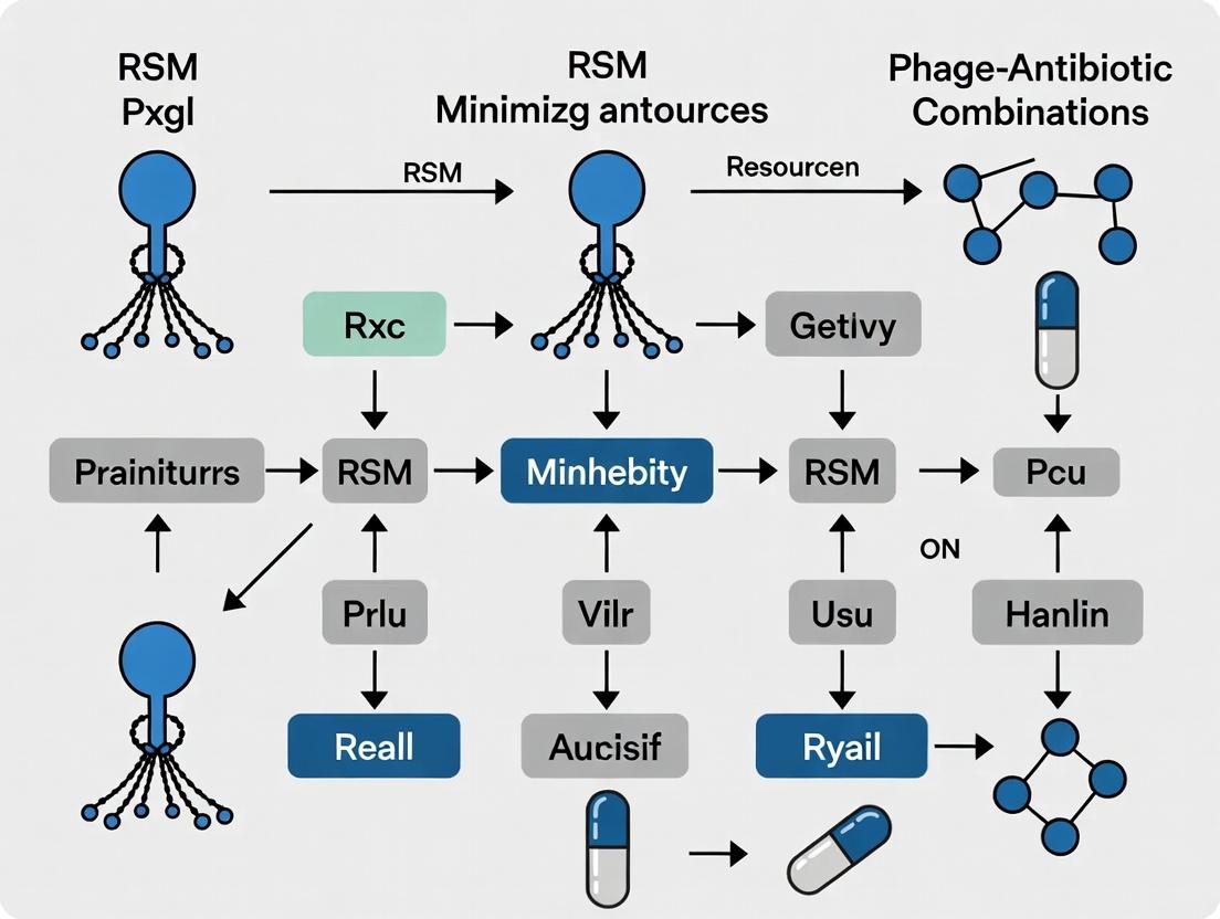
Visualizations
RSM Workflow for Synergy Optimization
Proposed Pathways in Phage-Antibiotic Synergy
The Scientist's Toolkit: Research Reagent Solutions
Table 2: Essential Materials for PAS Research with RSM
| Item | Function in Research | Example/Notes |
|---|---|---|
| Purified High-Titer Phage Lysate | Therapeutic agent; critical variable in RSM factor. | Purify via CsCl gradient; store in SM buffer at 4°C. Titer before each experiment. |
| Standardized Antibiotic Stock | Therapeutic agent; critical variable in RSM factor. | Use CLSI guidelines for preparation. Aliquot and store at -80°C. Verify MIC weekly. |
| Automated Liquid Handler | Enables precise, high-throughput setup of RSM-designed combination plates. | Critical for reproducibility in CCD experiments with many unique combinations. |
| Statistical Software with RSM Suite | For designing experiments and modeling synergy response surfaces. | e.g., Design-Expert, JMP, or R with rsm and DoE.wrapper packages. |
| Cell Density Meter (OD600) | To standardize bacterial inoculum, a key variable for assay reproducibility. | Ensure calibration and consistent measurement protocol. |
| Microtiter Plate Reader (with shaking) | To measure endpoint bacterial growth (OD) in high-throughput screens. | Shaking function improves aeration and growth consistency. |
| Lytic Phage Receptor Antiserum | Control for verifying phage activity is receptor-specific. | Use to block infection and confirm mechanism in synergy validation. |
Technical Support Center: Troubleshooting & FAQs
Frequently Asked Questions (FAQs)
Q1: My phage titer is dropping significantly after amplification. What could be the cause? A: This is often due to bacterial resistance development or poor host health. Use a fresh, low-passage bacterial culture. Check for the emergence of CRISPR-based or restriction-modification system defenses in your host by sequencing. Implement a cocktail of at least 2-3 phages with different receptors to delay resistance.
Q2: How do I confirm that observed bacterial killing is due to phage lytic activity and not a co-purified toxin? A: Perform a control experiment with a heat-inactivated (e.g., 65°C for 15 minutes) phage preparation. No killing should be observed. Additionally, perform a PCR or plaque assay on the supernatant of the killed culture to demonstrate phage replication, confirming active infectivity.
Q3: My phage-antibiotic combination (PAC) experiment shows no synergy or even antagonism. What variables should I check? A: Review the following parameters using the table below as a guide:
| Variable | Typical Issue | Recommended Adjustment |
|---|---|---|
| Timing | Phage & antibiotic added simultaneously | Stagger administration; try phage first (e.g., 60 min lead). |
| MOI | Too high (lysis from without) or too low | Titrate MOI from 0.01 to 10 in combination screens. |
| Antibiotic Class | Bacteriostatic vs. Lytic Phage | Pair lytic phages with bactericidal antibiotics (e.g., β-lactams, quinolones). |
| Bacterial Growth Phase | Stationary phase cells are less susceptible | Use mid-log phase cultures (OD600 ~0.3-0.5). |
| Rescue Effect | Phage lysis releases antibiotic pressure | Monitor regrowth; consider adding a second antibiotic. |
Q4: What is the best method to screen a large number of phage-antibiotic combinations with minimal resources? A: Utilize a Response Surface Methodology (RSM) design, such as a Central Composite Design. This allows you to systematically vary key factors (e.g., phage MOI, antibiotic concentration, time of addition) with a reduced number of experimental runs compared to a full factorial approach. Analyze outcomes (e.g., log CFU reduction) to model synergistic interactions and find optimal resource-minimizing ratios.
Q5: How can I mitigate endotoxin release during therapeutic phage application? A: Phage purification is critical. Use cesium chloride density gradient ultracentrifugation or column-based chromatography (e.g., size-exclusion) to separate phage particles from bacterial debris. Endotoxin removal kits (e.g., based on polymyxin B resin) can be used on purified lysates, followed by limulus amebocyte lysate (LAL) assay quantification.
Troubleshooting Guides
Issue: Inconsistent PAC Results in Microtiter Plate Assays
- Potential Cause 1: Evaporation in edge wells causing concentration artifacts.
- Solution: Use plate seals, incubate in humidified chambers, or only use inner 60 wells for critical assays.
- Potential Cause 2: Poor phage mixing leading to uneven MOI.
- Solution: Vortex phage stocks before use. Use multichannel pipettes with mixing steps when setting up combination plates.
- Protocol - Microtiter Checkerboard Assay:
- Prepare mid-log phase host bacteria in appropriate broth.
- In a 96-well plate, serially dilute antibiotic along the rows and phage lysate along the columns using a liquid handling robot or multichannel pipette for consistency.
- Add bacterial suspension to all wells for a final volume of 200 µL. Final bacterial density should be ~5 x 10^5 CFU/mL.
- Seal plate and incubate with shaking at host-optimal temperature for 16-24 hours.
- Measure OD600. Calculate synergy using models like the Zero Interaction Potency (ZIP) model or Bliss independence.
Issue: Rapid Development of Phage-Resistant Bacterial Mutants In Vitro
- Step 1: Characterize Resistance. Perform adsorption assays and efficiency of plating (EOP) on resistant isolates compared to parent strain. Sequence resistant mutants to identify receptor mutations.
- Step 2: Employ Evolutionary Traps. Use phages that exploit essential surface structures (e.g., protein involved in nutrient import). Resistance often comes with a fitness cost, making bacteria more susceptible to antibiotics or immune clearance.
- Step 3: Switch to Cocktails. Immediately re-challenge resistant mutants with a pre-prepared cocktail of phages targeting different receptors. This pre-emptive strategy is more effective than sequential addition.
- Protocol - Efficiency of Plating (EOP):
- Prepare serial 10-fold dilutions of your phage stock in SM buffer or broth.
- Mix 100 µL of each dilution with 100 µL of a fresh, dense bacterial culture (both target and resistant isolate).
- Add 3-5 mL of soft agar (0.5-0.7%), mix, and pour onto a base agar plate.
- Incubate overnight. Count plaques. EOP = (Plaque count on mutant / Plaque count on wild-type).
Research Reagent Solutions Toolkit
| Item | Function & Application |
|---|---|
| CsCl Gradient Solutions | Ultra-purification of phage particles via density gradient ultracentrifugation, removing host cell debris and endotoxins. |
| Polymyxin B Chromatography Resin | For endotoxin removal from purified phage preparations post-ultracentrifugation. |
| Automated Liquid Handler | Enables high-throughput, reproducible setup of complex PAC checkerboard assays and RSM design experiments, minimizing human error and resource use. |
| LAL Endotoxin Assay Kit | Quantifies endotoxin levels in final therapeutic phage preparations (target: <5 EU/kg/hr for IV administration). |
| qPCR with Propidium Monoazide (PMA) | Distinguishes between live and dead bacteria in PAC time-kill studies, as phages can amplify on dead cells. PMA dye penetrates only membrane-compromised cells. |
| 96-well Microtiter Plates with Oxygen-Permeable Seals | Facilitates high-throughput aerobic growth monitoring for PAC synergy screens while minimizing evaporation. |
Experimental Workflows and Pathways
Phage-Antibiotic Synergy (PAS) Technical Support Center
Welcome to the PAS Support Center. This resource, framed within a thesis on using Response Surface Methodology (RSM) to minimize resource expenditure in combination therapy research, provides troubleshooting and FAQs for experiments investigating enhanced bacterial killing.
FAQs & Troubleshooting Guides
Q1: We are not observing PAS in our checkerboard assay. The combination results appear merely additive. What could be wrong?
- A: This is a common issue. Please verify the following:
- Antibiotic Sub-MIC: PAS typically occurs at sub-inhibitory concentrations (sub-MIC) of the antibiotic. Confirm your antibiotic concentrations are truly below the MIC for the specific bacterial strain under your experimental conditions (e.g., broth, temperature). Re-run a MIC assay.
- Phage Multiplicity of Infection (MOI): Test a wider range of MOIs (e.g., 0.001, 0.01, 0.1, 1). Synergy may be lost at very high MOI.
- Treatment Timing: The order of addition can be critical. Try pre-treating bacteria with sub-MIC antibiotic for 30-60 minutes before adding phage.
- Bacterial Growth Phase: Use mid-log phase cultures. Stationary phase cells can be more refractory.
Q2: Our PAS effect is highly variable between replicate experiments. How can we improve reproducibility?
- A: Variability often stems from physiological state.
- Standardize Culture Conditions: Ensure precise control over inoculum size (use OD600 and confirm with CFU plating), media freshness, temperature, and aeration.
- Phage Stock Titer: Re-titer your phage stock immediately before the experiment. Phage stocks can lose potency.
- Antibiotic Stability: Check the stability of your antibiotic stock solution. Some antibiotics degrade in aqueous solution or with repeated freeze-thaw cycles.
- Implement RSM: Use a pilot RSM design (e.g., Central Composite) to model the interaction between key variables (e.g., antibiotic concentration, MOI, treatment timing) and identify the robust region for synergy, minimizing future experimental noise.
Q3: We want to investigate the mechanism of PAS. What is a reliable protocol to assess changes in phage receptor expression?
- A: A common mechanism is antibiotic-induced upregulation of phage receptors.
- Protocol: Quantitative Real-Time PCR (qRT-PCR) of Receptor Gene:
- Treatment: Grow target bacteria to mid-log phase. Treat with sub-MIC antibiotic (determined from Q1) for 60 mins. Include an untreated control.
- RNA Extraction: Harvest cells, stabilize RNA (e.g., using RNAprotect), and extract total RNA.
- cDNA Synthesis: Perform reverse transcription with a random hexamer primer.
- qPCR: Run SYBR Green-based qPCR with primers for the specific phage receptor gene (e.g., lamB for λ phage) and a housekeeping gene (e.g., rpoB).
- Analysis: Use the ΔΔCt method to calculate fold-change in receptor gene expression in antibiotic-treated vs. untreated cells.
- Protocol: Quantitative Real-Time PCR (qRT-PCR) of Receptor Gene:
Q4: How do we quantitatively measure and report the PAS effect for publication?
- A: Use standardized metrics. Calculate the following from time-kill curves or endpoint plating:
| Metric | Formula / Description | Interpretation |
|---|---|---|
| Δlog10CFU (Endpoint) | (log10CFUcombo - log10CFUphage) at time t | Direct measure of extra killing from the combo. PAS: Δlog > 0. |
| Synergy Score (Φ) | Φ = log10(NANP/NAP) where N=CFU. NA, NP, NAP are counts for antibiotic, phage, and combo. | Φ > 0 indicates synergy; Φ = 0, additive; Φ < 0, antagonism. |
| Time to Reach Detection Limit | The time (hours) for the combination to reduce CFU to a pre-set low level (e.g., 1 CFU/mL). | Demonstrates accelerated killing kinetics. |
Q5: How can RSM specifically optimize our PAS research with minimal resources?
- A: RSM reduces the total number of experiments needed to map the synergistic landscape.
- Workflow: Instead of a full factorial grid (e.g., 10x10 antibiotic x MOI combinations), design an RSM model with 10-15 strategically chosen experimental points.
- Outcome: The model will generate a 3D response surface predicting the synergy score (Φ) across a wide range of concentrations and MOIs, pinpointing the optimal combination and highlighting antagonistic zones to avoid.
The Scientist's Toolkit: Key Research Reagent Solutions
| Item | Function in PAS Research |
|---|---|
| Sub-MIC Antibiotic Stocks | Prepared from clinical-grade powder or analytical standard. Used to induce bacterial stress responses and modulate phage receptor expression without inhibiting growth. |
| High-Titer Phage Lysate (≥10^9 PFU/mL) | Purified and concentrated via cesium chloride gradient or PEG precipitation. Essential for accurate MOI calculation and strong signal in assays. |
| Phage Buffer (SM Buffer) | (50 mM Tris-HCl, 100 mM NaCl, 8 mM MgSO₄, pH 7.5). For phage storage and dilution to maintain viability and prevent osmotic shock. |
| Viable Cell Stain (e.g., Propidium Iodide) | Used in flow cytometry to distinguish between antibiotic-damaged (permeabilized) cells and healthy cells, correlating damage with phage infection efficiency. |
| cDNA Synthesis Kit | For converting extracted mRNA to cDNA in mechanistic studies analyzing gene expression changes under sub-MIC antibiotic pressure. |
RSM Software (e.g., Design-Expert, JMP, R rsm package) |
Critical for designing minimal experiment sets, building predictive models, and visualizing the synergy response surface. |
Experimental & Conceptual Diagrams
Diagram 1: Core PAS Mechanisms & Investigation Workflow
Diagram 2: RSM-Driven PAS Optimization Protocol
Empirical optimization in phage-antibiotic synergy (PAS) research traditionally involves testing numerous combinations one variable at a time (OFAT). This article, framed within a thesis on using Response Surface Methodology (RSM) to minimize resources, establishes a technical support center for researchers navigating these challenges.
Troubleshooting Guides & FAQs
FAQ: Experimental Design & Execution
Q1: My combinatorial assays show high variability, obscuring synergy detection. How can I improve reproducibility? A: High variability often stems from inconsistent phage titer or bacterial culture phase. Standardize your protocol:
- Phage Stock: Use double-agar overlay plaque assays for precise titer determination. Always use mid-log phase bacteria for propagation.
- Antibiotic Stock: Prepare fresh serial dilutions from a primary stock in the correct solvent (e.g., water, DMSO). Document MIC for the strain weekly.
- Bacterial Culture: Use cells harvested at the same optical density (OD600 ~0.5) from at least three independent overnight cultures.
Q2: I am overwhelmed by the number of combinations needed for a full factorial study. Is there a more efficient design? A: Yes. A full factorial design (e.g., 5 antibiotics × 5 phages × 5 concentrations = 125 trials) is resource-prohibitive. Implement a screening design first (e.g., fractional factorial or Plackett-Burman) to identify significant factors, then apply RSM (e.g., Central Composite Design) with fewer, strategically chosen data points to model the response surface. This can reduce experimental runs by 50-70%.
Q3: My RSM model has a poor fit (low R²). What are common causes? A: A low adjusted R² value suggests the model does not explain data variation well.
- Check 1: Ensure your experimental range includes the optimal region. Data points clustered in one corner provide poor information.
- Check 2: Include quadratic terms in your model to capture curvature in the response (common in biological systems).
- Check 3: Verify data for outliers or large measurement errors in key runs.
FAQ: Data Analysis & Interpretation
Q4: How do I statistically validate a synergistic interaction versus an additive one? A: Use quantitative models and hypothesis testing.
- Calculate the Fractional Inhibitory Concentration Index (FICI) for each combination.
- Use the RSM model to predict the response (e.g., log reduction in CFU/mL) for combinations.
- Statistically compare the observed effect of the combination against the predicted additive effect (via a t-test or using confidence intervals from the RSM model). Synergy is concluded if the combination effect is significantly greater.
Q5: How can I visualize complex multi-factor optimization results for a publication? A: Use contour (2D) and response surface (3D) plots generated from your fitted RSM model. These visually depict the relationship between two key factors (e.g., phage MOI and antibiotic concentration) on the response, with other factors held constant. The "stationary point" (peak or valley) indicates the predicted optimum.
Table 1: Resource Comparison of Optimization Methods for PAS Screening
| Method | Experimental Runs (Example: 3 Factors) | Time Estimate (Weeks) | Key Reagent Consumption (Relative Units) | Primary Statistical Output |
|---|---|---|---|---|
| One-Factor-at-a-Time (OFAT) | 15-30+ | 6-8 | 100% (Baseline) | Single-point optimum, no interaction data |
| Full Factorial Design | 27 (3³) | 8-10 | ~180% | Complete interaction model, resource-heavy |
| RSM (Central Composite) | 20 | 4-5 | ~65% | Predictive quadratic model with optimum region |
Table 2: Example Reagent & Material Requirements for a Standard PAS RSM Study
| Item | Specification/Function | Key Quality Control Step |
|---|---|---|
| Bacterial Strain | Target pathogen (e.g., Pseudomonas aeruginosa PAO1). | Check antibiotic susceptibility profile and phage receptor expression. |
| Phage Library | Purified, high-titer (>10¹⁰ PFU/mL) stocks of characterized phages. | Confirm purity via PCR of host genes and plaque morphology consistency. |
| Antibiotics | Clinical-grade powders of relevant drugs (e.g., Ciprofloxacin, Meropenem). | Verify solubility and prepare fresh stock solutions for each experiment. |
| Growth Medium | Cation-adjusted Mueller Hinton Broth (CAMHB) for antibiotics; specific broth for host growth. | Test for contaminants and performance against reference strains. |
| 96-Well Assay Plates | Cell culture-treated, sterile, with clear flat bottoms for OD reading. | Check for well-to-well consistency in background absorbance. |
| Automated Liquid Handler | For precise serial dilution and plate replication. | Calibrate pipetting volumes before each run with dye solution. |
Experimental Protocol: RSM-Optimized PAS Checkerboard Assay
Title: High-Throughput RSM Protocol for Phage-Antibiotic Synergy Screening.
Objective: To efficiently identify optimal combinations of one phage and one antibiotic that minimize bacterial viability using a Central Composite Design (CCD).
Materials: See Table 2.
Methodology:
- Design of Experiments (DoE):
- Define independent variables (e.g., X1: Phage Multiplicity of Infection (MOI), range 0.001-100; X2: Antibiotic concentration, range 0.25-4xMIC).
- Generate a CCD matrix using software (e.g., JMP, Design-Expert, R
rsmpackage). This includes factorial points, axial points, and center points (for error estimation).
Assay Setup:
- Prepare bacterial inoculum in CAMHB at ~5 × 10⁵ CFU/mL.
- According to the CCD matrix, dispense 50 µL of antibiotic at 2x the target concentration into designated wells of a 96-well plate.
- Add 50 µL of phage suspension at 2x the target MOI.
- Finally, add 100 µL of bacterial inoculum. The final volume is 200 µL, with 1x antibiotic and 1x phage MOI.
- Include controls: bacteria only, antibiotic only, phage only, and sterile medium.
Incubation & Reading:
- Seal plate and incubate at 37°C for 18-24 hours without shaking.
- Measure OD600 using a plate reader.
Data Analysis:
- Calculate percent inhibition:
[1 - (OD_sample / OD_bacteria_control)] * 100. - Input response data (% inhibition) into the DoE software.
- Fit a second-order polynomial model. Evaluate via ANOVA (check for significant model, lack-of-fit, and R²).
- Use the model's optimizer to find factor levels (MOI, [Antibiotic]) that maximize % inhibition.
- Calculate percent inhibition:
Visualizations
Title: RSM Optimization Workflow for PAS
Title: Proposed Pathways in Phage-Antibiotic Synergy
Introduction to Response Surface Methodology (RSM) as a Systematic Optimization Tool
Technical Support Center: Troubleshooting RSM Experiments in Phage-Antibiotic Synergy Research
This support center addresses common experimental and analytical challenges when applying RSM to optimize phage-antibiotic combination therapies, with the goal of minimizing resource expenditure (e.g., reagents, time, microbial biomass).
FAQs & Troubleshooting Guides
Q1: My Central Composite Design (CCD) experiments show high replication error for bacterial kill rates. How can I improve measurement consistency? A: High variability often stems from phage titer instability or bacterial growth phase inconsistency.
- Troubleshooting Steps:
- Standardize Inoculum: Always use bacteria from the same growth phase (mid-log phase recommended). Use optical density (OD600) with a calibrated spectrophotometer, and back-dilute from a fresh overnight culture.
- Phage Stock QC: Titrate phage stocks immediately before starting the CCD experiment. Use double-layer agar plaque assay in triplicate. Avoid repeated freeze-thaw cycles; aliquot stocks.
- Protocol: For the kill assay, mix standardized bacterial suspension (~10^5 CFU/mL), phage (at MOI specified by design), and antibiotic in a 96-well plate. Use a multichannel pipette for reagent dispensing. Include control wells (bacteria only, phage only, antibiotic only). Read OD600 every 30 minutes for 12-24 hours in a plate reader maintained at 37°C.
- Preventative Measure: Conduct a preliminary "lack-of-fit" study with 4-5 replicates of your center point before the full CCD. A standard deviation >15% of the response mean suggests process control issues.
Q2: The quadratic model from my RSM analysis has a significant "lack-of-fit" p-value (p < 0.05). What does this mean, and what should I do next? A: A significant lack-of-fit indicates your model (e.g., quadratic) does not adequately describe the relationship between factors and response. The data may have curvature not captured, or there are unexplored variables.
- Action Plan:
- Check for Outliers: Use studentized residual plots. Points with residuals > ±3 should be investigated for experimental error.
- Transform Response: If residual plots show a funnel pattern, apply a transformation (e.g., Log10, Square Root) to your response variable (e.g., kill rate) and re-fit the model.
- Consider Adding Terms: If using a two-factor design, you may need a cubic model or a more complex design like Box-Behnken with additional center points. However, in phage-antibiotic work, first ensure critical biological interactions (e.g., phage receptor expression affected by sub-lethal antibiotic) are not the missing variable.
- Verify Design Space: You may be operating near a steep biological cliff (e.g., complete resistance breakthrough). Expand or shift your experimental region.
Q3: How do I accurately determine "synergy" as a response variable in RSM? A: Synergy must be quantified as a continuous variable for RSM. Common metrics are summarized below.
| Synergy Metric | Calculation Formula | Advantage for RSM | Consideration |
|---|---|---|---|
| Loewe Additivity Index | Based on isobologram analysis. Requires full dose-response matrices. | Theoretically rigorous, gold standard. | Resource-intensive; many data points per run. |
| Bliss Independence Score | ΔE = Eab - (Ea + Eb - Ea*E_b) where E is fractional kill (0-1). | Easily calculated from single-agent controls. Computationally simple. | Assumes independent action; may overestimate synergy for shared targets. |
| Response Surface Fitting | Directly model kill rate (CFU/mL reduction) as a function of phage & antibiotic doses. | Directly outputs optimal combination from model. | Does not separate additive from synergistic effects. |
- Recommended Protocol: For initial RSM screens, use Bliss Score as the response. Measure kill (CFU/mL reduction after 24h) for combinations (A+B) and each agent alone (A, B). Calculate fractional kill (e.g., 1 - (CFUtreated / CFUcontrol)). Compute Bliss Score. A positive value indicates synergy.
Q4: My model suggests an optimal point at the edge of the design space. Can I trust this prediction? A: Predictions at the boundary are extrapolations and less reliable.
- Solution:
- Verify with Confirmation Runs: Perform 3-5 experimental replicates at the predicted optimal conditions. If the measured response falls within the model's prediction interval, it's partially validated.
- Expand Design: Use a "steepest ascent" approach. Create a new CCD centered on the promising edge point to explore the region beyond your initial space. This is systematic and resource-efficient.
Q5: How can I minimize resources when screening multiple phage-antibiotic pairs with RSM? A: Employ a sequential screening strategy.
- Workflow:
- First-Order Screening: Use a low-resolution fractional factorial design (e.g., 2^(k-1)) for 4-5 phage/antibiotic candidates to identify which factors (which phage, which antibiotic) significantly influence kill rate. This minimizes early-stage runs.
- Follow-Up RSM: Apply a full CCD or Box-Behnken design only to the top 1-2 candidate pairs identified in the initial screen, focusing resources on the most promising combinations.
Experimental Workflow Diagram
RSM Analysis & Model Diagnostics Pathway
The Scientist's Toolkit: Key Research Reagent Solutions
| Reagent / Material | Function in Phage-Antibiotic RSM Studies |
|---|---|
| High-Titer Phage Lysates (>10^10 PFU/mL) | Ensure consistent multiplicities of infection (MOI) across all design points. Purified by cesium chloride gradient or PEG precipitation. |
| Standardized Antibiotic Stocks | Prepared fresh from powder or frozen aliquots. Use CLSI guidelines for solvent. Critical for accurate dose in combination. |
| Cell Viability Stain (e.g., propidium iodide) | Alternative to CFU plating for rapid, high-throughput kill assessment via flow cytometry, enabling more design points. |
| Automated Liquid Handler | Dispenses precise volumes of phage, antibiotic, and media in 96/384-well plates, reducing human error in CCD execution. |
| Statistical Software (JMP, Design-Expert, R) | Required for designing CCD, analyzing ANOVA, fitting quadratic models, and generating optimization plots. |
| Robust Bacterial Glycerol Stocks | Master cell bank for all experiments to ensure genetically identical starting material, reducing biological noise. |
| 96-well Microtiter Plates (Optically Clear) | For high-throughput kill curve assays. Must be compatible with plate reader for kinetic OD600 reading. |
Designing Efficient RSM Experiments for Phage-Antibiotic Combinations: A Step-by-Step Protocol
Troubleshooting Guides & FAQs
FAQ 1: Defining and Optimizing MOI
Q: What is MOI, and how do I calculate the correct phage volume for my bacterial culture? A: Multiplicity of Infection (MOI) is the ratio of plaque-forming units (PFUs) of phage to colony-forming units (CFUs) of bacteria at the time of infection.
- Problem: Incorrect MOI leads to inconsistent lysis or failed experiments.
- Solution: Use the formula:
Volume of Phage Stock (µL) = (MOI × Number of Bacteria (CFUs)) / Phage Titer (PFU/mL). Always titer your phage stock and bacterial culture immediately before the experiment. For initial combination studies with antibiotics, test a range (e.g., MOI 0.1, 1, 10).
FAQ 2: Integrating Antibiotic Concentration
Q: How do I select a relevant antibiotic concentration to combine with phage therapy? A: The goal is often to use sub-inhibitory or minimally inhibitory concentrations to observe synergy.
- Problem: Using only the clinical breakpoint MIC may mask subtle synergistic or antagonistic effects.
- Solution: Perform a checkerboard assay combining serial dilutions of antibiotic with a range of phage MOIs. Refer to Table 1 for concentration guidelines based on common experimental aims.
Table 1: Antibiotic Concentration Selection Guide
| Experimental Aim | Recommended Concentration Range | Rationale |
|---|---|---|
| Screening for Synergy | 0.25x to 0.5x MIC | Identifies enhancement of sub-lethal effects. |
| Mimicking Sub-Therapeutic Dose | 0.5x to 1x MIC | Models scenarios where antibiotic levels are low. |
| Overcoming Resistance | 1x to 2x MIC | Tests if phage can restore antibiotic efficacy. |
| Time-Kill Assay Dynamics | 0.25x, 0.5x, 1x, 2x MIC | Captures a full dose-response relationship. |
FAQ 3: Determining Critical Timing of Administration
Q: Should I add the phage and antibiotic simultaneously, or stagger their addition? A: Timing is a critical experimental variable that can determine between synergy and antagonism.
- Problem: Simultaneous addition may not reflect therapeutic reality and can obscure mechanisms.
- Solution: Implement a timed-addition protocol. A common approach is to pre-treat with one agent (e.g., phage) for a specific duration (e.g., 30-60 minutes) before adding the second (e.g., antibiotic). This can model phage penetration before antibiotic stress. Test sequences in both orders.
FAQ 4: Interpreting Checkerboard Assay Results
Q: How do I quantify synergy from my phage-antibiotic checkerboard assay? A: Use quantitative metrics beyond visual inspection of plates.
- Problem: Subjective interpretation leads to non-reproducible claims of synergy.
- Solution: Calculate the Fractional Inhibitory Concentration Index (FICI). For phage-antibiotic combinations, adaptations like the Fractional Bactericidal Concentration Index (FBCI) may be more appropriate when using time-kill data. See the Experimental Protocol below.
FAQ 5: Managing Resource-Intensive Optimization
Q: How can I efficiently optimize these three factors (MOI, [Ab], Time) without an exhaustive, resource-heavy grid search? A: This is the core application of Response Surface Methodology (RSM).
- Problem: A full factorial experiment testing multiple levels of three factors requires an impractical number of runs.
- Solution: Employ a Central Composite Design (CCD) or Box-Behnken Design (BBD) within an RSM framework. These designs significantly reduce the number of required experimental runs while allowing you to model the interaction effects between MOI, antibiotic concentration, and timing. See the workflow diagram.
Experimental Protocols
Protocol 1: Phage-Antibiotic Checkerboard Assay in Microtiter Plates
Objective: To screen for synergistic interactions between phage and antibiotic across a matrix of concentrations.
- Prepare Bacteria: Grow the target bacterium to mid-log phase (OD600 ~0.3-0.5). Dilute in fresh broth to ~5 × 10^5 CFU/mL.
- Prepare Agents: Serially dilute the antibiotic (2X final highest concentration) across the rows of a 96-well plate. Serially dilute the phage stock (2X final highest titer) down the columns. Use broth as diluent.
- Inoculate: Add an equal volume of the bacterial suspension to each well. Final volume: 200 µL. Include controls: bacteria only, phage only, antibiotic only, broth sterility.
- Incubate & Read: Incubate statically at host temperature for 18-24 hours. Measure OD600.
- Analyze: Calculate the Fractional Inhibitory Concentration (FIC) for each agent in each well: FICab = (MIC of Ab in combo / MIC of Ab alone). FICphage = (MIC of phage in combo / MIC of phage alone). Summation ΣFIC = FICab + FICphage. Synergy: ΣFIC ≤ 0.5; Additivity: 0.5 < ΣFIC ≤ 1; Indifference: 1 < ΣFIC ≤ 4; Antagonism: ΣFIC > 4.
Protocol 2: Time-Kill Assay for Dynamic Synergy Assessment
Objective: To evaluate the bactericidal kinetics of phage-antibiotic combinations over time.
- Setup Cultures: In flasks or tubes, prepare 10 mL cultures containing: a) bacteria only (control), b) bacteria + antibiotic at selected concentration(s), c) bacteria + phage at selected MOI(s), d) bacteria + antibiotic + phage (combo). Use a starting inoculum of ~5 × 10^5 CFU/mL.
- Timed Addition: For staggered regimens, add the first agent (e.g., phage) and incubate with shaking. At the designated pre-treatment time (e.g., t=60 min), add the second agent (e.g., antibiotic).
- Sample: Remove aliquots (e.g., 100 µL) at predetermined timepoints (e.g., 0, 2, 4, 6, 8, 24h). Perform serial dilutions and plate for viable counts (CFU/mL).
- Analyze: Plot log10 CFU/mL versus time. Synergy is traditionally defined as a ≥2-log10 decrease in CFU/mL by the combination compared to the most effective single agent at 24h.
Protocol 3: Response Surface Methodology (RSM) Experimental Design
Objective: To model and optimize the three critical factors (MOI, [Antibiotic], Timing) with minimal experimental runs.
- Define Factors & Ranges:
- Factor A (MOI): Low (-1) = 0.1, High (+1) = 10
- Factor B ([Ab]): Low (-1) = 0.25x MIC, High (+1) = 1x MIC
- Factor C (Timing): Low (-1) = Antibiotic first (-60 min), High (+1) = Phage first (+60 min). 0 = simultaneous.
- Select Design: Use a Box-Behnken Design (BBD) requiring 15 runs (plus center point replicates).
- Execute Runs: Perform the combination experiments as per the BBD matrix using a Time-Kill assay (Protocol 2). The measured Response is the log10 reduction in CFU/mL at 24h.
- Model & Optimize: Use statistical software (e.g., JMP, Minitab, R) to fit a quadratic polynomial model to the data. The model will show the individual and interactive effects of the factors and predict the optimal combination for maximum killing.
Diagrams
RSM Optimization Workflow for Phage-Antibiotic Synergy
Phage & Antibiotic Checkerboard Assay Layout
The Scientist's Toolkit: Research Reagent Solutions
Table 2: Essential Materials for Phage-Antibiotic Combination Studies
| Item | Function & Critical Notes |
|---|---|
| High-Titer Phage Lysate (≥10^9 PFU/mL) | Provides sufficient titer for MOI-based experiments and avoids volume artifacts. Purify via CsCl gradient or PEG precipitation to remove bacterial debris/endotoxins. |
| Clinical/Biofilm Isolate Bacteria | Use relevant, well-characterized strains. Include standard ATCC controls. Maintain antibiotic susceptibility profiles. |
| Reference Standard Antibiotic | Use pharmaceutical-grade powder of known potency from a reputable supplier (e.g., Sigma, TOKU-E) to prepare fresh stock solutions. |
| Cation-Adjusted Mueller Hinton Broth (CAMHB) | Standard medium for antibiotic susceptibility testing. Ensures consistent cation concentrations (Ca2+, Mg2+) that affect aminoglycoside and polymyxin activity. |
| Soft Agar (0.4-0.7% Agar) | For double-layer agar overlay assays, the standard method for phage titer determination (plaque assay). |
| 96-Well & 24-Well Cell Culture Plates | For checkerboard (static) and time-kill (with shaking) assays, respectively. Use plates with lids to prevent evaporation. |
| Automated Liquid Handler | Critical for RSM. Enables precise, high-throughput dispensing of antibiotic/phage dilutions for complex design matrices, improving reproducibility. |
| Statistical Software (JMP, Minitab, R) | Required for designing RSM experiments and performing the subsequent regression analysis, ANOVA, and optimization. |
Troubleshooting Guides and FAQs
Q1: During screening, I have limited resources. Which design is more resource-efficient for initial RSM in phage-antibiotic synergy studies? A: The Box-Behnken Design (BBD) is generally more resource-efficient for initial Response Surface Methodology (RSM) in this context. BBD requires fewer experimental runs than a comparable Central Composite Design (CCD), conserving valuable phage stocks and antibiotics. For 3 factors, BBD requires 15 runs (12 factorial + 3 center points), while a full CCD requires 20 runs (8 factorial + 6 axial + 6 center points). This aligns with the thesis goal of minimizing resources.
Q2: I suspect a strong curvature in my response (e.g., phage titer optimization). Which design should I choose to better model this?
A: Choose the Central Composite Design. CCD includes axial points (star points) at a distance α from the center, which allows for efficient estimation of pure quadratic terms. This makes CCD superior for detecting and modeling strong curvature in the response surface, which is common in biological systems like phage replication dynamics.
Q3: My experimental region is constrained (e.g., antibiotic concentration cannot exceed a cytotoxic level). Which design adapts better?
A: A Face-Centered Central Composite Design (FCCD), where α=1, is often preferable for constrained regions as it keeps axial points on the faces of the cubic region. While BBD also avoids corner points and stays within a hypercube, a constrained CCD with appropriate α can be tailored more precisely to the exact feasible region of your combination therapy.
Q4: I need to perform sequential experimentation, starting with a factorial design. How do the designs integrate? A: CCD is inherently sequential. You can start with a 2^k factorial design (or fractional factorial), analyze results, and then augment it with axial and additional center points to form the full CCD. BBD is a standalone design and is not typically built sequentially from a factorial base. For a flexible, phased approach in resource-limited research, CCD is advantageous.
Q5: I'm getting a poor model fit (low R²) with my BBD. What could be the issue? A: BBD does not include corner points of the factor space. If the optimal region for phage-antibiotic synergy lies near a corner (e.g., very high phage MOI and a specific mid-range antibiotic concentration), BBD may have limited ability to capture it. Consider augmenting your design with corner point experiments or switching to a CCD in the next phase, which explicitly explores the entire cubic region.
Data Presentation: Design Comparison Table
Table 1: Quantitative Comparison of Central Composite Design (CCD) and Box-Behnken Design (BBD) for 3 Factors
| Feature | Central Composite Design (CCD) | Box-Behnken Design (BBD) |
|---|---|---|
| Total Runs (3 factors, typical) | 20 (8 cube + 6 axial + 6 center) | 15 (12 edge midpoints + 3 center) |
| Factorial Points | 2^k or 2^(k-1) (full/fractional) | None |
| Axial (Star) Points | Yes (2k points) | No |
| Center Points | Yes (typically 3-6) | Yes (typically 3) |
| Design Region | Spherical (Circumscribed) or Cubical (Face-Centered) | Spherical within a cube |
| Sequential from Factorial? | Yes, naturally augmentable | No, standalone |
| Efficiency for Quad. Model | Excellent | Very Good |
| Resource Efficiency | Lower (more runs) | Higher (fewer runs) |
| Ideal Use Case | When curvature is suspected; sequential studies | Initial RSM with tight resource constraints; avoiding extreme factor combinations |
Table 2: Example Experimental Run Comparison for a 3-Factor Phage-Antibiotic Study
| Design Type | Runs (N) | Factor A: Phage MOI | Factor B: Antibiotic Conc. (µg/mL) | Factor C: Time of Add. (hr post-inf.) |
|---|---|---|---|---|
| CCD (Face-Centered) | 20 | Levels: -1 (0.1), 0 (1), +1 (10) | Levels: -1 (0.5), 0 (5), +1 (50) | Levels: -1 (1), 0 (4), +1 (8) |
| BBD | 15 | Levels: -1 (0.1), 0 (1), +1 (10) | Levels: -1 (0.5), 0 (5), +1 (50) | Levels: -1 (1), 0 (4), +1 (8) |
Experimental Protocols
Protocol 1: Implementing a Central Composite Design for Phage-Antibiotic Synergy
- Define Factors & Ranges: Identify k critical factors (e.g., Multiplicity of Infection (MOI), antibiotic concentration, timing of administration). Set low (-1) and high (+1) levels based on prior knowledge.
- Choose CCD Type: For constrained resources and physical limits, select a Face-Centered CCD (FCCD, α=1). For a broader exploration, use a Circumscribed CCD (α=√k).
- Generate Design Matrix: Use statistical software (e.g., R, Design-Expert, Minitab) to create a randomized run order. For 3 factors, this yields 20 runs.
- Execute Experiments: Conduct the bacterial inhibition assays (e.g., plaque assay, OD600 measurement) as per the randomized matrix to minimize bias.
- Model & Analyze: Fit a second-order polynomial model (Y = β0 + ΣβiXi + ΣβiiXi² + ΣβijXiXj). Use ANOVA to assess significance.
- Optimize & Validate: Locate the optimal predicted factor combination for synergy (e.g., maximum bacterial killing). Perform 3-5 confirmatory experiments at this predicted optimum.
Protocol 2: Implementing a Box-Behnken Design for Resource-Limited Screening
- Define Factors & Ranges: As in Protocol 1.
- Generate BBD Matrix: For 3 factors, software will generate 15 experimental runs. Each factor is varied between three levels while holding others at their mid-point.
- Randomize & Execute: Randomize run order and perform the synergy assays.
- Model & Analyze: Fit the same second-order model as CCD. Note that BBD lacks corner points, so extrapolation to extremes should be cautious.
- Identify Trends: Use contour plots to identify a direction of improved response. This may guide a subsequent, more focused CCD if resources allow.
Mandatory Visualization
RSM Design Selection Workflow
CCD vs BBD Point Structure
The Scientist's Toolkit: Research Reagent Solutions
Table 3: Essential Materials for Phage-Antibiotic Combination RSM Studies
| Item | Function in RSM Experiment | Example/Note |
|---|---|---|
| Bacterial Host Strain | Target organism for phage infection and antibiotic action. | e.g., Clinical isolate of Pseudomonas aeruginosa. |
| Bacteriophage Stock | Biological agent; one of the key factors (e.g., MOI) in the optimization. | High-titer, purified lysate; titer must be accurately determined. |
| Antibiotic | Pharmaceutical agent; second key factor (concentration) in the combination. | Use a clinically relevant antibiotic (e.g., Ciprofloxacin). Prepare fresh serial dilutions. |
| Growth Medium | Supports bacterial growth for consistent assay conditions. | e.g., Mueller Hinton Broth, tailored to the specific bacterium. |
| Cell Viability Assay Kit | Quantifies the response variable (e.g., bacterial survival). | Resazurin (AlamarBlue) assay, ATP-based luminescence, or CFU plating. |
| Statistical Software | Generates design matrix, randomizes runs, and fits the RSM model. | R (rsm package), Design-Expert, JMP, Minitab. |
| Multi-well Plate Reader | Enables high-throughput measurement of the response for many design points. | For optical density (OD) or fluorescence-based viability assays. |
FAQs & Troubleshooting Guides
Q1: My time-kill kinetics data is highly variable between replicates. How can I define a robust response variable for my RSM model? A: High variability often stems from inconsistent initial inoculum preparation or sampling times. For RSM, consider deriving a summary metric from the time-kill curve.
- Protocol: Follow CLSI M26-A guidelines for time-kill studies. Precisely standardize the microbial growth phase (e.g., mid-log phase) and use controlled conditions for antibiotic and phage stock titers. Sample at fixed intervals (e.g., 0, 2, 4, 6, 8, 24h).
- Solution: Instead of using raw CFU/mL at a single time point, calculate the Area Under the Bacterial Killing Curve (AUBKC) or the Log Reduction at a critical time point (e.g., 24h). These integrated metrics are more robust response variables for RSM.
Q2: When testing phage-antibiotic synergy, should I use a synergy score (e.g., ΔE) or a direct kill metric like log reduction as my Y-variable? A: The choice depends on your RSM objective. To minimize resources, a direct kill metric is often more efficient.
- Issue: Calculating a synergy score (e.g., ΔE = E(combination) – [E(phage alone) + E(antibiotic alone)]) requires running three separate assays per data point, tripling resources.
- Recommendation: Directly use Log Reduction at 24h for the combination as your primary Y-variable. Your RSM model will then directly predict the combination's efficacy based on input factors (e.g., MOI, antibiotic concentration, time of addition), which is more resource-effective.
Q3: How do I handle a log reduction value when the combination causes complete eradication (no colonies detected)? A: This is a "censored data" issue common in antimicrobial studies. You cannot take log10(0).
- Protocol: Implement a limit of detection (LOD) correction.
- Solution: If your plating volume is 100 µL, your practical LOD is 10 CFU/mL. For complete eradication, assign a value equal to Log10(Initial Inoculum) - Log10(LOD). For example, if you start with 1x10^6 CFU/mL, your maximum log reduction value is 6 - 1 = 5 log10. Use this corrected value in your RSM analysis.
Q4: My response variable data does not meet the normality assumption for RSM regression. What should I do? A: This is expected for bounded data like log reduction. A transformation is necessary.
- Troubleshooting Step: Apply a Box-Cox transformation to your Y-variable (Log Reduction values) to stabilize variance and improve normality before building your polynomial model. Most statistical software (R, Design-Expert, JMP) includes this feature.
Experimental Protocols Summary
| Protocol Name | Key Steps | Primary Output | RSM Y-Variable Recommendation |
|---|---|---|---|
| Standard Time-Kill Kinetics | 1. Prepare ~1x10^6 CFU/mL inoculum. 2. Add treatment (phage, antibiotic, combo). 3. Sample at t=0,2,4,6,8,24h. 4. Serially dilute & plate for CFU count. | CFU/mL over time curve. | Area Under the Killing Curve (AUBKC) or Log Reduction at 24h. |
| Checkerboard Assay (for Reference) | 1. Set up 2D serial dilutions of phage & antibiotic. 2. Add standardized inoculum. 3. Incubate 18-24h. 4. Read optical density. | Fractional Inhibitory Concentration (FIC) Index. | FIC Index (less ideal for RSM due to multiple assays). |
| Single-Time-Point Synergy Log Reduction | 1. Prepare treatments: control, phage alone, antibiotic alone, combination. 2. Use a single, optimized combination ratio. 3. Incubate 18-24h. 4. Perform final viable count. | Log10 Reduction for each group. | Log10 Reduction (Combo). Calculate synergy score only if needed for validation. |
The Scientist's Toolkit: Research Reagent Solutions
| Item | Function & Rationale |
|---|---|
| Cation-Adjusted Mueller Hinton Broth (CAMHB) | Standard medium for antibiotic susceptibility testing, ensures consistent cation levels affecting antibiotic activity. |
| Phage Storage Buffer (SM Buffer) | Long-term, stable storage of phage stocks, preventing titer loss which is critical for accurate MOI calculation. |
| Triphenyl Tetrazolium Chloride (TTC) or Resazurin | Metabolic dye used in plate assays to visualize bacterial growth and inhibition endpoints clearly. |
| Automated Colony Counter | Reduces human error and increases throughput and consistency in CFU enumeration from time-kill assays. |
| Statistical Software with RSM Module | Essential for designing experiments (e.g., Central Composite Design) and analyzing complex response surfaces (e.g., JMP, Design-Expert, R rsm package). |
Visualization: Experimental Workflow for RSM Response Variable
Title: RSM Workflow for Efficient Response Variable Collection
Visualization: Decision Pathway for Y-Variable Selection
Title: Choosing a Response Variable for RSM in Synergy Studies
Technical Support Center: Troubleshooting & FAQs
FAQ 1: My central composite design (CCD) model for phage-antibiotic synergy is not significant (p > 0.05). What are the primary causes and solutions?
Answer: Common causes include incorrect factor range selection, excessive noise overwhelming signal, or insufficient model degrees of freedom. First, verify your factor ranges (e.g., phage MOI: 0.1-10, antibiotic concentration: 0.5-2x MIC) are appropriate via preliminary screening. For minimum-run designs, ensure center points are replicated (at least 3) to estimate pure error. If lack of fit is significant, consider transforming your response (e.g., log10 transformation for bacterial CFU counts).
Experimental Protocol for Diagnostic Check:
- Fit your RSM model (e.g., quadratic).
- Perform ANOVA and note p-value for the model and lack-of-fit test.
- Examine residual plots (Residuals vs. Predicted, Normal Q-Q).
- If a pattern exists in residuals vs. predicted, apply a Box-Cox transformation to your response data.
- Re-fit the model and re-evaluate significance.
FAQ 2: How do I validate a predictive model when I have no additional replicates for a separate validation set?
Answer: Leverage internal validation methods. For small designs, use PRESS (Predicted Residual Error Sum of Squares) statistics and cross-validation. A high R²-Predicted (e.g., > 0.7) indicates good predictive capability. Calculate the Adequate Precision ratio; a ratio > 4 is desirable, indicating the model signal is strong relative to noise.
Experimental Protocol for Internal Validation:
- After model fitting, calculate PRESS (statistical software usually provides this).
- Compute R²-Predicted = 1 - (PRESS / Total Sum of Squares).
- Compute Adequate Precision = (Max Yˆ - Min Yˆ) / √(Variance of Predicted Values).
- If metrics are poor, refine the model by removing non-significant terms (p > 0.1) or adding axial points if the design allows.
FAQ 3: My contour plot shows an optimum outside my tested experimental region. What should I do?
Answer: This indicates the true optimum may lie beyond your current factor boundaries. Do not extrapolate. Conduct a subsequent Steepest Ascent/Descent experiment to guide the new design region.
Experimental Protocol for Steepest Ascent:
- From your current model, take the first-order derivative (the linear coefficients) to determine the path of steepest ascent.
- Define a step size for your primary factor (e.g., 0.5x MIC for antibiotic).
- Run new experiments along this path, measuring the response (e.g., log reduction in biofilm).
- Once the response decreases, you have passed the optimum. Use the point of maximum response as the new center point for an augmented or new RSM design.
Data Summary Tables
Table 1: Comparison of Common Minimum-Run RSM Designs for Combination Therapy Screening
| Design Type | Total Runs (k=2 factors) | Total Runs (k=3 factors) | Can Estimate Full Quadratic? | Recommended Use Case |
|---|---|---|---|---|
| Central Composite (CCD) - Circumscribed | 13 (8 + 5) | 20 (14 + 6) | Yes | Standard, well-characterized regions. |
| Box-Behnken (BBD) | 13 | 15 | Yes | Efficient, avoids extreme factor levels. |
| 3-Level Factorial (Reduced) | 9-10 | 15-17 | Yes | When axial points are impractical. |
| Optimal (D-Optimal) | 6-10 | 10-15 | Yes * | Constrained regions or resource-limited cases. |
Note: D-Optimal designs require pre-specification of the model form and may have lower predictability than CCD/BBD.
Table 2: Typical Reagent & Instrument Requirements for Phage-Antibiotic RSM Studies
| Item Name | Function/Description | Example Specification |
|---|---|---|
| Bacteriophage Stock | Therapeutic agent; titer must be precisely quantified. | High-titer lysate (>10^9 PFU/mL), purified, host range validated. |
| Antibiotic Standard | Co-therapeutic agent; prepare fresh serial dilutions. | Clinical-grade powder, dissolved in appropriate solvent (e.g., DMSO, water). |
| Mueller Hinton Broth (MHB) | Standardized medium for checkerboard and time-kill assays. | Cation-adjusted for antibiotic testing. |
| 96-Well Microtiter Plate | Platform for high-throughput synergy screening. | Tissue culture-treated, flat-bottom, sterile. |
| Automated Liquid Handler | For precise, reproducible dispensing of agents in small volumes. | Capable of dispensing 2-50 µL with low variance (<5%). |
| Plate Reader (OD600 & Fluorescence) | To monitor bacterial growth (OD) and viability/response markers. | Incubating reader with shaking, capable of kinetic reads. |
| Colony Counter (Manual or Automated) | To quantify bacterial load (CFU/mL) for endpoint validation. | Standard for plating serial dilutions. |
The Scientist's Toolkit: Research Reagent Solutions
| Essential Material | Primary Function in Minimum-Run RSM |
|---|---|
| Phage Propagation & Purification Kits | To generate high-titer, endotoxin-reduced phage stocks for consistent dosing. |
| Pre-dosed Antibiotic Plates or Strips | For rapid, reproducible gradient antibiotic concentration setup (e.g., Etest strips in adapted format). |
| Cell Viability Stain (e.g., resazurin) | A fluorescent metabolic indicator for high-throughput readout of bacterial inhibition. |
| Biofilm Crystal Violet Assay Kit | For quantifying biofilm biomass, a common response in chronic infection models. |
| Statistical Software (e.g., JMP, Design-Expert, R) | Essential for designing minimum-run experiments, analyzing RSM data, and generating predictive models/plots. |
Experimental Workflow & Pathway Diagrams
Title: Minimum-Run RSM Optimization Workflow for Combination Therapy
Title: Target Pathway for Phage-Antibiotic Synergy Optimization
Troubleshooting Guides & FAQs
FAQ 1: My ANOVA for my quadratic RSM model shows a significant Lack-of-Fit. What does this mean and how should I proceed? A significant Lack-of-Fit p-value (typically <0.05) indicates your chosen polynomial model (e.g., quadratic) does not adequately describe the relationship between your factors and the response. This is critical for reliable optimization in phage-antibiotic synergy studies. To resolve this:
- Check for Outliers: Review experimental runs for measurement or execution errors.
- Consider a Higher-Order Model: Your system may require a cubic or quartic term. However, this demands more experimental runs.
- Verify Factor Ranges: Ensure your chosen ranges for antibiotic concentration, phage titer, and incubation time adequately capture the response surface.
- Inspect Replication Error: Ensure pure error from replicated center points is not excessively small.
FAQ 2: When interpreting my polynomial equation coefficients, why is a factor's linear effect insignificant while its quadratic effect is highly significant? This is common in optimization experiments where the response reaches a peak or valley within the experimental domain. An insignificant linear coefficient suggests the response is at a plateau or turning point with respect to that factor. The significant quadratic coefficient confirms the curvature (a maximum or minimum) is present. For minimizing resources, this peak/valley is precisely the optimal region you are searching for.
FAQ 3: The "Predicted R²" and "Adjusted R²" in my RSM output are not in reasonable agreement. What is the issue? A large gap between Adjusted R² and Predicted R² (commonly >0.2) suggests your model may be overfit. It incorporates too many terms (like higher-order interactions) that fit the noise in your specific dataset but will not predict new observations well. Simplify the model by removing statistically insignificant terms (consider hierarchical model building) or collect more data to improve estimation.
FAQ 4: How do I choose between a full quadratic model and a reduced model when analyzing my Central Composite Design (CCD) data? Always perform a sequential model reduction analysis. Start with a linear model, then add interaction terms, then quadratic terms. Use ANOVA to test if the addition of each set of terms significantly improves the model. The principle of parsimony is key: choose the simplest model with an insignificant Lack-of-Fit and high predictive power (Predicted R²). This ensures a robust model for identifying minimal effective resource combinations.
Table 1: Key ANOVA Metrics and Their Interpretation in RSM for Phage-Antibiotic Research
| Metric | Ideal Value/Outcome | Interpretation for Resource Minimization |
|---|---|---|
| Model p-value | < 0.05 | The model is statistically significant. The factors (e.g., concentrations, time) do influence the response (e.g., biofilm inhibition, bacterial kill rate). |
| Lack-of-Fit p-value | > 0.05 | The model fits the data well. No significant unexplained variance remains, giving confidence in optimization predictions. |
| Adjusted R² | Close to 1.0 | The proportion of variation in the response explained by the model, adjusted for the number of terms. Values >0.9 indicate excellent explanatory power. |
| Predicted R² | Close to Adjusted R² | Measures the model's ability to predict new data. Agreement with Adjusted R² indicates the model is not overfit and is reliable for finding optimal conditions. |
| Adequate Precision | > 4 | Measures the signal-to-noise ratio. A ratio >4 indicates an adequate model for navigating the design space to find the minimum effective doses. |
Experimental Protocol: Generating an RSM Model for Synergy Optimization
Objective: To model the combined effect of phage titer (PFU/mL) and sub-inhibitory antibiotic concentration (µg/mL) on the reduction of bacterial biofilm biomass, with the goal of identifying the minimal resource combination for maximum efficacy.
Methodology:
- Experimental Design: A face-centered Central Composite Design (CCD) with 3 center points is employed. Two factors are studied over three levels (-1, 0, +1).
- Factor Ranges:
- Phage Titer (A): 10⁶, 10⁷, 10⁸ PFU/mL (log scale).
- Antibiotic Conc. (B): 0.25x, 0.5x, 0.75x the MIC (µg/mL).
- Procedure: For each of the 11 design runs (including center points in triplicate), incubate a standardized biofilm in a 96-well plate with the prescribed combination for 24 hours. Remove planktonic cells, stain the adherent biofilm with crystal violet, solubilize with acetic acid, and measure absorbance at 595 nm. Calculate percentage inhibition relative to an untreated control.
- Analysis: Input the data (Factors A, B; Response: % Inhibition) into statistical software (e.g., Design-Expert, Minitab, R). Fit a second-order polynomial model:
Y = β₀ + β₁A + β₂B + β₁₂AB + β₁₁A² + β₂₂B² + ε. Perform ANOVA to evaluate model significance, lack-of-fit, and individual term significance. - Optimization: Use the software's numerical and graphical optimization tools to find the factor settings that maximize inhibition while minimizing both phage titer and antibiotic concentration.
Visualizations
Title: RSM Model Fitting & Validation Workflow
Title: ANOVA Sum of Squares Decomposition
The Scientist's Toolkit: Research Reagent Solutions
Table 2: Essential Materials for RSM in Phage-Antibiotic Synergy Studies
| Item | Function in the Experiment |
|---|---|
| 96-Well Polystyrene Microtiter Plates | Standardized platform for high-throughput biofilm cultivation and treatment under defined conditions. |
| Crystal Violet Stain (0.1% w/v) | A basic dye that binds to negatively charged surface molecules and polysaccharides in the biofilm matrix, enabling quantitative biomass assessment. |
| Glacial Acetic Acid (33% v/v) | Solvent for dissolving crystal violet stain bound to the biofilm, creating a homogeneous solution for spectrophotometric reading. |
| Phage Buffer (SM Buffer or PBS-Mg²⁺) | Provides the appropriate ionic strength and magnesium ions to maintain phage stability and prevent adsorption to tube walls during serial dilution. |
| Mueller Hinton Broth (MHB) or TSB with Supplements | A standardized, nutrient-rich growth medium suitable for both antibiotic susceptibility testing and supporting robust biofilm growth. |
| Microplate Reader (with 595 nm filter) | Instrument for rapid, accurate measurement of the absorbance of the solubilized crystal violet, correlating to biofilm biomass. |
| Statistical Software (e.g., Design-Expert, JMP, R with 'rsm' package) | Critical for designing efficient RSM experiments, performing complex ANOVA, fitting polynomial models, and generating contour plots for optimization. |
Solving Common RSM Challenges in Phage-Antibiotic Assays: From Model Lack-of-Fit to Resource Limits
Addressing Non-Linear and Interactive Effects in Biological Systems
Technical Support Center: Troubleshooting Response Surface Methodology (RSM) in Phage-Antibiotic Synergy Studies
This support center provides targeted guidance for researchers employing RSM to model the complex, non-linear interactions between phages and antibiotics, with the goal of minimizing experimental resources.
Frequently Asked Questions (FAQs) & Troubleshooting
Q1: My RSM model for phage-antibiotic combination efficacy shows a poor fit (low R²). What are the primary causes and solutions? A: A low R² value often indicates the model is not capturing the system's complexity.
- Cause 1: The experimental region (range of phage MOI and antibiotic concentration) may be too narrow or miss the optimal synergistic zone.
- Solution: Perform a scouting experiment to identify a broader range. Consider a steeper gradient for the antibiotic if resistance is rapid.
- Cause 2: Significant non-linear or interaction effects exist that your current polynomial model (e.g., quadratic) cannot fit.
- Solution: Increase the model order (e.g., to a cubic model) or transform your response variable (e.g., log transformation of bacterial density). Confirm you have sufficient center points to detect curvature.
- Cause 3: Excessive uncontrolled biological variability (e.g., bacterial growth phase, phage stock titer variance).
- Solution: Standardize pre-culture protocols. Use internal controls in every assay plate. Perform phage titer determination immediately before combination experiments.
Q2: How do I handle a "lack of fit" that is statistically significant in my RSM analysis? A: A significant lack of fit means your model is systematically incorrect despite a possibly high R².
- Action 1: Verify your experimental error is minimal. Replicate center point conditions to obtain a pure error estimate. High pure error suggests technical issues.
- Action 2: The biological interaction may require a mechanistic model component. Augment your RSM design with time-course data. Model the rate of bacterial killing (dN/dt) as the response, not just endpoint CFU/mL.
- Action 3: Include a key categorical factor you may have overlooked. Example: Bacterial physiological state (exponential vs. stationary phase) drastically changes phage adsorption and antibiotic efficacy. Incorporate it as a two-level categorical factor in your RSM design.
Q3: My predicted optimal combination from the RSM model fails validation in a follow-up experiment. Why? A: This is often due to overfitting or model extrapolation.
- Diagnosis: Check the model's prediction interval at the optimal point. A wide interval indicates low confidence. Examine the optimization desirability function—it may be too sensitive to one response (e.g., efficacy) and ignore another (e.g., resistance suppression).
- Protocol for Robust Validation:
- Prepare the predicted optimal phage-antibiotic combination.
- In parallel, prepare combinations at the vertices of the confidence interval region around the optimum (slightly higher/lower doses).
- Run the validation experiment using a dynamic time-kill assay, not just a single timepoint.
- Compare the area under the bacterial kill curve (AUC) for all treatments. The true robust optimum should perform best across the AUC metric.
Q4: What is the most efficient RSM design to start with for a novel phage-antibiotic pair to conserve resources? A: A Central Composite Design (CCD) or Box-Behnken Design (BBD) is standard. For extreme resource minimization, a Fractional Factorial Design augmented with center points is recommended for initial screening.
- Recommended Initial Protocol:
- Factors: Phage MOI (0.01, 1), Antibiotic Concentration (0.25x, 1x MIC), Time of Antibiotic Addition (0, 2h post-phage).
- Design: Use a 2³⁻¹ fractional factorial design (4 runs) plus 3 center point replicates (Phage MOI 0.1, Abx 0.5x MIC, Addition at 1h). Total = 7 initial experiments.
- Response: Measure log-reduction in CFU/mL at 8h and 24h.
- Analysis: Identify which main effects and two-factor interactions are significant. This informs the region for a more detailed, focused CCD.
Table 1: Comparison of RSM Designs for Phage-Antibiotic Combination Studies
| Design Type | Runs (3 Factors) | Can Estimate Full Quadratic? | Optimal for | Resource Efficiency |
|---|---|---|---|---|
| Full Factorial + Center | 17+ | Yes | Final optimization | Low |
| Central Composite (CCD) | 15-20 | Yes | Building a comprehensive model | Medium |
| Box-Behnken (BBD) | 15 | Yes | When extreme points are risky | Medium |
| Fractional Factorial + Center (Screening) | 7-9 | No | Initial factor screening | Very High |
Table 2: Common Response Variables & Their Trade-offs
| Response Variable | Measurement | Pros | Cons | Recommended Model |
|---|---|---|---|---|
| Endpoint Log CFU/mL | Plate counts at fixed time | Simple, absolute measure | Misses dynamics, high variance | Polynomial (Quadratic) |
| Area Under Curve (AUC) | Integration of time-kill curve | Captures total effect, robust | More labor-intensive | Polynomial or Nonlinear |
| Time to 99% Kill (T99) | Time from intervention | Mechanistically relevant | Can be infinite if ineffective | Survival Analysis Models |
| Resistance Emergence Rate | Resistant colony counts | Critical long-term outcome | Requires specialized plating | Generalized Linear Model |
Experimental Protocols
Protocol 1: Dynamic Time-Kill Assay for RSM Response Generation Objective: Generate robust response data (AUC, T99) for RSM modeling.
- Prepare: Grow target bacterium to mid-exponential phase (OD₆₀₀ ~0.3-0.4). Dilute to ~1x10⁶ CFU/mL in fresh media.
- Dose: In a 96-well plate or tube, add phage and antibiotic at concentrations defined by your RSM design matrix. Include mono-therapy and growth controls.
- Incubate & Sample: Incubate with shaking. Take 20µL samples at t=0, 2, 4, 6, 8, 12, 24h.
- Quantify: Serially dilute and spot-plate samples (or use automated colony counters) to determine CFU/mL. For phage-only wells, use a phage-inactivating agar overlay.
- Calculate Response: Plot log(CFU/mL) vs. time. Calculate AUC using the trapezoidal rule. T99 is interpolated from the curve.
Protocol 2: Validation of Predicted Optimal Combination Objective: Confirm the performance of the RSM-optimized combination in a biologically relevant model.
- Test Articles: Prepare the exact optimal combination, the vehicle control, and mono-therapies at the optimized doses.
- Inoculum: Use a higher starting density (e.g., 1x10⁷ CFU/mL) or incorporate a small percentage of pre-formed antibiotic-resistant mutants to stress-test the combination.
- Extended Duration: Run the time-kill assay for 48-72h, sampling every 6-12h after 24h.
- Key Metric: The optimized combination should maintain suppression (>3-4 log reduction) at 48h, where mono-therapies often fail due to resistance.
Visualizations
Title: RSM Workflow for Phage-Antibiotic Optimization
Title: Non-Linear Interaction Network in Phage-Antibiotic Therapy
The Scientist's Toolkit: Research Reagent Solutions
Table 3: Essential Materials for RSM in Phage-Antibiotic Research
| Item | Function & Specific Role in RSM Context | Example/Note |
|---|---|---|
| Automated Liquid Handler | Enables precise, high-throughput dispensing of phage & antibiotic gradients for RSM design matrices. Minimizes human error. | Beckman Coulter Biomek, Hamilton STAR. |
| Phage Titer Quick Assay Kit | Rapid, standardized quantification of phage stock concentration before each experiment. Critical for accurate MOI in design. | SpeedyPhage, qPCR-based kits. |
| Cell Density Meter (OD) | Standardizes initial bacterial inoculum density, a major source of uncontrolled variability in response. | McFarland Densitometer. |
| Automated Colony Counter | Accurately quantifies CFU/mL from time-kill assays. Essential for generating reliable, low-variance response data. | Protocol 3M, OpenCFU. |
| Statistical Software with RSM | Fits complex polynomial models, performs lack-of-fit tests, and generates optimization plots. | JMP, Design-Expert, R (rsm package). |
| 96-well Time-Kill Assay Plates | Allow simultaneous, small-volume testing of multiple combination conditions with kinetic reading. | Breathable sealed plates. |
| Phage-Neutralizing Agar | Allows accurate CFU counting in phage-containing samples by inactivating phages post-sampling. | Contains chelating agents (e.g., citrate). |
| Reference Strain Panel | Includes antibiotic-resistant and phage-resistant mutants. Used to stress-test and validate robustness of optimized combinations. | ATCC or clinically derived strains. |
Handling High Variability in Phage Titration and Bacterial Growth Assays
Technical Support Center
Troubleshooting Guide
Q1: Why are my phage plaque assays showing high variability in plaque counts between replicates? A: High variability often stems from inconsistent top agar preparation or bacterial lawn density. Ensure the molten top agar is held at a consistent 45-55°C before mixing with the host bacteria. The host culture should be in mid-exponential phase (OD600 ~0.3-0.5) and diluted to a standardized concentration. Mix the phage-bacterium-top agar suspension by gentle vortexing for 2-3 seconds before pouring immediately onto the base agar plate. Allow plates to solidify on a perfectly level surface.
Q2: What causes inconsistent bacterial growth curves in antibiotic combination assays? A: Inconsistency typically originates from antibiotic stock solution degradation, inoculum size variation, or poor aeration in microtiter plates. Prepare fresh antibiotic stocks monthly, store aliquots at -80°C, and avoid freeze-thaw cycles. Standardize the starting inoculum precisely to 5 x 10^5 CFU/mL. Use microtiter plates with oxygen-permeable membranes and ensure consistent shaking speed (e.g., 200 rpm) in the incubator to maintain uniform aeration.
Q3: How can I reduce variability in phage titer determination when using the double agar overlay method? A: Implement a standardized phage adsorption step. Follow this protocol: Mix 100 µL of a log-phase bacterial culture with 100 µL of appropriate phage dilutions. Incubate at the host's optimal growth temperature for 5 minutes to allow adsorption. Add this mixture directly to 3-5 mL of molten top agar (held at 48°C), mix gently, and pour. This controlled adsorption period minimizes variability compared to mixing all components in the top agar directly.
Q4: Why do I see "lawn failures" or confluent lysis instead of discrete plaques? A: This indicates an incorrect phage-to-bacteria ratio (MOI too high) or bacterial culture that is too dense or in the wrong growth phase. Titer the phage stock accurately and perform serial dilutions to achieve an MOI of approximately 0.001-0.01 for the assay. Use a fresh bacterial culture in the early to mid-exponential phase. If the problem persists, the phage stock may contain a high proportion of temperate or mutant phages; re-isolate plaques to clonally purify the stock.
Q5: How do I handle high background noise in optical density readings for bacterial growth with phage+antibiotic combinations? A: Cell debris from phage lysis can scatter light. Include control wells containing phage-only (no bacteria) and lysed bacteria at each time point to subtract background. Alternatively, shift from endpoint OD measurements to real-time, kinetic monitoring. Use a plate reader that can measure fluorescence (e.g., using a viability dye like resazurin) in parallel with OD600, as fluorescence is less affected by debris.
Frequently Asked Questions (FAQs)
Q: What is the optimal temperature and duration for storing phage stocks to maintain stable titers? A: For long-term storage, add a sterile final concentration of 50% glycerol or 10% DMSO to your high-titer phage lysate. Aliquot and store at -80°C. Avoid storage at -20°C. Under these conditions, most phage titers remain stable for years. For short-term (a few weeks), store purified phage in SM Buffer at 4°C in the dark.
Q: How many technical replicates are statistically recommended for phage plaque assays? A: Due to inherent procedural variability, a minimum of three biological replicates, each with three technical replicates (plates), is recommended. The coefficient of variation (CV) for plaque counts should ideally be below 15%. If CV exceeds 25%, the experimental protocol requires optimization.
Q: Which growth medium is best for combined phage-antibiotic synergy assays? A: Use a chemically defined medium (e.g., MHB-II for antibiotics) whenever possible, as it reduces batch-to-batch variability. Supplement with calcium (1mM CaCl2) and magnesium (1mM MgSO4) if your phage requires divalent cations for adsorption. Avoid media with high phosphate concentrations that can precipitate these cations.
Q: How do I standardize the initial bacterial state for combination assays? A: Always start from a single colony, grow overnight, then subculture into fresh medium to reach the target early-exponential phase. Standardize by optical density (OD600) AND confirm by plating for colony-forming units (CFU/mL). The correlation between OD and CFU/mL should be established for each bacterial strain.
Q: Can automation help reduce variability? A: Yes. Using automated liquid handlers for serial dilutions of phages and antibiotics, and robotic plate readers for consistent OD measurements, significantly reduces human error and operational variability, especially critical for Response Surface Methodology (RSM) experimental setups.
Table 1: Common Sources of Variability and Their Impact
| Source of Variability | Typical Coefficient of Variation (CV) | Recommended Mitigation Strategy |
|---|---|---|
| Phage Serial Dilution (manual) | 20-35% | Use automated liquid handlers or reverse dilution series |
| Top Agar Pouring & Solidification | 15-30% | Standardize volume, temperature, and level surface |
| Bacterial Inoculum Preparation | 10-25% | Calibrate OD600 to CFU/mL; use mid-exponential phase |
| Microtiter Plate OD Reading | 5-15% | Use same plate reader model; correct for background/debris |
| Antibiotic Stock Concentration | 5-20% | Use weight/volume preparation; verify potency with QC strain |
Table 2: Comparison of Phage Titer Methods
| Method | Precision (CV) | Time | Cost | Best Use Case |
|---|---|---|---|---|
| Double Agar Overlay (Plaque Assay) | 10-25% | 18-24 hrs | Low | Gold standard, viable count |
| Spot Test (Semi-Quantitative) | 30-50% | 18-24 hrs | Very Low | Rapid titer estimation |
| qPCR (Genome Copies) | 5-10% | 2-3 hrs | High | Total particles (viable+non-viable) |
| Fluorescence Microscopy | 15-40% | 1-2 hrs | Medium | Direct visualization, rapid |
Detailed Experimental Protocols
Protocol 1: Standardized Double Agar Overlay for Phage Titration
- Host Preparation: Grow host bacteria in appropriate broth to mid-exponential phase (OD600 0.3-0.5). Dilute to a concentration of ~10^8 CFU/mL (as pre-calibrated).
- Phage Dilution: Perform ten-fold serial dilutions of phage stock in SM buffer or sterile broth. Use a fresh pipette tip for each dilution or perform a "reverse dilution" series to minimize carryover error.
- Adsorption Mix: Combine 100 µL of diluted phage with 100 µL of prepared host cells in a sterile tube. Incubate for 5-7 minutes at host growth temperature.
- Top Agar Mixing: Add the 200 µL adsorption mix to 3-5 mL of molten top agar (0.5-0.7% agar) tempered to 48°C in a sterile culture tube. Vortex gently for 2-3 seconds.
- Pouring: Immediately pour the mixture onto a pre-warmed, dried base agar (1.5% agar) plate. Swiftly tilt the plate to ensure even coverage.
- Solidification & Incubation: Let plates solidify on a level surface for 15 minutes. Invert and incubate at the optimal temperature for 12-18 hours.
- Counting: Count plaques on plates containing 30-300 plaques. Calculate PFU/mL: (Plaque count) / (Dilution factor x Volume of diluted phage plated).
Protocol 2: Robust Growth Curve Assay for Phage-Antibiotic Combinations
- Inoculum Standardization: From an overnight culture, subculture bacteria to an OD600 of 0.05 in fresh medium. Grow to OD600 0.3 (mid-exponential). Dilute in fresh medium to a final concentration of 5 x 10^5 CFU/mL (confirm by plating).
- Plate Setup: In a sterile 96-well flat-bottom plate, add 98 µL of standardized bacterial suspension per well.
- Compound Addition: Add 1 µL of antibiotic at 100x the desired final concentration. Add 1 µL of phage suspension at 100x the desired MOI. For combination wells, add 1 µL of each. Include controls: bacteria only, antibiotic only, phage only, and medium blank.
- Assay Execution: Seal the plate with a breathable, adhesive membrane. Place in a pre-warmed (37°C) plate reader. Measure OD600 (and optionally fluorescence) every 15-30 minutes for 16-24 hours, with continuous orbital shaking.
- Data Processing: Subtract the average of the medium blank wells from all readings. Plot OD600 vs. time. Calculate area under the curve (AUC) or time to reach a threshold OD for quantitative comparison.
Diagrams
Plaque Assay Workflow for Low Variability
Impact of Assay Variability on RSM for Resource Minimization
The Scientist's Toolkit: Research Reagent Solutions
Table 3: Essential Materials for Robust Phage-Bacteria Assays
| Item | Function & Rationale | Recommendation |
|---|---|---|
| Chemically Defined Growth Medium (e.g., M9, CDM) | Eliminates batch-to-batch variability from complex media (like LB). Essential for reproducible antibiotic kinetics. | Prepare from base chemicals; filter sterilize. |
| SM Buffer (or Phage Dilution Buffer) | Provides stable ionic environment for phage storage and dilution. Prevents adsorption to tube walls. | 100 mM NaCl, 8 mM MgSO4, 50 mM Tris-Cl (pH 7.5), 0.01% gelatin. |
| High-Purity Agarose (for Top Agar) | Low gelling temperature and high purity reduce toxicity to bacteria and improve plaque clarity. | Use molecular biology-grade agarose at 0.5-0.7%. |
| Oxygen-Permeable Plate Seals | Allows adequate aeration during growth curve assays in microtiter plates, preventing anoxic conditions. | Use breathable polyester or adhesive membranes. |
| Resazurin Viability Dye | Fluorescent indicator of metabolic activity. Provides a secondary, debris-insensitive growth metric alongside OD. | Add at low concentration (e.g., 44 µM) at start of assay. |
| Automated Colony Counter / Image Analyzer | Removes subjectivity from plaque or colony counting, increasing speed and repeatability. | Use software with adjustable sensitivity for plaque detection. |
| Calibrated OD600 Standard (e.g., Silica Microspheres) | Allows normalization between different spectrophotometers or plate readers for accurate inoculum prep. | Use a commercial standard or prepared McFarland standards. |
Technical Support Center: Troubleshooting RSM Designs in Phage-Antibiotic Research
FAQs & Troubleshooting Guides
Q1: I am designing a Central Composite Design (CCD) for a phage-antibiotic synergy study but have a severely limited number of phage stocks. What is the minimum viable run number I can use?
A: For a two-factor system (e.g., Phage MOI and Antibiotic Concentration), a standard face-centered CCD requires 13 runs. You can reduce this to a Fractional Factorial or a highly constrained Optimal (Custom) Design. Using a D-Optimal design algorithm, you can achieve a valid model with as few as 6-8 runs. The key is to ensure your design spans the factor space adequately and includes center points for pure error estimation.
Table 1: Comparison of RSM Design Runs for a 2-Factor Experiment
| Design Type | Total Runs | Factorial Points | Axial Points | Center Points | Resource Efficiency |
|---|---|---|---|---|---|
| Full Face-Centered CCD | 13 | 4 (2²) | 4 | 5 | Low |
| Minimal CCD | 9 | 4 | 4 | 1 | Medium |
| D-Optimal Design | 6-8 | Varies (algorithm-selected) | - | At least 2 | High |
Q2: My initial screening results show no significant interaction in a Fractional Factorial design. Can I skip the axial runs for Response Surface Methodology (RSM)?
A: No. If your goal is to model curvature and find an optimum (e.g., optimal synergistic combination), axial points are essential. They allow you to estimate quadratic terms. Without them, you can only fit a linear or interaction model, which may incorrectly suggest no optimum exists within your design space. Consider a hybrid approach: use your screening data, then augment it with strategically chosen axial and center points to build the full RSM model.
Q3: How do I validate a model from a reduced-run design when I have no resources for a full confirmation experiment?
A: Internal validation is critical. Use these methods:
- Leverage Center Points: Replicate center points (3-5) within your design to estimate pure error and lack-of-fit.
- Data Splitting: Use a leave-one-out or k-fold cross-validation technique on your existing data. While not ideal for tiny datasets, it can indicate model stability.
- Leverage Statistics: Rely on Adjusted R² and Predicted R². A difference of less than 0.2 between R² and Predicted R² suggests reasonable predictive power. Use the model to predict the response at your center point—if the prediction interval contains the observed average, it's a good sign.
Q4: What specific software or package is best for generating D-Optimal designs with complex constraints in biological assays?
A: For phage-antibiotic work where factors have practical constraints (e.g., antibiotic concentration cannot exceed a sub-inhibitory level, phage MOI has an upper limit due to stock titer), dedicated statistical software is needed.
- JMP Pro and Design-Expert are leading commercial options with intuitive interfaces for custom constraint setting.
- R with the
skprandDoE.wrapperpackages is a powerful, free alternative. You can specify linear inequality constraints to define your feasible region algorithmically.
Experimental Protocol: Implementing a Reduced-Run D-Optimal Design for Phage-Antibiotic Synergy
Objective: To model the synergistic reduction of bacterial biofilm (measured by OD595) using a combination of a novel phage and a sub-MIC antibiotic.
1. Define Factors and Constraints:
- Factor A (Phage Titer): 10³ to 10⁷ PFU/mL (log10 scale: 3 to 7).
- Factor B (Antibiotic Concentration): 0.125x to 0.5x MIC (e.g., 2 to 8 µg/mL).
- Constraint: Total reagent volume per well ≤ 200 µL.
2. Generate the Design:
- Using JMP or R's
skprpackage, generate a D-Optimal design for a quadratic model. - Specify 8 total experimental runs, including 2 replicates at the candidate center point (Phage: 10⁵ PFU/mL, Antibiotic: 0.25x MIC).
- Export the randomized run order to prevent bias.
3. Experimental Execution:
- Prepare a 96-well plate with a standard bacterial biofilm-forming strain.
- According to the design matrix, add calculated volumes of phage lysate and antibiotic solution in triplicate wells.
- Include controls: Bacteria only, phage only, antibiotic only.
- Incubate statically for 24-48 hours.
- Wash, stain biofilm with crystal violet, elute with acetic acid, and measure OD595.
4. Analysis & Validation:
- Fit a second-order polynomial model to the mean OD595 response.
- Check ANOVA (focus on significant quadratic terms).
- Generate a 2D contour plot to identify the "valley" of minimal biofilm OD.
- The predicted optimum can be considered a candidate for a single confirmatory run if resources allow, using the model's prediction interval as a validation benchmark.
Visualization: RSM Workflow for Resource-Limited Studies
Title: RSM Optimization Workflow with Minimal Runs
Title: Example 8-Run D-Optimal Design for Two Factors
The Scientist's Toolkit: Research Reagent Solutions
Table 2: Essential Materials for Phage-Antibiotic RSM Studies
| Reagent/Item | Function in RSM Optimization | Key Consideration for Resource Limitation |
|---|---|---|
| Phage Lysate (High Titer Stock) | The primary viral therapeutic agent. Factor A in the design. | Critical. Purify and concentrate to create a single, large master stock to ensure consistency across all runs. Aliquot to avoid freeze-thaw cycles. |
| Sub-MIC Antibiotic Solution | The co-therapeutic agent. Factor B in the design. | Prepare a single, sterile stock solution at the highest required concentration (e.g., 10x). Perform serial dilution across the plate as per the design matrix. |
| Crystal Violet Stain (0.1%) | Quantifies total biofilm biomass, the common response variable (OD595). | Can be prepared in-house in bulk. Re-use for non-contaminated plates if filtered. |
| 96-Well Microtiter Plates (Flat-Bottom) | Standard platform for biofilm assays and high-throughput synergy screening. | Source untreated plates for optimal biofilm attachment. Consider reusing plates for pilot designs if properly sterilized (e.g., UV treatment). |
| Automated Liquid Handler | Ensures precise, reproducible dispensing of small volumes of phage/antibiotic across many wells. | Major Efficiency Gain. Reduces pipetting error and reagent waste. If unavailable, use calibrated multi-channel pipettes with sterile tips. |
| Statistical Software (JMP, R, Design-Expert) | Generates optimal design matrices, randomizes run order, and performs RSM analysis. | Essential for reduced-run designs. Open-source R packages (rsm, skpr) provide a cost-free, powerful alternative. |
Interpreting Contour Plots and 3D Response Surfaces to Identify Optimal Zones
Troubleshooting Guides & FAQs
Q1: My contour plot shows concentric, circular contours, but no clear "ridge" or optimum. What does this mean and how should I proceed? A1: This indicates a flat or near-stationary region in your response surface. The factors you are testing within the chosen range have minimal interactive effect on the response (e.g., phage kill rate). You should expand your experimental design space to include higher/lower levels of your critical factors (e.g., antibiotic concentration, MOI, incubation time) to locate the region where the slope changes.
Q2: The optimal zone identified in the 3D surface plot is at the very edge of my experimental region. Is this result valid? A2: This is a common issue. An optimum on the boundary suggests the true optimum may lie outside your current tested levels. It is not invalid, but it is incomplete. You must perform a subsequent "canonical analysis" or "ridge analysis" and design a new experiment (e.g., using the method of steepest ascent) to move the design center toward the predicted optimal region and re-run RSM.
Q3: How do I distinguish between a "saddle point" and a true "maximum/minimum" on a 3D response surface? A3: A saddle point appears as a ridge where the surface curves up in one direction and down in another. Use the eigenvalues from your regression analysis:
- All eigenvalues negative: True Maximum.
- All eigenvalues positive: True Minimum.
- Mixed positive & negative eigenvalues: Saddle point. Your goal is then to identify the direction of maximum increase (for synergy) or decrease (for resource use) along the ridge.
Q4: My model has a high R² but a non-significant lack-of-fit test. Can I still use it to identify an optimal zone? A4: A high R² with a significant lack-of-fit (p < 0.05) means your model does not adequately describe the data—there is systematic variation it cannot explain. Do not use it for optimization. You likely need a higher-order model (e.g., cubic) or must investigate for outliers or missing influential factors.
Q5: In a phage-antibiotic combination study, how do I overlay multiple response surfaces (e.g., for efficacy and cost) to find a compromise optimum?
A5: Use the desirability function approach (D). It transforms each predicted response (Y_i) into an individual desirability (d_i, scale 0-1). The overall composite desirability (D) is the geometric mean of all d_i. The optimal zone is where D is maximized, visualized as a contour plot of D over your factors.
Key Experimental Protocol: Central Composite Design (CCD) for Phage-Antibiotic Synergy
- Define Variables & Levels: Select independent variables (e.g.,
A: Phage MOI [0.01, 1],B: Antibiotic Concentration [0.5, 2x MIC]). Set low (-1), center (0), and high (+1) coded levels. - Design Matrix: Construct a CCD with:
- Factorial Points: 2^k runs (e.g., 4 runs for 2 factors) covering all ±1 combinations.
- Axial Points: 2k runs (e.g., 4 runs) at ±α (alpha, distance from center). Alpha value is chosen for rotatability.
- Center Points: 3-6 replicates at (0,0) to estimate pure error.
- Run Experiment: Execute all design points in random order to minimize bias. Measure responses (e.g., log CFU reduction, synergy score via Bliss independence, total cost).
- Model Fitting: Fit a second-order polynomial model using least squares regression:
Y = β₀ + β₁A + β₂B + β₁₁A² + β₂₂B² + β₁₂AB + ε. - Generate & Interpret Surfaces: Use statistical software (e.g., R, Design-Expert) to produce contour and 3D surface plots from the fitted model. Identify coordinates of stationary point and optimal zone.
Data Presentation
Table 1: Example CCD Design Matrix and Hypothetical Response Data for Phage-Antibiotic Synergy
| Run Order | Coded A (MOI) | Coded B (Abx) | Actual MOI | Actual Abx (x MIC) | Response: Log Reduction (CFU/mL) |
|---|---|---|---|---|---|
| 7 | -1 (0.01) | -1 (0.5) | 0.01 | 0.5 | 2.1 |
| 12 | +1 (1) | -1 (0.5) | 1.0 | 0.5 | 3.8 |
| 3 | -1 (0.01) | +1 (2) | 0.01 | 2.0 | 3.5 |
| 9 | +1 (1) | +1 (2) | 1.0 | 2.0 | 6.2 |
| 5 | -α (0.005) | 0 (1.25) | 0.005 | 1.25 | 2.0 |
| 10 | +α (1.41) | 0 (1.25) | 1.41 | 1.25 | 4.5 |
| 1 | 0 (0.5) | -α (0.25) | 0.5 | 0.25 | 2.8 |
| 8 | 0 (0.5) | +α (2.5) | 0.5 | 2.5 | 4.9 |
| 2,4,6,11 | 0 (0.5) | 0 (1.25) | 0.5 | 1.25 | 3.9, 4.1, 3.8, 4.0 |
Table 2: Key Reagent Solutions for Phage-Antibiotic RSM Studies
| Reagent / Material | Function in Experiment |
|---|---|
| Standardized Phage Lysate (High Titer, PFU/mL known) | Provides consistent infectious units; critical for accurate MOI calculation in combination studies. |
| MIC-Calibrated Antibiotic Stock | Ensures precise, reproducible sub-inhibitory to inhibitory concentrations relative to the target pathogen's MIC. |
| Neutralizing Broth | Stops phage/antibiotic action at precise timepoints for accurate CFU enumeration, preventing carryover effect. |
| Automated Colony Counter Software | Provides objective, high-throughput quantification of bacterial survival (CFU) as the primary response metric. |
| 96-Well Microtiter Plates with Lid | Enables high-throughput, reproducible setup of multiple combination conditions with minimal reagent use. |
Mandatory Visualizations
RSM Optimization Workflow for Phage-Antibiotic Studies
Relationship Between RSM Factors, Model, and Visual Outputs
Validating Model Predictions with Confirmatory Experiments
Technical Support Center: Troubleshooting RSM-Guided Phage-Antibiotic Combination Experiments
This support center assists researchers in implementing and validating Response Surface Methodology (RSM) models for optimizing phage-antibiotic synergy (PAS) while minimizing resource use. The guides address common experimental pitfalls.
Frequently Asked Questions (FAQs) & Troubleshooting
Q1: My confirmatory experiment results show a significant deviation from the RSM model's prediction for bacterial inhibition. What are the primary sources of this discrepancy? A: Discrepancies often arise from:
- Model Overfitting: The RSM model may have been fitted to a limited or noisy dataset. Verify your initial DOE had adequate replication and center points.
- Biological Variability: Phage stock titer decay or changes in bacterial growth phase between DOE and confirmation runs. Always use freshly propagated, titered phage stocks and standardized overnight bacterial cultures (OD~0.5) for confirmation.
- Uncontrolled Environmental Factors: Minor temperature fluctuations or incubation time deviations can impact synergy. Ensure strict environmental control.
Q2: During validation, my phage-antibiotic combination shows less synergy than predicted. How should I systematically troubleshoot? A: Follow this isolation protocol:
- Re-test Single Agents: Confirm the Minimum Inhibitory Concentration (MIC) of the antibiotic and the phage's Efficiency of Plating (EOP) on the target strain have not changed.
- Check Interaction Timing: Synergy in PAS is often timing-dependent. Repeat the confirmatory run testing different administration sequences (e.g., phage first, antibiotic first, simultaneous).
- Assay Sensitivity: Ensure your viability assay (e.g., CFU counting) is sufficiently sensitive to detect the expected log-reduction. Include technical replicates.
Q3: The optimal combination point from my RSM model is economically or technically infeasible to scale. What are my options? A: The RSM model provides a continuous surface. You can:
- Navigate the Contour Plot: Identify a new, feasible coordinate (e.g., lower antibiotic dose with a slightly higher phage titer) that lies within the same "desirable" prediction region (e.g., >99% inhibition).
- Apply a Desirability Function: Re-analyze your RSM data with a desirability function that penalizes high resource cost, generating a new pragmatic optimum.
Q4: How do I statistically determine if my confirmatory experiment validates the RSM model? A: Perform a lack-of-fit test. Compare the results of your confirmatory runs (n>=3) at the optimal conditions to the model's prediction interval. The model is validated if the experimental mean falls within the 95% prediction interval. See Table 1.
Table 1: Statistical Validation of a Hypothetical RSM Model Prediction for PAS
| Metric | RSM Model Prediction (Mean ± 95% PI) | Confirmatory Experiment (Mean ± SD, n=4) | Within Prediction Interval? | Conclusion |
|---|---|---|---|---|
| Log Reduction (CFU/mL) | 5.2 ± 0.8 | 4.9 ± 0.3 | Yes | Model Validated |
| Synergy Index (SI) | 0.15 ± 0.07 | 0.22 ± 0.05 | Yes | Model Validated |
Detailed Experimental Protocols for Key Validation Experiments
Protocol 1: Confirmatory Checkerboard Assay for Phage-Antibiotic Synergy Validation Purpose: To experimentally verify the synergistic interaction predicted by the RSM model at the identified optimum. Methodology:
- Prepare a 96-well microtiter plate with Mueller-Hinton Broth (MHB).
- Axis 1: Serially dilute the antibiotic (e.g., Ciprofloxacin) along the rows to cover 0.25x to 2x the predicted optimal concentration.
- Axis 2: Serially dilute the phage lysate along the columns to cover 0.25x to 2x the predicted optimal MOI (Multiplicity of Infection).
- Inoculate each well with ~5 x 10^5 CFU/mL of the target bacterial strain from a mid-log phase culture.
- Incubate statically at 37°C for 18-24 hours.
- Measure OD600. Calculate the Fractional Inhibitory Concentration Index (FICI). FICI ≤ 0.5 confirms synergy.
Protocol 2: Time-Kill Curve Assay for Dynamic Validation Purpose: To validate the predicted bactericidal kinetics of the optimal combination over time. Methodology:
- Prepare four flasks with MHB: a) Control (bacteria only), b) Antibiotic only (at predicted optimum), c) Phage only (at predicted optimum MOI), d) Combination.
- Inoculate each flask with ~10^6 CFU/mL of bacteria.
- Incubate at 37°C with shaking.
- Sample each flask at t = 0, 2, 4, 6, 8, and 24 hours.
- Serially dilute samples in saline and spot-plate on appropriate agar for CFU enumeration (for bacterial counts) and use a double-layer agar assay (for phage counts).
- Plot Log10(CFU/mL) vs. Time. Synergy is confirmed if the combination causes a ≥2-log10 greater reduction in CFU/mL at 24h than the most effective single agent.
Visualizations
Title: RSM Model Validation and Refinement Workflow
Title: Proposed Synergistic Pathways in Phage-Antibiotic Combinations
The Scientist's Toolkit: Key Research Reagent Solutions
Table 2: Essential Materials for PAS RSM Validation Studies
| Item | Function in Validation | Example/Notes |
|---|---|---|
| Phage Propagation Host | To produce high-titer, contaminant-free phage stocks for confirmatory assays. | Use a susceptible, well-characterized bacterial strain; not the target clinical isolate. |
| Reference Antibiotic | The antibiotic used in the RSM model. Critical for accurate dose confirmation. | Use pharmaceutical-grade powder of known potency from a reliable supplier (e.g., Sigma). |
| Cell Viability Assay Kit | To accurately measure bactericidal effects of combinations. | ATP-based luminescence kits provide rapid results; CFU plating remains the gold standard. |
| Automated Liquid Handler | To ensure precision and reproducibility in setting up checkerboard and DOE plates. | Minimizes human error in dispensing small volumes of phage/antibiotic. |
| Statistical Software | To perform lack-of-fit tests and generate prediction intervals for validation. | JMP, Design-Expert, or R (with rsm and DoE.base packages). |
Benchmarking RSM Against Traditional Methods: Efficiency, Predictivity, and Translational Value
Technical Support Center: Troubleshooting & FAQs
This support center provides guidance for researchers conducting comparative analyses of Response Surface Methodology (RSM) and Checkerboard Assay techniques within phage-antibiotic synergy (PAS) research, aligned with the thesis goal of minimizing resource use.
Frequently Asked Questions (FAQs)
Q1: Our RSM model shows a lack of fit (p-value < 0.05). What steps should we take to improve the model for our phage-antibiotic combination data? A: A significant lack of fit indicates the model does not adequately describe the observed data. First, verify you have included sufficient axial (star) points in your Central Composite Design (CCD) to capture curvature. Ensure you have replicated center points (minimum 3-5) to estimate pure error. Consider transforming your response variable (e.g., log reduction in CFU/mL) if variance is non-constant. Finally, evaluate if adding higher-order terms (e.g., cubic) is necessary by exploring a Box-Behnken design, which avoids extreme factor combinations that may be infeasible with biological agents.
Q2: In the Checkerboard Assay, how do we handle the "skip" wells where phage alone causes complete lysis, making antibiotic effect uninterpretable? A: This is a common issue. The standard protocol is to treat these wells as having a Fractional Inhibitory Concentration Index (FICi) of 0 for the antibiotic, as its contribution is negligible in the presence of complete phage-mediated killing. However, for calculating the ΣFIC, you must use the lowest concentration of antibiotic that does show an observable effect in combination with that phage dose. Documenting the "skip" phenomenon is itself a valuable data point on phage potency.
Q3: When comparing resource use, what specific metrics should we track to quantitatively demonstrate RSM's efficiency? A: Systematically track the following for a direct comparison:
| Resource Metric | Checkerboard Assay (8x8 Grid) | RSM (CCD with 3 Factors) | Notes |
|---|---|---|---|
| Total Wells Used | 64 | 20-30 (e.g., 2³ CCD + 6 axial + 6 center) | RSM reduces plate use by >50%. |
| Antibiotic Dilutions | 8 | 5-7 distinct levels | RSM uses broader, optimized ranges. |
| Phage Dilutions | 8 | 5-7 distinct levels | RSM uses broader, optimized ranges. |
| Replicate Capacity | Limited (often single) | Built-in (center points) | RSM inherently provides error estimates. |
| Data Output | Single FICi (synergy/antagonism) | Predictive model of response surface | RSM quantifies interaction magnitude and optima. |
Q4: How can we validate the optimal phage-antibiotic combination predicted by our RSM model? A: Conduct a confirmatory experiment using the precise optimal concentrations of phage and antibiotic predicted by the model (e.g., the coordinates of the stationary point). Use a higher-n replicate (n≥6) than in the screening design. Compare the observed response (e.g., log CFU reduction) to the model's predicted value and its confidence interval. A successful validation occurs when the observed value falls within the 95% prediction interval of the model.
Q5: The Checkerboard Assay gives a ΣFIC of 0.75, suggesting "indifference." Can RSM provide more actionable insight from this result? A: Yes. While the Checkerboard classifies the interaction, RSM can map the response landscape. An area of "indifference" in a checkerboard may conceal a localized synergistic region that the grid-based assay missed. RSM's continuous model can identify this region, revealing a specific ratio or concentration window where the combination performs better than either agent alone, even if the overall ΣFIC is not <0.5.
Experimental Protocols
Protocol 1: Checkerboard Assay for Phage-Antibiotic Synergy
- Prepare Solutions: Serially dilute the antibiotic (e.g., Ciprofloxacin) along the x-axis of a 96-well microtiter plate (typically 1:2 dilutions in 50µL broth). Serially dilute the phage stock along the y-axis (1:10 dilutions in 50µL broth).
- Inoculate: Add 100µL of a mid-log phase bacterial suspension (~5 x 10⁵ CFU/mL) to each well. Include control wells for growth (bacteria only), antibiotic only, phage only, and sterility (broth only).
- Incubate: Incubate statically at 37°C for 18-24 hours.
- Read & Calculate: Measure OD600. The Minimum Inhibitory Concentration (MIC) for each agent alone is determined. The Fractional Inhibitory Concentration (FIC) for each agent in combination is calculated as: (MIC of drug in combination / MIC of drug alone). The ΣFIC = FICantibiotic + FICphage. Interpret: ΣFIC ≤ 0.5 = synergy; >0.5 to ≤4 = indifference; >4 = antagonism.
Protocol 2: Response Surface Methodology (RSM) using a Central Composite Design (CCD)
- Define Factors & Ranges: Select independent variables (e.g., [Antibiotic] (µg/mL), [Phage] (PFU/mL), Time of Addition (hr)). Define low (-1) and high (+1) levels based on preliminary data.
- Design Experiments: Construct a CCD comprising: a) Factorial points (2^k, k=factors): all combinations of ±1 levels. b) Axial (star) points at distance α from center (α=√k for rotatability). c) Center points (n≥3) for error estimation.
- Execute Runs: Perform all experiments in randomized order to minimize bias. Measure response (e.g., Log₁₀ CFU reduction after 24h).
- Model & Analyze: Fit data to a second-order polynomial model: Y = β₀ + ΣβiXi + ΣβiiXi² + ΣβijXiX_j. Use statistical software (e.g., Design-Expert, R) for regression analysis, ANOVA, and optimization to find the combination that minimizes bacterial density.
Visualizations
Diagram 1: Checkerboard Assay Workflow (100 chars)
Diagram 2: RSM Iterative Optimization Cycle (92 chars)
Diagram 3: Data Detail Comparison (78 chars)
The Scientist's Toolkit: Research Reagent Solutions
| Item | Function in PAS Research | Example / Specification |
|---|---|---|
| Cation-Adjusted Mueller Hinton Broth (CAMHB) | Standard medium for antibiotic susceptibility testing, ensures consistent cation concentrations for aminoglycoside/polymyxin activity. | Prepared per CLSI guidelines. |
| Phage Storage Buffer (SM Buffer) | Long-term storage and dilution of phage stocks. Contains gelatin for stability and MgSO₄ for infectivity maintenance. | 50 mM Tris-HCl, 100 mM NaCl, 8 mM MgSO₄, 0.01% gelatin, pH 7.5. |
| Resazurin Dye (AlamarBlue) | Metabolic indicator for high-throughput viability checks. A colorimetric/fluorimetric alternative to OD600, especially for slow-growing bacteria. | 0.015% w/v solution in PBS, filter-sterilized. |
| Automated Liquid Handler | Enables precise, high-throughput serial dilutions for both checkerboard and RSM plate setups, minimizing human error and variability. | e.g., Integra Viaflo, Tecan D300e. |
| Statistical Software with DoE Module | Essential for designing efficient RSM experiments (CCD, Box-Behnken) and performing regression analysis & optimization. | e.g., JMP, Design-Expert, R (rsm package). |
| 96-Well Microtiter Plates (Flat-Bottom) | Standard vessel for broth microdilution assays. Optically clear for absorbance reading. | Tissue-culture treated, non-pyrogenic. |
| Multichannel Pipettes & Reagent Reservoirs | For rapid, parallel dispensing of bacterial inoculum across assay plates. | 8- or 12-channel, sterile. |
Technical Support Center & Troubleshooting
FAQ 1: In our RSM-PAS experiment against P. aeruginosa, the phage titer drops precipitously after antibiotic addition, negating synergy. What could be the cause?
- Answer: This is often due to antibiotic concentration exceeding the sub-inhibitory threshold. Even "sub-MIC" levels can stress bacteria enough to repress phage receptor expression or induce prophages that lyse your therapeutic phage. Troubleshooting Steps: 1) Verify the exact MIC for your specific bacterial strain and growth medium via broth microdilution. 2) In your RSM design, ensure the antibiotic factor's low level is at least 0.25xMIC, not 0.5xMIC. 3) Pre-measure phage adsorption rates at your chosen antibiotic levels before the full experiment.
FAQ 2: When applying the RSM-optimized PAS protocol to a new clinical isolate of S. aureus, the predicted synergy is not achieved. How should we proceed?
- Answer: RSM models are strain-specific. The optimized factor levels (phage MOI, antibiotic concentration, timing of addition) are not directly transferable. Troubleshooting Steps: 1) Run a preliminary factorial screen (e.g., a Plackett-Burman design) with the new isolate to identify which factors are significant. 2) Use the data from this screen to inform a new, smaller Central Composite Design (CCD) for this specific strain. 3) Validate the new model with a confirmation run.
FAQ 3: Our RSM model for time-kill curve analysis shows a poor fit (low R² adjusted). What are the likely sources of error?
- Answer: This typically points to excessive experimental noise or an incorrect model. Troubleshooting Steps: 1) Check technical replicates for consistency—high variance inflates error. 2) Ensure your response (e.g., log CFU reduction) is measured at a standardized time point. 3) Consider transforming your response variable (e.g., log transformation). 4) Your design space may include a sharp inflection point; adding axial points to a Box-Behnken design may improve the model.
FAQ 4: During combinatorial treatment, how do we distinguish between true synergy and simple additive effects for data input into the RSM?
- Answer: You must calculate a quantitative synergy metric. Troubleshooting Protocol: Use the Δlog CFU/mL reduction at 24h. Calculate the Bliss Independence or Zero Interaction Potency (ZIP) model score.
- Sample Calculation (Bliss): Effect(combination)observed = Log reduction (Phage + Antibiotic).
- Effect(combination)expected = Effect(phage alone) + Effect(antibiotic alone) - (Effect(phage alone) * Effect(antibiotic alone)).
- Synergy Score = Effectobserved - Effectexpected. A positive score indicates synergy. This score should be used as a response in your RSM.
Key Experimental Protocol: RSM-Optimized Time-Kill Curve Assay
Objective: To generate data for fitting an RSM model that predicts optimal phage-antibiotic synergy (PAS) conditions.
Methodology:
- Experimental Design: Construct a Central Composite Design (CCD) with three core factors: Phage Multiplicity of Infection (MOI, 0.001 to 10), Antibiotic Concentration (as a fraction of MIC, 0.1x to 0.5x), and Time of Antibiotic Addition (post-phage adsorption, 0 to 60 minutes).
- Inoculum Preparation: Grow target bacteria (P. aeruginosa PAO1 or S. aureus ATCC 29213) to mid-log phase (OD600 ~0.3-0.4). Dilute to ~5 x 10⁵ CFU/mL in fresh cation-adjusted Mueller-Hinton Broth (CAMHB).
- Combinatorial Treatment: In a 96-well plate, add 100µL of bacterial suspension per well. According to the CCD matrix, first add phage at the specified MOI, incubate at 37°C with shaking. At the designated time point, add the specified concentration of antibiotic (e.g., Ciprofloxacin for Pa, Tobramycin for Pa, or Oxacillin for Sa).
- Time-Kill Analysis: Sample aliquots from each well at T=0h (pre-treatment), 2h, 6h, and 24h. Serially dilute in PBS and spot-plate on appropriate agar for CFU enumeration. Include phage-only, antibiotic-only, and growth control wells.
- Response Calculation: Calculate the primary response: Log₁₀ CFU/mL reduction at 24h compared to the initial inoculum. Calculate secondary synergy metrics (e.g., Bliss score) as described in FAQ 4.
- Model Fitting & Optimization: Input data into RSM software (e.g., Design-Expert, JMP, R). Fit a second-order polynomial model. Use the optimizer to find factor levels that maximize log reduction or synergy score.
Table 1: Example RSM-Optimized Conditions for PAS Efficacy (Log CFU Reduction at 24h)
| Pathogen | Optimized Factor Levels (CCD Output) | Predicted Response | Validated Experimental Result |
|---|---|---|---|
| P. aeruginosa | MOI: 0.1, [Tobramycin]: 0.3x MIC, Time of Addition: 20 min post-phage | 5.8 log reduction | 5.6 ± 0.3 log reduction |
| S. aureus | MOI: 1.0, [Oxacillin]: 0.2x MIC, Time of Addition: 0 min (simultaneous) | 4.2 log reduction | 4.0 ± 0.4 log reduction |
Table 2: Key Resource Comparison for PAS-RSM Studies
| Component | Pseudomonas aeruginosa PAS Study | Staphylococcus aureus PAS Study | Rationale |
|---|---|---|---|
| Standard Medium | Cation-Adjusted Mueller Hinton Broth (CAMHB) | CAMHB + 2% NaCl (for MRSA) | Ensures reproducible antibiotic activity; NaCl induces mecA expression. |
| Common Antibiotic | Tobramycin / Ciprofloxacin | Oxacillin / Vancomycin | Clinically relevant; different modes of action (protein synth., cell wall). |
| Synergy Metric | Bliss Independence / ΔE Model | Zero Interaction Potency (ZIP) | ZIP accounts for dose-response curves, often better for antibiotic combos. |
| Critical Control | Mg²⁺/Ca²⁺ chelator control for phages | Anti-phage serum control for adsorption | Confirms phage activity is specific and not due to cation-mediated lysis. |
The Scientist's Toolkit: Research Reagent Solutions
| Item | Function in RSM-PAS Experiments |
|---|---|
| Cation-Adjusted Mueller Hinton Broth (CAMHB) | Standardized medium for reproducible antibiotic susceptibility testing; essential for accurate MIC determination. |
| Phage Buffer (SM Buffer) | Stable storage and dilution buffer for phage stocks, containing gelatin to prevent surface adhesion. |
| Anti-Phage Serum | Used in control experiments to neutralize free phage, allowing differentiation between killing by phage vs. antibiotic or other factors. |
| Resazurin Dye (AlamarBlue) | Metabolic indicator for rapid, high-throughput screening of bacterial viability in preliminary factorial design experiments. |
| Sterile-Filtered PBS with Mg²⁺/Ca²⁺ | For phage dilution and washing steps; divalent cations are often crucial for phage adsorption and stability. |
Visualizations
Diagram 1: RSM-PAS Optimization Workflow
Diagram 2: PAS Mechanism Against Resistant Bacteria
Technical Support Center
Frequently Asked Questions (FAQs)
Q1: Our Checkerboard Assay for phage-antibiotic synergy (PAS) testing is consuming excessive reagents and plates. How can we reduce this while maintaining data quality? A: Implement a streamlined Fractional Inhibitory Concentration Index (FICI) determination protocol using Response Surface Methodology (RSM). Instead of a full 8x8 matrix, a central composite design (CCD) requires only 13-20 combinations per replicate to model the entire interaction surface. This typically results in a 60-75% reduction in culture media, antibiotics, and phage stock consumed per assay.
Q2: The traditional time-kill curve assay for PAS is labor-intensive and time-consuming. Are there efficient alternatives? A: Yes. Utilize automated, microplate-based spectrophotometry with frequent, automated OD600 readings. Coupled with RSM experimental design, this can reduce hands-on labor by ~50% and compress the data collection timeline from a 24-hour manual process to primarily instrument time. Data analysis via pre-built scripts for synergy modeling further saves time.
Q3: We encounter high variability in phage titer determination (plaque assays), wasting agar plates and time. Any troubleshooting tips? A: High variability often stems from inconsistent top agar temperature or host cell growth phase. Standardize by:
- Using host cells in mid-log phase (OD600 ~0.5-0.6).
- Holding prepared top agar at a constant 48°C in a dry bath incubator for no more than 2 hours.
- Implementing a single-plate, multi-dilution "drop assay" method where multiple serial dilutions are spotted on a single pre-poured base agar plate. This can reduce agar and plate usage by 80% per titer determination.
Q4: How can we optimize our phage propagation protocols to maximize yield while minimizing reagent use? A: Apply RSM to optimize the Multiplicity of Infection (MOI), time of infection, and host cell density. A simple face-centered cube design with these three factors will identify the optimal combination for high titer lysates, preventing wastage from sub-optimal, guess-based protocols. This systematic approach can increase titers by 1-2 logs, reducing the number of propagation runs needed.
Q5: Our data analysis for synergy is fragmented across different software, increasing labor. Is there a consolidated solution?
A: For RSM-based analysis, use integrated statistical software (e.g., Design-Expert, JMP, or R with the rsm package). These tools consolidate design-of-experiment, regression modeling, statistical validation, and contour plot generation into one workflow, reducing analysis time by ~40% and minimizing error-prone data transfer.
Troubleshooting Guides
Issue: Inconsistent or No Plaque Formation in Drop Assays.
- Possible Cause 1: Top Agar Too Hot. >55°C will kill host cells.
- Solution: Calibrate the dry bath. Cool top agar to 48°C before mixing with bacteria.
- Possible Cause 2: Host Cell Lawn Overgrowth.
- Solution: Reduce the incubation time post-adsorption before adding top agar, or decrease the concentration of cells in the top agar mix.
- Possible Cause 3: Phase Adsorption Issues.
- Solution: Include 10 mM MgSO₄ and 5 mM CaCl₂ in the media to facilitate phage adsorption during the pre-spotting incubation step.
Issue: Poor Model Fit (Low R²) in RSM Analysis of PAS Data.
- Possible Cause 1: Insufficient Replication at Center Points.
- Solution: Include a minimum of 3-5 replicates at the center point of your design to better estimate pure error and model adequacy.
- Possible Cause 2: Incorrect Scale for Factors (e.g., Antibiotic Concentration).
- Solution: Transform your factors (e.g., log10 transformation for concentration) to create a more linear and fittable response surface within the experimental region.
Issue: High Background in MIC/MBC Determination with Phages Present.
- Possible Cause: Phage-Induced Lysis Interfering with Antibiotic Readout.
- Solution: Use a cell viability dye (e.g., resazurin) or ATP-based luminescence assay instead of, or in conjunction with, OD readings. This provides a direct measure of metabolic activity unaffected by lysed cell debris.
Table 1: Comparison of Traditional vs. RSM-Optimized Protocols in PAS Research
| Experimental Component | Traditional Method | RSM-Optimized Method | Estimated Resource Saving |
|---|---|---|---|
| Checkerboard Assay (per run) | 64 wells (8x8 matrix) | 16-20 wells (CCD design) | ~70-75% reagents, plates |
| Time-Kill Curve Labor | Extensive manual sampling & plating | Automated plate reader + scripting | ~50% hands-on labor |
| Phage Titer Determination | 3-6 full agar plates | 1 plate (drop assay) | ~80% plates/agar |
| Propagation Optimization | Sequential one-factor-at-a-time | Concurrent multi-factor RSM | ~60% time & culture media |
| Data Analysis & Modeling | Multiple software packages | Single integrated platform | ~40% analysis time |
Experimental Protocols
Protocol 1: RSM-Optimized Microtiter Checkerboard for FICI Determination
- Design: Using software, generate a Central Composite Design (CCD) with two factors: Antibiotic concentration (log2 scale) and Phage titer (log10 PFU/mL). Include 5 center points.
- Inoculation: In a 96-well cell culture plate, prepare broth medium containing ~5 x 10⁵ CFU/mL of target bacteria in each designated well.
- Dosing: Add antibiotic and phage stock to each well according to the CCD plan. Include growth control (no agent) and sterility control wells.
- Incubation & Reading: Incubate at 37°C for 18-24h in a plate reader, taking OD600 readings every 15-60 minutes.
- Analysis: Calculate %inhibition at 18h. Input data into RSM software to fit a quadratic model and generate a 2D contour plot of inhibition. The synergistic region is identified where the observed inhibition is significantly greater than the predicted additive effect.
Protocol 2: Efficient Single-Plate Phage Drop Titer Assay
- Prepare Base Layer: Pour 20-25 mL of standard agar into a 90-100mm petri dish. Let solidify.
- Prepare Host Cells: Grow host bacteria to mid-log phase (OD600 ~0.5). Keep on ice.
- Prepare Top Agar: Melt and hold soft agar (0.5-0.7%) at 48°C.
- Serially Dilute Phage: Perform 10-fold serial dilutions of phage lysate in SM buffer or medium in a 96-well plate.
- Mix and Plate: Mix 100µL of host cells with 3mL of molten top agar and pour evenly over the base agar.
- Spot Dilutions: Once top layer solidifies, spot 5-10µL of each phage dilution onto defined, labeled sectors of the plate. Let spots dry.
- Incubate & Count: Incubate plate right-side-up overnight. Count plaques in the highest countable dilution spot and calculate titer: PFU/mL = (Plaques / (Dilution factor x Spot Volume (mL)).
Visualizations
Diagram 1: RSM Workflow for PAS Optimization
Diagram 2: Phage-Antibiotic Synergy (PAS) Signaling & Effects
The Scientist's Toolkit: Research Reagent Solutions
Table 2: Key Reagents and Materials for Efficient PAS Research
| Item | Function/Application | Notes for Resource Efficiency |
|---|---|---|
| 96-Well Cell Culture Microplates (Flat-Bottom) | High-throughput checkerboard assays, growth curves. | Use plates compatible with your plate reader. Re-use plates for non-sterile steps like serial dilutions. |
| Automated Microplate Spectrophotometer | Automated, high-frequency OD600 monitoring for time-kill kinetics. | Drastically reduces manual labor and enables simultaneous testing of multiple RSM-designed conditions. |
| Resazurin Sodium Salt | Cell viability indicator for metabolic endpoint assays. | More accurate than OD in presence of lysis; use at low concentration (0.01-0.1 mg/mL) to save reagent. |
| SM Buffer (or Phage Dilution Buffer) | Stable storage and dilution of phage stocks. | Prepare in bulk and autoclave for consistent, long-term use. Contains gelatin for phage stabilization. |
| Dry Bath Incubator with Block | Precise temperature control for top agar. | Maintaining top agar at 48°C ±1°C is critical for reproducible plaque assays, reducing repeats. |
| Statistical Software (e.g., JMP, Design-Expert, R) | Design of Experiments (DoE) and RSM analysis. | Essential for designing minimal, informative experiments and modeling synergy, saving countless trial runs. |
| Pre-poured, Dried Base Agar Plates | For plaque/drop assays. | Pour in bulk, dry, and store sealed at 4°C for consistent quality over weeks, saving daily preparation time. |
Technical Support Center
Troubleshooting Guides & FAQs
FAQ 1: In Vitro to In Vivo Correlation (IVIVC)
- Q: Our in vitro RSM-optimized phage-antibiotic combination shows excellent synergy, but efficacy drops significantly in the preliminary mouse model. What are the primary factors to check?
- A: This is a common translational challenge. First, verify the pharmacokinetic/pharmacodynamic (PK/PD) parameters. The dosing regimen from in vitro (static concentration over time) does not replicate in vivo PK (changing concentrations). Use the table below to compare critical parameters. Second, check the bacterial load and growth phase at treatment initiation in vivo versus your in vitro model. Finally, assess potential immune system interference or rapid clearance of phage.
FAQ 2: RSM Model Validation Failure
- Q: The Response Surface Methodology (RSM) model predictions for optimal synergy do not match experimental validation results in the checkerboard assay. How should we proceed?
- A: This indicates a lack of model fit. 1) Re-examine your experimental design: Ensure your central composite or Box-Behnken design points were executed with precise technical replicates. 2) Check for outliers: Use diagnostic plots (e.g., residual vs. predicted) to identify and investigate aberrant data points. 3) Consider model terms: The interaction between phage MOI and antibiotic concentration may be more complex; you may need a higher-order polynomial model. 4) Verify assay conditions: Ensure consistency in bacterial strain passage number, culture medium, and incubation time between the RSM design experiments and the validation assay.
FAQ 3: Phage Titer Instability
- Q: Phage titers drop unexpectedly during preparation or storage, complicating the application of precise RSM-defined doses. How can this be mitigated?
- A: Phage stability is critical for reproducible research. Implement these protocols: Always store phage lysates in SM buffer or with stabilizers (e.g., gelatin) at 4°C for short-term use. For long-term storage, aliquot and freeze at -80°C, avoiding repeated freeze-thaw cycles. Titrate your phage stock immediately before starting a key experiment to confirm the input multiplicity of infection (MOI). Use the table below for recommended storage conditions.
FAQ 4: Measuring Synergy in Animal Models
- Q: What is the most resource-efficient method to preliminarily validate synergy from our RSM model in a murine infection model?
- A: To minimize animals and resources while assessing predictive power, employ a well-designed pilot study with clear endpoints. Use a single, relevant infection route (e.g., IP for septicemia, IT for pneumonia). Compare three key groups: 1) Vehicle control, 2) Optimal antibiotic monotherapy (dose from RSM), 3) RSM-optimized combination (phage + antibiotic). Primary endpoint: Bacterial burden (CFU/organ) at 24h post-treatment. Sample size can be small (n=3-4 per group) for this preliminary validation. Include phage-only group if phage PK is unknown.
Data Presentation
Table 1: Critical Parameters for Translating In Vitro to In Vivo Efficacy
| Parameter | In Vitro Model (e.g., Static Checkerboard) | Preliminary In Vivo Validation (e.g., Murine Sepsis) | Troubleshooting Action |
|---|---|---|---|
| Drug Exposure | Constant Concentration | Dynamic (PK: Cmax, T½, AUC) | Model in vivo PK to simulate time-kill. Adjust dosing frequency. |
| Bacterial State | Log-phase, High Inoculum | Variable phase, Inoculum in tissue | Match in vitro inoculum to expected in vivo burden. Test combination on stationary phase cells. |
| Immune Factors | Absent | Present (Phagocytosis, Inflammation) | Use immunocompromised mice for initial phage efficacy studies. |
| Endpoint Metric | FIC Index, ΔlogCFU at 24h | ΔlogCFU/organ, Survival, Bioluminescence | Correlate in vitro ΔlogCFU with in vivo ΔlogCFU for IVIVC. |
Table 2: Reagent Stability & Storage Guidelines
| Reagent | Recommended Storage Buffer | Short-term (1 week) | Long-term (>1 month) | Key Consideration |
|---|---|---|---|---|
| Phage Lysate (High Titer) | SM Buffer + 1% Gelatin | 4°C | -80°C (Aliquoted) | Avoid repeated freeze-thaw; re-titer after thaw. |
| Antibiotic Stock Solution | As per manufacturer (e.g., DMSO, water) | -20°C | -80°C | Check for precipitation after thawing. |
| Bacterial Glycerol Stock | LB + 15-25% Glycerol | -80°C | -80°C | Ensure single-use aliquots to prevent genetic drift. |
Experimental Protocols
Protocol 1: Resource-Minimized Checkerboard Assay for RSM Input Objective: To generate Fractional Inhibitory Concentration (FIC) index data for RSM modeling of phage-antibiotic synergy. Methodology:
- Prepare Agents: Dilute antibiotic in culture medium to 4x the highest test concentration. Dilute phage stock in SM buffer to 4x the highest tested MOI.
- Plate Setup: In a 96-well plate, perform 2D serial dilutions. Combine 50µL of antibiotic dilution (2x final) with 50µL of phage dilution (2x final) in each well.
- Inoculation: Add 100µL of bacterial suspension (prepared at 2x10^5 CFU/mL in fresh medium) to each well. Final volume: 200µL. Include growth and sterility controls.
- Incubation & Reading: Incubate statically at 37°C for 18-24h. Measure optical density (OD600) or use resazurin viability dye.
- Analysis: Calculate FIC Index = (MICantibiotic in combination / MICantibiotic alone) + (MICphage in combination / MICphage alone). Synergy: FIC ≤ 0.5.
Protocol 2: Pilot In Vivo Validation of RSM-Optimized Combination Objective: To preliminarily assess the efficacy predicted by the in vitro RSM model in a murine acute infection model. Methodology:
- Infection Model: Induce infection (e.g., intraperitoneal septicemia) with a defined LD50-70 dose of the target pathogen.
- Treatment Groups & Randomization: 1 hour post-infection, randomize mice (n=3-4/group) into: (i) Vehicle control, (ii) Antibiotic monotherapy (dose approximating human-equivalent AUC/MIC), (iii) RSM-optimized combination (phage + antibiotic).
- Dosing & Administration: Administer treatments via appropriate routes (IP, IV, IN). Phage should be given shortly before or concurrently with the antibiotic.
- Endpoint Sampling: At 24h post-treatment, euthanize animals. Harvest target organs (spleen, liver, blood). Homogenize and serially dilute for CFU enumeration.
- Analysis: Calculate mean log10 CFU/organ per group. A ≥1-log reduction in the combination group vs. the best monotherapy suggests translational synergy, supporting the RSM model's predictive power.
The Scientist's Toolkit: Research Reagent Solutions
| Item | Function & Relevance to RSM in Phage-Antibiotic Research |
|---|---|
| Automated Liquid Handler | Enables high-precision, reproducible setup of complex RSM design matrices (e.g., checkerboards) and serial dilutions, minimizing human error and resource waste. |
| Resazurin Viability Dye | A cost-effective, non-destructive alternative to CFU plating for rapid endpoint determination in high-throughput synergy screens, saving time and materials. |
| SM Buffer | The standard storage and dilution buffer for phage, maintaining stability and preventing adsorption to container surfaces, ensuring accurate dose delivery. |
| Pathogen-Specific Bioluminescent Strain | Allows for real-time, non-invasive monitoring of bacterial burden in live animals during preliminary validation, reducing the number of animals needed per study. |
| Statistical Software (e.g., Design-Expert, R) | Essential for generating RSM experimental designs, performing regression analysis, building predictive models, and optimizing combination parameters. |
| Microbial Glycerol Stock System | Ensates long-term genetic stability of bacterial and phage stocks, which is critical for reproducible experiments over an extended RSM research project. |
Visualizations
Title: RSM-Driven Workflow for Predicting Combination Efficacy
Title: Potential Synergistic Mechanisms of Phage-Antibiotic Action
Troubleshooting Guides and FAQs
Q1: My RSM model shows a good fit (high R²) but fails to predict new experimental validation points accurately. What could be wrong? A: This is a classic sign of model overfitting or an incorrectly specified model structure. Response Surface Methodology (RSM) assumes a continuous, quadratic relationship. In phage-antibiotic systems, biological thresholds (e.g., phage receptor saturation, antibiotic resistance breakpoints) can create sharp, discontinuous responses that a quadratic polynomial cannot capture.
- Troubleshooting Steps:
- Examine Residual Plots: Plot residuals vs. predicted values. A random scatter indicates a good fit. Patterns (e.g., funnel shape, curves) suggest model inadequacy.
- Lack-of-Fit Test: Statistically check if the model error is significantly larger than the pure error from replicate runs. A significant lack-of-fit means the model form is wrong.
- Expand Design Space: If the experimental region is near a biological cliff (e.g., just below the MIC), include points beyond the suspected threshold to detect the discontinuity.
- Consider Alternative Models: Shift to non-linear models (e.g., mechanistic, neural networks) or split the data into regimes for separate RSM analysis.
Q2: The interaction effects between phage MOI and antibiotic concentration in my central composite design are statistically insignificant, but biologically we know synergy exists. Why? A: RSM may fail to detect known interactions due to:
- Incorrect Scale or Range: The chosen levels for phage (e.g., MOI 0.1 to 10) and antibiotic (e.g., 0.25x to 4x MIC) may not span the synergistic window. The synergy might occur in a very narrow, non-linear band.
- High Experimental Noise: Biological variability in phage plating or bacterial survival assays can obscure real effects. The signal-to-noise ratio may be too low for RSM to discern.
- Time-Dependent Interactions: Synergy may be highly dependent on timing (e.g., phage administered before antibiotic). Standard RSM designs often treat factors as simultaneous, missing critical kinetic relationships.
Q3: My optimization goal is to minimize both bacterial load and treatment cost, but RSM provides a single "optimal" point that is too expensive. How can I explore trade-offs? A: RSM's desirability function can combine goals, but its single optimum may not be practical. You should perform a multi-objective optimization analysis.
- Protocol: Use the RSM model outputs to generate a Pareto Front.
- From your fitted RSM models for Bacterial Load (Y1) and Cost (Y2), use a numerical optimizer or grid search across the design space.
- Calculate thousands of combinations of input factors (phage dose, antibiotic dose).
- Filter and identify all non-dominated points: where you cannot improve one objective (e.g., lower bacteria) without worsening the other (e.g., higher cost).
- Present this Pareto Front as a set of viable optimal solutions for decision-makers.
Q4: When replicating center points in my Box-Behnken design, I get wildly different results for phage plaque counts. Is RSM still valid? A: High pure error at center points invalidates a core RSM assumption—that random error is homoscedastic and relatively small. This noise can stem from:
- Inherent Biological Variability: Phage-bacteria encounter rates are stochastic.
- Protocol Issue: Inconsistent phage storage, bacterial growth phase, or agar overlay technique.
- Action: First, stabilize your experimental protocol with rigorous standardization. If high inherent variability remains, RSM's precise mathematical optimization may not be suitable. You may need to switch to a more robust, less precise optimization framework or dramatically increase replicates.
Key Research Reagent Solutions
| Item | Function in Phage-Antibiotic RSM Studies |
|---|---|
| Phage Lysate (High-Titer, Purified) | Essential agent. Must be titered precisely (PFU/mL) for accurate MOI calculation in design factors. Purification removes bacterial debris that can confound results. |
| Standardized Bacterial Inoculum | Target organism in a defined growth phase (typically mid-log). Critical for reproducible interaction studies with both antimicrobials. |
| Clinical Antibiotic Reference Powder | Used to prepare fresh, precise stock solutions. Old stocks can degrade, adding uncontrolled error to the antibiotic concentration factor. |
| Cell Viability Stain (e.g., SYTOX, PI) | Allows for high-throughput, time-resolved measurement of bacterial killing, generating rich response data for dynamic models. |
| 96-Well Microtiter Plates with Lids | Standardized platform for running the many experiments of an RSM design matrix, ensuring consistent volume and surface area. |
| Automated Liquid Handler | Crucial for accurately dispensing small volumes of phage and antibiotic across dozens of design points and replicates, minimizing dispensing error. |
| Statistical Software (e.g., JMP, Design-Expert, R) | Used to generate the experimental design, fit the quadratic models, perform ANOVA, and generate optimization plots. |
Table 1: Common RSM Design Types and Their Limitations in Phage-Antibiotic Context
| Design Type | Pros | Cons for Phage-Antibiotic Studies |
|---|---|---|
| Central Composite | Excellent for full quadratic fit, estimable lack-of-fit. | High number of runs required (expensive in resource-intensive biology). Axial points (extreme values) may be biologically impossible (e.g., negative MOI). |
| Box-Behnken | Fewer runs than CCD; avoids extreme corner points. | Cannot include extreme factor combinations where potent synergy/antagonism may occur. Poor for predicting behavior at factor boundaries. |
| 3ᵏ Full Factorial | Captures all interaction effects. | Number of runs explodes with factors (e.g., 3 factors=27 runs, 4 factors=81 runs). Impractical for lengthy kinetic assays or limited phage stock. |
Table 2: Scenarios Where RSM is Likely Unsuitable
| Scenario | Reason for RSM Limitation | Recommended Alternative Approach |
|---|---|---|
| Discontinuous "All-or-Nothing" Killing | Quadratic models cannot fit threshold/step functions. | Use logistic regression or mechanistic models that account for thresholds (e.g., pharmacodynamic Emax models). |
| Strong Time-Dependence | Standard RSM treats time as a fixed endpoint, not a dynamic factor. | Employ time-series modeling (e.g., ODE systems) or incorporate time as a separate dimension in the design. |
| High Stochastic Noise | RSM assumes random error is small relative to effect. | Adopt robust optimization techniques or shift focus to qualitative "screening" (e.g., using Plackett-Burman design first). |
| More than 4-5 Key Factors | Quadratic model coefficients grow exponentially, requiring prohibitive experimental runs. | Use definitive screening design (DSD) or sequential approach: screen key factors first, then apply RSM. |
Experimental Protocol: Validating RSM Model Adequacy
Protocol: Lack-of-Fit Test and External Validation Objective: To statistically and empirically test if a fitted RSM model is adequate for predicting phage-antibiotic combination outcomes.
- Design and Execution: Perform a standard RSM design (e.g., Box-Behnken) with a minimum of 3-5 replicated center points. Record the response (e.g., log10 CFU reduction at 24h).
- Model Fitting: Fit a full quadratic polynomial model using least squares regression.
- Internal Lack-of-Fit Test:
- The software partitions the residual sum of squares into "Pure Error" (from replicates) and "Lack-of-Fit" (from model inadequacy).
- An F-test compares Lack-of-Fit mean square to Pure Error mean square. A p-value < 0.05 indicates significant lack-of-fit, meaning the model is inadequate.
- External Validation:
- Generate 5-10 new random factor combinations within the design space that were not part of the original design matrix.
- Conduct experiments at these new validation points.
- Calculate the Prediction Error (Actual Value – Predicted Value) for each.
- Compute R²prediction: A value below 0.5 suggests the model has poor predictive power and is not suitable for optimization.
Visualizations
Title: RSM Failure Due to Model-Biology Mismatch
Title: RSM Suitability Decision Flowchart for Researchers
Conclusion
Response Surface Methodology offers a powerful, statistically rigorous framework for optimizing phage-antibiotic combinations while consciously minimizing precious laboratory resources. By moving beyond one-factor-at-a-time or exhaustive checkerboard approaches, RSM efficiently models complex interactions, identifies true synergy zones, and predicts optimal dosing ratios with fewer experiments. This methodology accelerates the preclinical pipeline, making the exploration of combination therapies more feasible for resource-limited labs. The future of this field lies in integrating RSM with mechanistic pharmacokinetic/pharmacodynamic (PK/PD) models and adaptive machine learning algorithms, ultimately guiding the design of robust clinical trials. Embracing such efficient design strategies is crucial for rapidly translating promising phage-antibiotic synergies into tangible therapeutic options against the escalating threat of antimicrobial resistance.