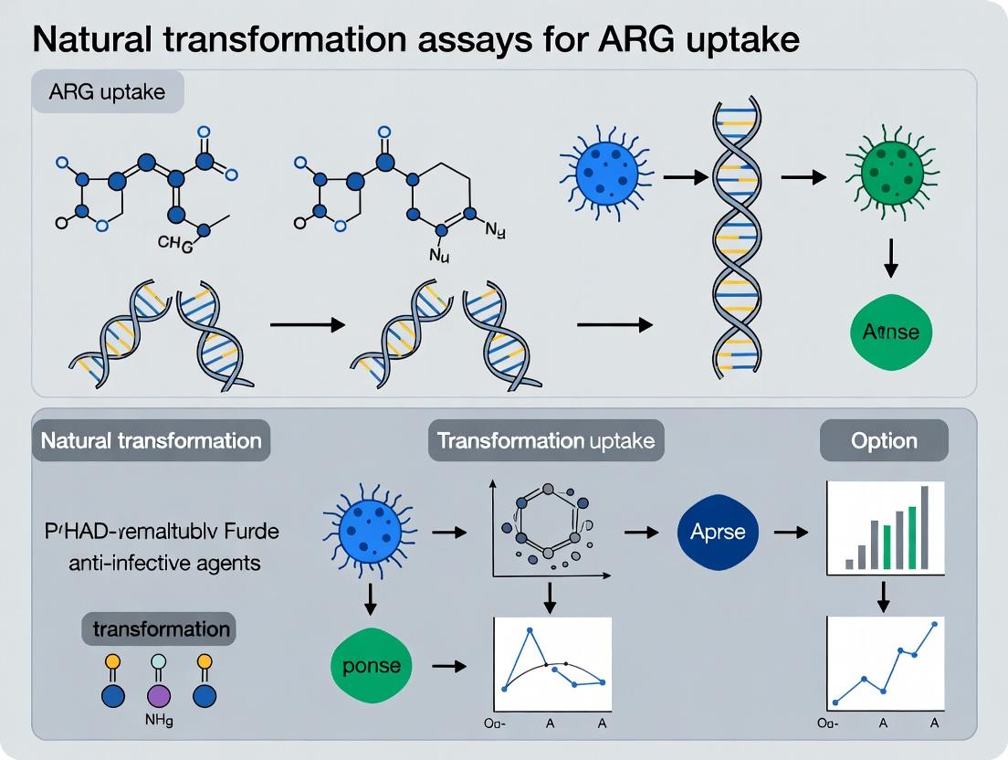
Natural Transformation Assays for ARG Uptake: Methods, Protocols, and Applications in Antimicrobial Resistance Research
This comprehensive guide explores natural transformation assays as critical tools for studying the horizontal gene transfer of antimicrobial resistance genes (ARGs).
Natural Transformation Assays for ARG Uptake: Methods, Protocols, and Applications in Antimicrobial Resistance Research
Abstract
This comprehensive guide explores natural transformation assays as critical tools for studying the horizontal gene transfer of antimicrobial resistance genes (ARGs). Tailored for researchers and drug development professionals, the article details the foundational biology of natural competence, provides step-by-step methodological protocols for in vitro and in vivo assays, addresses common troubleshooting and optimization challenges, and compares validation techniques. It serves as a practical resource for quantifying and characterizing ARG uptake dynamics to understand and combat the spread of antimicrobial resistance.
Understanding Natural Competence: The Biological Basis of ARG Uptake
Natural transformation is a genetically programmed, energy-dependent process by which competent bacteria actively take up free extracellular DNA (eDNA) and incorporate it into their genome. Within the critical context of antibiotic resistance gene (ARG) dissemination, natural transformation serves as a direct pathway for the uptake of ARG-bearing eDNA from environmental reservoirs (e.g., wastewater, soil biofilms, gut microbiomes). This mechanism bypasses the need for donor cells, enabling the acquisition of resistance even from lysed bacterial populations. Research employing natural transformation assays is pivotal for quantifying the transfer frequencies of specific ARGs under various environmental and clinical stressors, thereby informing risk assessments and mitigation strategies.
Core Mechanism and Signaling Pathways
Natural transformation is a multi-stage process regulated by complex signaling networks that respond to environmental cues such as nutrient limitation, cell density, and DNA availability.
Diagram 1: Core Mechanism of Natural Transformation
Diagram 2: Key Competence Regulation Pathway (Streptococcus pneumoniae)
Application Notes: Key Parameters Influencing ARG Uptake
Natural transformation efficiency for ARG acquisition is not constant; it is modulated by a confluence of factors. The following table summarizes quantitative data from recent studies (2022-2024) on key influencing parameters.
Table 1: Environmental & Physiological Parameters Affecting ARG Transformation Frequency
| Parameter | Experimental Model | Effect on Transformation Frequency (vs. Control) | Key Implication for ARG Spread |
|---|---|---|---|
| Sub-inhibitory Antibiotic (Tetracycline, 1/4 MIC) | Acinetobacter baylyi | ~10^3 fold increase (Nweke et al., 2023) | Antibiotic pollution can directly stimulate competence and HGT. |
| DNA Concentration (plasmid with bla_{NDM-1}) | Acinetobacter baumannii | Saturation curve: Max ~5×10^-4 CFU/recipient at ≥500 ng/mL (Wang et al., 2022) | ARG density in environment dictates transfer risk. |
| Species/Mating Pair | Vibrio cholerae (donor DNA) -> Various spp. | Varies: 10^-6 (V. cholerae) to <10^-9 (E. coli) (Ellison et al., 2023) | Phylogenetic barriers exist but are not absolute. |
| Temperature | Pseudomonas stutzeri | Optimal at 30°C; ~50% reduction at 25°C or 37°C (Trend et al., 2024) | Climate/season may modulate environmental HGT rates. |
| Metal ions (Ca^2+) | Streptococcus pneumoniae | Essential; No transformation in Ca^2+-free media (Standard protocol) | Ionic composition of niches (e.g., respiratory tract) is critical. |
Detailed Experimental Protocols
Protocol 1: Standard Quantitative Natural Transformation Assay for Acinetobacter baylyi ADP1 (Liquid Medium) Objective: To quantify the transformation frequency of a chromosomal or plasmid-borne ARG into a competent recipient. Materials: See "The Scientist's Toolkit" below. Procedure: 1. Competence Induction: Grow recipient strain (e.g., ADP1 ΔcomEC as negative control, wild-type as experimental) in 5 mL LB at 30°C to mid-exponential phase (OD600 ~0.4-0.6). 2. DNA Preparation: Purify donor DNA (genomic DNA from an ARG-bearing strain or plasmid DNA) and quantify. Prepare a dilution series (e.g., 0.1 µg/mL to 10 µg/mL final concentration). 3. Transformation Reaction: Mix 100 µL of recipient culture with 1-10 µL of donor DNA in a sterile microcentrifuge tube. Include a no-DNA control. Incubate statically for 90 minutes at 30°C. 4. Selection: Plate the entire reaction mixture, or appropriate serial dilutions, onto selective agar plates containing the relevant antibiotic for the ARG. Also plate onto non-selective agar for total viable count (TVC). 5. Incubation & Calculation: Incubate plates at 30°C for 24-48 hours. Count colonies. Transformation Frequency = (CFU on selective plate) / (TVC from non-selective plate).
Protocol 2: High-Throughput Microplate-Based Competence Induction Assay (Promoter-GFP Fusion) Objective: To screen chemical libraries or environmental samples for compounds that induce/repress the competence regulon. Materials: 96-well black microplates, plate reader (fluorescence, OD), strain with P_{comX}-gfp transcriptional fusion. Procedure: 1. Inoculate reporter strain into fresh medium in a 96-well plate. Add test compounds to respective wells. Include known inducer (positive control) and medium only (negative control). 2. Incubate plate in a plate reader at 37°C with continuous shaking. Measure OD600 (growth) and GFP fluorescence (ex/em ~485/520 nm) every 15-30 minutes for 12-16 hours. 3. Normalize GFP fluorescence to OD600 for each time point. Analyze the peak fluorescence/OD ratio or area under the curve to quantify induction level relative to controls.
Diagram 3: Standard Transformation Assay Workflow
The Scientist's Toolkit: Key Research Reagent Solutions
Table 2: Essential Materials for Natural Transformation Assays
| Item/Reagent | Function & Application | Example/Note |
|---|---|---|
| Competent Model Strain | Genetically tractable, reliably transformable species for mechanistic studies. | Acinetobacter baylyi ADP1, Bacillus subtilis 168, Streptococcus pneumoniae CP1500. |
| Defined Competence-Inducing Media | Provides reproducible, controlled conditions for competence development. | MIV (for V. cholerae), CSP-supplemented CAT medium (for S. pneumoniae). |
| Purified Donor DNA | Substrate for transformation; quality and concentration are critical variables. | Genomic DNA from ARG donor, PCR-amplified ARG cassettes, or plasmid DNA. |
| Selective Agar Plates | Allows selective outgrowth of successful transformants carrying the ARG. | LB or defined agar supplemented with specific antibiotic (e.g., carbenicillin for bla_{TEM-1}). |
| Competence-Specific Reporter Strain | Enables monitoring of competence regulation without selection. | Strain with fluorescence (gfp/lux) under control of a competence-specific promoter (e.g., comX). |
| ComEC / ComA Mutant Strains | Essential negative controls to distinguish transformation from other HGT events. | ΔcomEC (DNA import defect) should yield zero transformants. |
The Role of Natural Competence in the Spread of Antimicrobial Resistance
Application Notes
Natural competence, the genetically programmed ability of bacteria to take up extracellular DNA, is a significant driver of horizontal gene transfer (HGT) and the dissemination of antimicrobial resistance genes (ARGs). This process allows competent bacteria to integrate exogenous DNA into their genome, rapidly acquiring new traits, including resistance, without the need for mobile genetic elements. Research within the broader thesis on natural transformation assays aims to quantify this phenomenon, identify environmental and genetic regulators, and assess its contribution to the AMR crisis in clinical and environmental settings.
Key Quantitative Findings: Recent studies highlight the prevalence and efficiency of natural transformation. The following table summarizes critical data on transformation frequencies and ARG uptake across key bacterial species.
Table 1: Documented Natural Transformation Frequencies for ARG Uptake
| Bacterial Species | ARG Acquired | Substrate (DNA form) | Avg. Transformation Frequency (transformants/μg DNA/recipient) | Key Condition(s) | Reference (Year) |
|---|---|---|---|---|---|
| Acinetobacter baylyi | blaOXA-23 | Linear fragment | 5.4 x 10^-4 | Late exponential phase, 30°C | Cooper et al. (2023) |
| Streptococcus pneumoniae | mef(E), tet(M) | Chromosomal DNA | 2.1 x 10^-5 | Competence peptide induction, pH 7.8 | Wüthrich et al. (2022) |
| Neisseria gonorrhoeae | penA mosaic | Genomic DNA | 8.7 x 10^-6 | Microaerobic, 37°C | Liao et al. (2024) |
| Pseudomonas stutzeri | qnrB | Plasmid | 3.2 x 10^-7 | Biofilm state, low nutrient | Sharma & Finkel (2023) |
| Vibrio cholerae | sul2, strAB | Chitin-associated DNA | 1.8 x 10^-5 | Chitin surface, natural seawater | Pinto et al. (2023) |
Environmental & Clinical Relevance: Natural transformation is not confined to the laboratory. Conditions inducing competence, such as antibiotic stress, DNA damage, nutrient limitation, and biofilm formation, are frequently encountered in host organisms, wastewater, and soil. This facilitates the transfer of ARGs between commensals and pathogens, complicating infection treatment and accelerating the emergence of multidrug-resistant strains.
Experimental Protocols
Protocol 1: Standard Quantitative Natural Transformation Assay forAcinetobacterspp.
Objective: To quantify the uptake and genomic integration of an ARG-containing DNA fragment into a naturally competent recipient strain.
Research Reagent Solutions & Essential Materials:
| Item | Function/Brief Explanation |
|---|---|
| Competent Recipient Strain (e.g., A. baylyi ADP1 ΔcomEC) | Isogenic mutant defective in DNA uptake; serves as negative control. |
| Purified Donor DNA (Linear PCR amplicon, ~2kb) | Contains target ARG flanked by homology arms for recombination. |
| LB Broth & Agar (with/without selective antibiotic) | For cell growth and selection of transformants. |
| DNase I (1 mg/mL stock) | Control treatment to confirm transformation is DNA-dependent. |
| Competence-Inducing Buffer (CIB): 5 mM MgCl2, 5 mM CaCl2, 0.5% BSA, 10 mM Tris-HCl pH 7.5 | Divalent cations stabilize DNA and facilitate membrane passage. |
| Selective Antibiotic (e.g., Imipenem for blaOXA-23) | Selects for transformants that have acquired functional resistance. |
| Glass Beads (for spreading) | For even distribution of cells on agar plates. |
Methodology:
- Culture Preparation: Grow the wild-type competent recipient strain and the negative control mutant overnight in LB at 30°C with shaking.
- Competence Induction: Sub-culture 1:100 into fresh LB. Grow to mid-late exponential phase (OD600 ~0.6-0.8), a known competence window for many species.
- Transformation Reaction: Aliquot 100 μL of cells into sterile microcentrifuge tubes.
- Test: Cells + 100 ng purified donor DNA.
- DNase Control: Cells + 100 ng DNA + 1 μL DNase I (incubate 15 min at 37°C before plating).
- DNA-only Control: Donor DNA plated alone on selective media.
- Cell-only Control: Cells without donor DNA. Incubate mixtures for 90 minutes at 30°C without shaking.
- Selection & Enumeration: Serially dilute reactions in CIB. Plate dilutions onto both non-selective LB agar (for total viable count) and LB agar containing the relevant antibiotic (for transformant count). Use glass beads for spreading.
- Incubation & Calculation: Incubate plates for 24-48 hours at 30°C. Count colonies. Transformation Frequency = (Number of transformants on selective plate) / (Total viable cells on non-selective plate). Normalize per microgram of DNA if concentration varies.
Protocol 2: Biofilm-Associated Natural Transformation Assay
Objective: To assess ARG uptake under biofilm conditions, which mimic many natural environments.
Research Reagent Solutions & Essential Materials:
| Item | Function/Brief Explanation |
|---|---|
| Flow-cell or 96-well Polystyrene Microtiter Plate | Substrate for biofilm growth. |
| Minimal Media with Low Nutrients (e.g., M63) | Mimics environmental conditions and can induce competence. |
| Fluorescently-labeled Donor DNA (e.g., Cy3-dCTP labeled) | Allows visualization of DNA uptake within the biofilm matrix via microscopy. |
| Concanavalin A-Tetramethylrhodamine (ConA-TMR) | Stain for biofilm extracellular polymeric substance (EPS). |
| Confocal Laser Scanning Microscope (CLSM) | For high-resolution 3D imaging of biofilm and DNA localization. |
Methodology:
- Biofilm Establishment: Inoculate flow-cells or microtiter wells with the bacterial strain in minimal media. Incubate statically for 24-72 hours to allow biofilm formation.
- DNA Exposure: Gently introduce a solution containing both fluorescently-labeled DNA (500 ng/mL) and unlabeled ARG-containing DNA (100 ng/mL) into the system. Incubate for 2-6 hours.
- Control Setup: Include samples with DNase I-treated DNA mixture.
- Biofilm Fixation & Staining: Fix biofilms with 4% paraformaldehyde. Stain EPS with ConA-TMR (100 μg/mL) for 30 min. Wash gently.
- Imaging & Analysis: Visualize using CLSM. Z-stack images will show co-localization of fluorescent DNA (green) within bacterial microcolonies (stained red for EPS). Process images to quantify fluorescence intensity per biomass volume.
- Viability & Transformation Assessment: In parallel wells, after DNA exposure, disrupt biofilm by sonication/vortexing with beads. Plate serial dilutions on selective and non-selective media as in Protocol 1 to determine transformation frequency within the biofilm population.
Diagrams
Title: Natural Competence Pathway for ARG Uptake
Title: Workflow: Quantitative Natural Transformation Assay
Key Bacterial Species Known for Natural Competence (e.g., Streptococcus, Acinetobacter, Neisseria).
This document provides essential application notes and detailed protocols for working with key naturally competent bacterial species, specifically Streptococcus pneumoniae, Acinetobacter baylyi (and related pathogens), and Neisseria gonorrhoeae. Within the broader thesis on "Natural transformation assays for ARG (Antibiotic Resistance Gene) uptake research," these protocols are foundational for quantifying and understanding the horizontal transfer of genetic material, a primary driver of antibiotic resistance dissemination.
Application Notes: Species-Specific Competence Physiology & Relevance to ARG Uptake
| Species | Inducing Signal/ Condition | Primary DNA Uptake Specificity | Optimal Growth for Competence | Key Regulator(s) | Relevance to ARG Research |
|---|---|---|---|---|---|
| Streptococcus pneumoniae | Competence-Stimulating Peptide (CSP), Quorum Sensing | Non-specific, but prefers S. pneumoniae DNA (uptake sequence: 5'-AGCAGTCTGAAGC-3') | Early stationary phase in CAT medium (pH ~8.0) | ComABCDE, ComX | Model organism for Gram+ ARG transfer (e.g., pbp genes, ermB, tetM). |
| Acinetobacter baylyi (ADP1) | Nutritional Starvation (e.g., Lactate minimal medium) | Virtually non-specific (highly promiscuous) | Late exponential/stationary phase in minimal medium | ComP, ComE, CRP | Ideal for environmental DNA scavenging studies and tracking ARG (e.g., blaOXA) uptake from complex samples. |
| Neisseria gonorrhoeae | Microaerobic conditions, cAMP, contact with epithelial cells? | Highly specific via 10-bp DNA Uptake Sequence (DUS: 5'-GCCGTCTGAA-3') | Log-phase on GC agar + Kellogg's supplements | TfoX, CRP, RegF | Critical for studying ARG acquisition in pathogens (e.g., penA mosaicism, tetM acquisition). |
Table 1: Comparative physiology of natural competence in key bacterial species.
Core Protocol: Standard Natural Transformation Assay for ARG Uptake
This generalized protocol can be adapted for the species in Table 1.
Objective: To quantify the uptake and functional integration of exogenous antibiotic resistance DNA into a competent bacterial recipient.
Research Reagent Solutions Toolkit
| Item | Function | Example/Specification |
|---|---|---|
| Competence-Inducing Medium | Provides chemical/physical signals to trigger competence state. | CAT medium (S. pneumoniae), Lactate Minimal Medium (A. baylyi), GC Base + Supplements (N. gonorrhoeae). |
| Purified Donor DNA | Source of ARG for uptake. | Genomic DNA from resistant strain, or PCR-amplified ARG cassette. Typically 0.1-1 µg/mL final concentration. |
| DNase I (Sterile) | Control enzyme to degrade extracellular DNA, confirming transformation is internalization-dependent. | 10-100 µg/mL, added to control tubes after DNA incubation. |
| Selection Agar Plates | Allows growth only of transformants that acquired the ARG. | Contains antibiotic at predetermined MIC breakpoint for recipient strain. |
| Competence-Specific Reporter | Optional, for monitoring competence development. | Plasmid with promoter of key competence gene (e.g., comX, tfoX) fused to luciferase or GFP. |
Procedure:
- Culture Preparation: Grow the recipient bacterial strain to the optimal phase for competence (see Table 1) in the appropriate competence-inducing medium.
- Competence Induction: Divide culture into aliquots. For S. pneumoniae, add synthetic CSP (100 ng/mL final).
- Transformation Reaction:
- To experimental tube(s), add purified donor DNA containing the ARG. Mix gently.
- To negative control tube, add an equal volume of buffer or DNase I-treated DNA.
- To DNase control tube, incubate DNA with culture for 5 min, then add DNase I to degrade non-internalized DNA.
- Incubation: Incubate reactions at species-optimal temperature (usually 37°C) for 30-90 minutes to allow DNA uptake and recombination.
- Termination & Plating: Halt transformation by dilution in cold medium or by adding DNase I. Pellet cells, resuspend in fresh medium, and plate serial dilutions onto non-selective (for total CFU count) and antibiotic-containing selective agar plates.
- Calculation: Incubate plates for 24-48 hours. Transformation Frequency = (CFU on selective plate) / (Total CFU on non-selective plate).
Protocol Variation: High-Throughput ARG Uptake Screen inA. baylyi
Objective: To screen environmental or synthetic DNA samples for functional ARGs using A. baylyi ADP1's promiscuous competence.
Workflow:
- Grow A. baylyi to late exponential phase in rich (LB) medium.
- Wash cells twice and resuspend in lactate minimal medium to induce starvation/competence.
- Dispense 96-well plate with: test DNA samples, positive control (known ARG DNA), negative control (no DNA).
- Add bacterial suspension to each well. Incubate statically for 2 hours at 30°C.
- Using a replicator or multichannel pipette, spot the mixtures onto large LB agar plates containing the antibiotic of interest.
- After growth, count resistant colonies. Transformation frequency can be normalized to DNA concentration.
Visualization of Competence Pathways and Workflows
Title: S. pneumoniae Competence Quorum Sensing Pathway (76 chars)
Title: Generic Natural Transformation Assay Workflow (56 chars)
Regulatory Networks and Environmental Triggers for Competence Development
Within the broader thesis on Natural transformation assays for ARG (Antibiotic Resistance Gene) uptake research, understanding competence development is fundamental. Competence is the genetically programmed physiological state in which bacteria can uptake extracellular DNA, a primary route for ARG dissemination. This document details the regulatory networks controlling competence and the environmental triggers that induce it, providing application notes and protocols for studying these processes in model transformable species like Streptococcus pneumoniae, Bacillus subtilis, and Vibrio cholerae.
Core Regulatory Networks & Key Quantitative Data
Competence networks are species-specific but often involve phosphorylay systems, peptide pheromones, and transcriptional regulators. Core quantitative parameters for major models are summarized below.
Table 1: Key Competence Regulatory Components and Dynamics
| Species | Master Regulator | Key Inducing Signal | Peak Competence Window (min post-induction) | Estimated % of Competent Cells in Population | Reference Year |
|---|---|---|---|---|---|
| Streptococcus pneumoniae | ComX (SigX) | Competence-Stimulating Peptide (CSP) | 10-20 | ~100% (in controlled conditions) | 2023 |
| Bacillus subtilis | ComK | Nutrient depletion, cell density | 90-180 | ~10-20% | 2022 |
| Vibrio cholerae | TfoX, QstR | Chitin, carbon source shift | 240-360 | Variable, up to ~30% | 2023 |
| Haemophilus influenzae | Sxy | cAMP via purine starvation | 30-60 | High | 2021 |
| Neisseria gonorrhoeae | CrgA, IHF | Unknown (constitutive?) | Constitutive | ~100% (in log phase) | 2022 |
Table 2: Environmental Triggers and Their Experimentally Determined Thresholds
| Trigger | Relevant Species | Typical Experimental Concentration/ Condition | Primary Sensor/ Receptor |
|---|---|---|---|
| Synthetic CSP (CSP-1) | S. pneumoniae (Rv304) | 50-100 ng/mL | ComD (Histidine Kinase) |
| Chitin Oligosaccharides | V. cholerae | 0.5% (w/v) colloidal chitin | ChiS (Sensor Kinase) |
| Cell Density (Quorum) | S. pneumoniae, B. subtilis | ~10^7 - 10^8 CFU/mL | ComD, ComP (Histidine Kinases) |
| Antibiotic Stress (e.g., Mitomycin C) | S. pneumoniae | Sub-inhibitory, e.g., 0.05 µg/mL | Linked to SOS response? |
| Nutrient Limitation (Starvation) | B. subtilis | M9 minimal medium, stationary phase | Multiple metabolic sensors |
Detailed Experimental Protocols
Protocol 3.1: Inducing and Monitoring Competence inStreptococcus pneumoniae
Objective: To synchronously induce competence via synthetic CSP and measure transformation frequency. Materials: See Scientist's Toolkit (Section 5). Procedure:
- Culture Growth: Grow strain of interest (e.g., R800 or D39 derivative) in C+Y medium at 37°C, 5% CO₂ to an OD₅₉₀ ~0.03-0.05.
- Competence Induction: Split culture. To the experimental sample, add synthetic CSP-1 to a final concentration of 100 ng/mL. The control sample receives an equal volume of solvent (e.g., 0.1% acetic acid).
- Incubation: Incubate cultures for 10 minutes.
- Transformation Assay: Add 1 µg of donor DNA (containing a selectable marker, e.g., rpsL1 for streptomycin resistance) to 1 mL of competent culture. For a no-DNA control, add TE buffer.
- Uptake Phase: Incubate for 20 minutes at 30°C to allow DNA uptake/integration.
- Quenching: Add 100 U of recombinant DNase I (commercial solution) to degrade non-internalized DNA. Incubate for 10 minutes at 37°C.
- Outgrowth: Dilute cultures 1:10 in fresh pre-warmed C+Y medium. Incubate for 2 hours to allow expression of antibiotic resistance.
- Plating: Plate serial dilutions on non-selective (total viable count) and selective agar (e.g., containing 200 µg/mL streptomycin).
- Calculation: Transformation Frequency = (CFU/mL on selective agar) / (CFU/mL on non-selective agar).
Protocol 3.2: Assessing Competence viacomX-sfgfpTranscriptional Reporter inB. subtilis
Objective: To quantify the dynamics and heterogeneity of competence development using fluorescence. Procedure:
- Strain Preparation: Use a B. subtilis strain harboring a transcriptional fusion of the comX promoter to a stable GFP variant (e.g., sfgfp) at an ectopic locus (amyE).
- Induction Setup: Inoculate strain in competence medium (e.g., Modified competence medium, MM). Grow with shaking at 37°C. Monitor OD₆₀₀.
- Sampling: At T₀ (early exponential, OD₆₀₀ ~0.1) and every 30 minutes thereafter for 4 hours, sample 1 mL of culture.
- Flow Cytometry: a. Fix cells immediately with 1% formaldehyde for 15 min on ice. Wash with PBS. b. Resuspend in PBS and analyze using a flow cytometer with a 488 nm laser and 530/30 nm BP filter. c. Collect data for at least 50,000 events per sample.
- Data Analysis: Gate on live cell population using FSC/SSC. Determine the mean fluorescence intensity (MFI) and the percentage of cells above a fluorescence threshold (determined from a ΔcomK negative control strain).
Visualizations (Pathways & Workflows)
Title: S. pneumoniae Competence Regulatory Pathway
Title: General Natural Transformation Assay Workflow
The Scientist's Toolkit
Table 3: Essential Research Reagent Solutions for Competence Studies
| Item | Function & Application | Example/Notes |
|---|---|---|
| Synthetic CSP Peptides | Chemically defined inducer for S. pneumoniae competence. Eliminates variability of auto-induction. | CSP-1 (EMRLSKFFRDFILQRKK). Aliquot in 0.1% acetic acid, store at -80°C. |
| Colloidal Chitin | Natural substrate to induce competence in V. cholerae and other chitinolytic species. | Prepare from crab shells; use at 0.5% w/v in induction medium. |
| Competence-Specific Media (C+Y, MM) | Chemically defined media that supports growth and reproducible competence development. | C+Y for S. pneumoniae; Modified Competence Medium (MM) for B. subtilis. |
| DNase I (Recombinant) | Enzymatically degrades extracellular DNA after uptake phase, critical for accurate transformation frequency calculation. | Use at 100 U/mL for 10 min; ensures only internalized DNA is scored. |
| Fluorescent Transcriptional Reporters | Reporters (e.g., PcomX-sfGFP) enable real-time, single-cell monitoring of competence gene expression. | Integrated at ectopic locus; analyzed via flow cytometry or microscopy. |
| qPCR Primers for comX, recA, etc. | Quantify absolute levels of competence gene transcripts to correlate with transformability. | Use 16S rRNA as endogenous control. SYBR Green or probe-based assays. |
| Selective Agar with Antibiotics | For selection of transformants that have acquired ARG markers from donor DNA. | Concentration must be pre-tested for the specific strain and resistance marker. |
Within the broader thesis on Natural Transformation Assays for Antibiotic Resistance Gene (ARG) Uptake Research, understanding the precise molecular pathway from extracellular DNA capture to stable genomic inheritance is paramount. This pathway is a key driver of horizontal gene transfer (HGT), facilitating the spread of antimicrobial resistance (AMR) in bacterial populations. These Application Notes detail the experimental protocols and reagents required to dissect this transformation pathway, providing a framework for researchers to quantify and inhibit ARG acquisition.
Natural transformation is a regulated, multi-stage process enabling competent bacteria to take up free DNA and integrate it into their genome. The pathway consists of four primary phases:
- Competence Development: A physiological state triggered by quorum-sensing or stress response signals, leading to the expression of DNA uptake machinery.
- DNA Binding & Uptake: Surface-exposed proteins bind double-stranded DNA (dsDNA), which is then cleaved and transported as single-stranded DNA (ssDNA) into the cytoplasm.
- Cytoplasmic Protection & Transport: The ssDNA is bound by protective proteins (e.g., SSB, RecA) and shuttled to the replication fork or homologous site.
- Genomic Integration: Via homologous recombination (HR), the incoming ssDNA is integrated into the chromosome, resulting in a transformed genotype.
Key Research Reagent Solutions
Essential materials for studying the natural transformation pathway are listed below.
Table 1: The Scientist's Toolkit for Transformation Pathway Research
| Reagent/Material | Function in Research |
|---|---|
| Competence-Inducing Peptides (e.g., CSP for S. pneumoniae) | Synthetic peptides used to artificially induce the competence state in laboratory cultures for synchronized transformation experiments. |
| Fluorescently-Labeled DNA Probes (e.g., Cy3-dsDNA) | Allow visualization and quantification of DNA binding to the cell surface and uptake kinetics using flow cytometry or fluorescence microscopy. |
| Recombinant RecA Protein | Used in in vitro assays to study the kinetics of strand invasion and homologous pairing during the integration phase. |
| Homologous Donor DNA | Purified DNA fragment containing an ARG (e.g., ermB, blaZ) flanked by regions homologous to the recipient genome. Essential for measuring transformation frequency. |
| Selective Agar Plates (Antibiotic-Containing) | Used to plate transformation mixtures and select for clones that have successfully integrated the ARG. Critical for quantifying transformation efficiency. |
| SSB (Single-Stranded Binding) Protein Mutants | Proteins with altered affinity for ssDNA used to elucidate the role of cytoplasmic protection in transformation success. |
| qPCR Primers for com Genes | Primers targeting competence-specific genes (e.g., comE, comX) to monitor competence development at the transcriptional level. |
| Membrane Integrity Dyes (e.g., PI) | Distinguish between competent (permeable) and non-competent subpopulations in a culture. |
Experimental Protocols
Protocol 4.1: Induction of Competence and Quantitative Transformation Assay
Objective: To measure the frequency of ARG uptake and genomic integration in a model naturally competent bacterium (e.g., Streptococcus pneumoniae).
Materials:
- Competence-Inducing Peptide (CIP) stock solution
- Recipient bacterial strain (ARS)
- Donor DNA fragment (ARG flanked by ~1kb homology arms)
- Selective antibiotic plates
- Non-selective control plates
- CAT medium (pH 8.0, pre-warmed)
Procedure:
- Culture Growth: Grow the recipient strain to mid-exponential phase (OD₆₀₀ ~0.05).
- Competence Induction: Divide culture. To the experimental tube, add CIP at 100 ng/mL. The control tube receives no peptide.
- Transformation: 10 minutes post-induction, add 100-500 ng of purified donor DNA to both tubes. Incubate at 37°C for 30 minutes.
- Recombination Arrest: Add 10 U of DNase I to degrade non-internalized DNA. Incubate for 10 minutes.
- Recovery & Selection: Dilute cultures and plate on both non-selective and antibiotic-selective agar plates. Incubate for 24-48 hours.
- Calculation:
- Transformation Frequency = (CFU/mL on selective plate) / (CFU/mL on non-selective plate).
- Fold Induction = (Frequency with CIP) / (Frequency without CIP).
Protocol 4.2: Visualization of DNA Uptake via Flow Cytometry
Objective: To quantify the proportion of a bacterial population that binds and internalizes fluorescent DNA during competence.
Materials:
- Cy3-labeled dsDNA (homologous or heterologous)
- Competent bacterial culture (induced vs. non-induced)
- Flow cytometry buffer (PBS + 0.1% BSA)
- Membrane-impermeable DNA quencher (e.g., Trypan Blue)
Procedure:
- Sample Preparation: Induce competence as in Protocol 4.1. Aliquot 1 mL of induced and non-induced culture.
- DNA Binding: Add 50 nM Cy3-dsDNA to each aliquot. Incubate in the dark at 30°C for 10 minutes.
- Uptake vs. Binding Discrimination: Split each sample into two tubes. To one tube, add Trypan Blue (0.2 mg/mL) to quench extracellular/surface-bound fluorescence.
- Analysis: Immediately analyze all samples via flow cytometry, detecting fluorescence in the Cy3 channel (~550/570 nm excitation/emission).
- Gating: Gate the population of cells with high internalized fluorescence (Trypan Blue-treated, Cy3+). This represents the competent, DNA-importing subpopulation.
Table 2: Representative Transformation Frequencies for Key Pathogens
| Bacterial Species | Inducing Signal | Donor DNA (ARG) | Avg. Transformation Frequency (Range) | Key Reference (Example) |
|---|---|---|---|---|
| Streptococcus pneumoniae | Competence-Stimulating Peptide (CSP) | ermB (Homologous) | 1 x 10⁻³ (1 x 10⁻⁴ – 5 x 10⁻³) | Johnston et al., 2014 |
| Neisseria gonorrhoeae | Microaerobic Conditions | penA (Mosaic) | 5 x 10⁻⁵ (1 x 10⁻⁶ – 1 x 10⁻⁴) | Hamilton & Dillard, 2006 |
| Acinetobacter baylyi (ADP1) | Stationary Phase / Nutrient Starvation | aadB (Homologous) | 2 x 10⁻⁴ (1 x 10⁻⁵ – 1 x 10⁻³) | de Vries & Wackernagel, 2002 |
| Haemophilus influenzae | Cyclic AMP | cat (Specific USS sequence) | 1 x 10⁻² (1 x 10⁻³ – 5 x 10⁻²) | Goodgal, 1982 |
Table 3: Flow Cytometry Data: DNA Uptake Kinetics in S. pneumoniae
| Time Post-CSP (min) | % Cells Cy3+ (Surface Bound) | % Cells Cy3+ (Internalized, Quenched) | Mean Fluorescence Intensity (Internalized) |
|---|---|---|---|
| 0 (No CSP) | 1.2 ± 0.3 | 0.1 ± 0.05 | 102 ± 15 |
| 10 | 45.5 ± 5.2 | 28.7 ± 4.1 | 1850 ± 210 |
| 20 | 32.1 ± 3.8 | 35.2 ± 3.9 | 2250 ± 190 |
| 30 | 15.4 ± 2.1 | 18.5 ± 2.5 | 1650 ± 175 |
Pathway & Workflow Visualizations
Natural Transformation Pathway from Induction to Integration
Experimental Workflow for Quantitative Transformation Assay
Step-by-Step Protocols: Designing and Executing ARG Uptake Assays
Within the context of natural transformation assays for antibiotic resistance gene (ARG) uptake research, the precise selection of donor DNA, recipient competent cells, and selection markers is fundamental. This protocol details the core components and methodologies for establishing robust assays to study horizontal gene transfer mechanisms, critical for understanding the dissemination of antimicrobial resistance.
Donor DNA: Selection and Preparation
Donor DNA serves as the substrate for uptake and integration. Key considerations include size, purity, concentration, and genetic markers.
DNA Source and Type
- Genomic DNA (gDNA): Ideal for studying transformation with homologous sequences. Represents a natural scenario where chromosomal ARGs are acquired.
- Plasmid DNA: Used to study the acquisition of extrachromosomal genetic elements. Crucial for assessing plasmid-borne ARG spread.
- PCR Amplicons: Defined linear fragments containing the ARG of interest and flanking regions. Allows precise control over homology arms.
Key Parameters for Donor DNA
Quantitative parameters significantly impact transformation efficiency (TE).
Table 1: Donor DNA Preparation Parameters and Impact on Transformation Efficiency
| Parameter | Optimal Range (Typical) | Effect on Transformation | Protocol Consideration |
|---|---|---|---|
| Concentration | 10 ng/µL - 1 µg/µL | TE increases with concentration until saturation. High conc. can inhibit. | Perform a dose-response curve (0-2 µg) to determine optimum. |
| Purity (A260/A280) | 1.8 - 2.0 | Impurities (phenol, salts) inhibit uptake. | Use gel extraction or commercial clean-up kits. Ethanol precipitation is standard. |
| Size (gDNA) | >20 kb (for homology) | Larger fragments increase homologous recombination probability. | Gentle isolation (e.g., using lysozyme/proteinase K, no vortexing) to avoid shearing. |
| Size (Amplicon) | 1-5 kb (including homology arms) | Must encompass the ARG and sufficient flanking homology (~500-1000 bp each side). | Design primers with high-fidelity polymerase to minimize mutations. |
| State | Linear (for chromosomal integration) / Circular (for plasmid maintenance) | Linear DNA requires homology; circular plasmid may replicate autonomously. | For gDNA, verify fragmentation by pulse-field or standard gel electrophoresis. |
Protocol 1.1: Preparation of Purified Genomic Donor DNA from an ARG-bearing Strain
Objective: Isolate high-molecular-weight, pure genomic DNA for natural transformation assays.
- Culture Donor Strain: Grow the donor bacterium (e.g., Acinetobacter baylyi ADP1, Streptococcus pneumoniae, or Neisseria gonorrhoeae) harboring the ARG to mid-exponential phase (OD600 ~0.5-0.8) in appropriate media.
- Harvest Cells: Pellet 1.5 mL of culture at 8,000 x g for 2 min. Resuspend in 500 µL of TE buffer (pH 8.0).
- Cell Lysis: Add 30 µL of 10% SDS and 3 µL of Proteinase K (20 mg/mL). Incubate at 56°C for 1-2 hours until clear.
- DNA Precipitation: Add 500 µL of phenol:chloroform:isoamyl alcohol (25:24:1). Mix gently by inversion for 10 min. Centrifuge at 12,000 x g for 10 min at 4°C. Transfer the upper aqueous phase to a new tube.
- Purification: Add 0.6 volumes of isopropanol and 0.1 volumes of 3M sodium acetate (pH 5.2). Mix gently to precipitate DNA. Spool out DNA with a pipette tip or glass rod.
- Wash and Resuspend: Wash the DNA in 70% ethanol, air-dry briefly, and resuspend in 100 µL of nuclease-free TE buffer. Incubate at 4°C overnight to ensure complete resuspension.
- Quantification & Storage: Measure concentration and purity via spectrophotometry. Verify size on a 0.8% agarose gel. Store at -20°C.
Competent Cells: Induction and Characterization
Competence is a physiological state enabling active DNA uptake. It can be natural or artificially induced.
Selection of Model Organisms
- High Natural Competence: Bacillus subtilis, S. pneumoniae, N. gonorrhoeae, Helicobacter pylori, A. baylyi.
- Inducible Competence: Escherichia coli (via chemical/electroporation) is used for cloning but is not naturally competent. Its use in ARG uptake studies is for control or plasmid propagation.
Critical Factors for Competence Development
Table 2: Factors Influencing Competence Development and Efficiency
| Factor | Impact on Competence | Standard Condition / Method for Induction |
|---|---|---|
| Growth Phase | Often transient, peaking in mid-late exponential phase. | Monitor OD600 closely. For B. subtilis, competence peaks at the end of exponential growth (OD600 ~0.8-1.0 in competence medium). |
| Nutritional Status | Starvation for carbon, nitrogen, or phosphorus can induce competence. | Use competence-specific media (e.g., MIV for Vibrio cholerae, MII for N. gonorrhoeae). |
| Cell Density (Quorum Sensing) | Essential for some species (S. pneumoniae, B. subtilis). | Use appropriate starting density and ensure proper aeration during pre-culture. |
| Temperature | Optimal growth temperature is typically required. | 37°C for most human pathogens; 30°C for some environmental isolates. |
| Inducing Peptides/Signals | Required for competence pheromone systems. | Add synthetic competence-stimulating peptide (CSP) for Streptococci at 50-200 ng/mL. |
Protocol 2.1: Induction of Natural Competence inStreptococcus pneumoniae
Objective: Prepare a culture of S. pneumoniae highly competent for DNA uptake.
- Pre-culture: Inoculate S. pneumoniae from a frozen stock onto a blood agar plate. Incubate overnight at 37°C with 5% CO₂.
- Starter Culture: Pick a single colony and inoculate 5 mL of C medium (a casein hydrolysate-based semi-defined medium) supplemented with 0.2% yeast extract. Grow to OD550 ~0.1 at 37°C without CO₂.
- Competence Induction: Dilute the starter culture 100-fold into fresh pre-warmed C medium. Grow until OD550 reaches 0.04-0.05.
- Pheromone Addition: Add synthetic competence-stimulating peptide (CSP-1 at 100 ng/mL final concentration). Continue incubation for 10-15 minutes. Cells are now maximally competent.
- Transformation: Immediately add donor DNA (10-100 ng of PCR amplicon or gDNA) to 1 mL of competent cells. Incubate for 30-60 minutes at 37°C to allow for uptake and integration.
Selection Markers: Design and Application
Selection markers enable the isolation of transformants that have successfully acquired the donor ARG.
Types of Selection Markers
- Antibiotic Resistance: The most direct marker for ARG uptake studies (e.g., Kanamycin, Ampicillin, Chloramphenicol resistance). Selects directly for the acquired trait.
- Auxotrophic Markers: Complementation of a metabolic deficiency in the recipient (e.g., leuB, trpE). Useful when studying ARGs without an inherent selectable phenotype.
- Fluorescent or Chromogenic Reporters: Genes like gfp, rfp, or lacZ can be fused to the ARG or placed downstream to visualize uptake events.
Selection Scheme Design
- Direct Selection: Plate transformation mix on media containing the antibiotic to which the donor ARG confers resistance.
- Counterselection: Use an antibiotic or condition to which the recipient is sensitive but the donor is resistant, or vice versa, to eliminate one population.
- Dual/Marker Rescue: Use a two-step selection where the ARG is linked to a second, distinct marker to confirm integration.
Table 3: Common Selection Markers and Their Applications in ARG Uptake Assays
| Marker Gene | Resistance/Function | Typical Working Concentration (in media) | Notes for Natural Transformation Assays |
|---|---|---|---|
| aph(3')-IIIa (KanR) | Kanamycin / Neomycin | 50 µg/mL (for E. coli), 250-500 µg/mL (for Gram+) | Common in Gram+ and Gram- cassettes. Verify recipient's innate sensitivity. |
| bla (AmpR) | Ampicillin / Amoxicillin | 100 µg/mL (for E. coli) | Common in plasmid studies. Ineffective for many natural producers of β-lactamases. |
| cat (CmR) | Chloramphenicol | 5-20 µg/mL (for E. coli), 5 µg/mL (for S. pneumoniae) | Useful for low-background selection; ensure recipient is sensitive. |
| erm (EryR) | Erythromycin | 1 µg/mL (for S. pneumoniae), 150 µg/mL (for E. coli) | Common in Gram+ systems. Can be used for inducible gene expression. |
| rpsL (StrR) | Streptomycin | Conferring resistance via specific point mutation. | Useful for allelic exchange when recipient has a wild-type (sensitive) rpsL gene. |
Protocol 3.1: Selection and Confirmation of Transformants
Objective: Isolate and verify clones that have acquired the ARG via natural transformation.
- Post-Uptake Recovery: After the transformation incubation (Protocol 2.1, Step 5), add 1 mL of rich recovery medium (e.g., Todd-Hewitt broth with 0.5% yeast extract for S. pneumoniae). Incubate for 1-2 hours at permissive temperature to allow expression of the resistance marker.
- Plating for Selection: Plate appropriate dilutions (e.g., 100 µL of undiluted and 10⁻¹) onto agar plates containing the selective antibiotic at the predetermined concentration. Also plate onto non-selective agar to determine total viable count.
- Incubation: Incubate plates under optimal growth conditions for 24-48 hours.
- Confirmation: Pick putative transformant colonies. Re-streak onto fresh selective plates. Confirm by:
- Colony PCR: Using primers specific to the acquired ARG.
- Antibiotic Sensitivity Profile: Test resistance to the target antibiotic and related ones.
- Sequencing: Sanger sequence the amplified ARG to confirm identity and lack of unintended mutations.
Diagrams
Diagram Title: Natural Transformation Workflow for ARG Uptake
Diagram Title: Competence Regulation in S. pneumoniae
The Scientist's Toolkit: Research Reagent Solutions
Table 4: Essential Materials for Natural Transformation Assays
| Item / Reagent | Function / Purpose in ARG Uptake Assays | Example Product / Specification |
|---|---|---|
| High-Fidelity DNA Polymerase | Amplify donor ARG amplicons with minimal error rates for precise homology-directed recombination. | Q5 High-Fidelity (NEB), Phusion (Thermo Scientific). |
| Gel Extraction Kit | Purify donor DNA (PCR amplicons, digested fragments) from agarose gels to remove primers and non-specific products. | QIAquick Gel Extraction Kit (Qiagen), Monarch Gel Extraction Kit (NEB). |
| Genomic DNA Isolation Kit | Isate pure, high-molecular-weight genomic DNA from donor strains with minimal shearing. | DNeasy Blood & Tissue Kit (Qiagen), MasterPure DNA Purification Kit (Lucigen). |
| Competence-Stimulating Peptide (CSP) | Chemically defined peptide to induce natural competence in streptococci and related species. | Custom synthetic peptide (>95% purity), resuspended in sterile water or DMSO. |
| Defined Competence Media | Nutritionally limited media to induce competence by mimicking starvation conditions. | MIV for V. cholerae, MII for Neisseria, C medium for S. pneumoniae. |
| Selective Agar Antibiotics | Solid media formulation for the selective outgrowth of transformants. | Prepared from stock solutions (e.g., 50 mg/mL Kanamycin in water, filter sterilized). Added to autoclaved, cooled agar. |
| Recovery Broth | Nutrient-rich, non-selective liquid media allowing expression of newly acquired resistance genes post-uptake. | Todd-Hewitt Broth with Yeast Extract (for Streptococci), LB Broth (for E. coli). |
| Colony PCR Master Mix | Rapid screening of putative transformant colonies for the presence of the acquired ARG. | OneTaq Quick-Load Master Mix (NEB), DreamTaq Green PCR Master Mix (Thermo Scientific). |
| RecA Protein / Antibody | Study the role of homologous recombination in integration. RecA is essential for strand invasion. | Recombinant RecA protein (for in vitro assays), anti-RecA antibody (for Western blot). |
Within the broader thesis on Natural transformation assays for Antibiotic Resistance Gene (ARG) uptake research, the Standard In Vitro Transformation Assay serves as a foundational methodology. It quantitatively measures the frequency of horizontal gene transfer via natural transformation, a critical route for ARG dissemination among bacterial populations. This protocol details the steps to induce competence, expose cells to extracellular DNA, and select for transformants, enabling researchers to study factors influencing ARG acquisition.
Key Research Reagent Solutions
The following table details essential materials for performing a standard in vitro transformation assay.
| Reagent / Material | Function in the Assay |
|---|---|
| Competence-Inducing Medium | A chemically defined medium (e.g., MIV, BHI+Ca/Mg) that triggers the physiological state of competence in the target bacterial strain, allowing DNA uptake. |
| Purified Donor DNA | Contains the antibiotic resistance marker or ARG of interest. Must be high-quality, double-stranded genomic or plasmid DNA. Concentration is critical. |
| Selective Agar Plates | Solid media containing the appropriate antibiotic to selectively allow only transformed cells (those that have taken up and expressed the ARG) to grow into colonies. |
| Non-Selective Agar Plates | Used for determining the total viable count of the competent cell population prior to selection, enabling transformation frequency calculation. |
| DNase I | Enzyme used in control reactions to degrade free DNA, confirming that antibiotic resistance arises from DNA uptake and not from spontaneous mutation or DNA adherence. |
| Competent Cells (Negative Control) | Cells processed identically but not exposed to donor DNA. Essential for assessing the background level of antibiotic-resistant mutants. |
Detailed Experimental Protocol
Part 1: Preparation of Competent Cells
- Day 1 - Starter Culture: Inoculate 5 mL of appropriate non-inducing growth medium (e.g., LB) with a single colony of the recipient bacterial strain. Incubate overnight at optimal growth temperature with shaking.
- Day 2 - Competence Induction:
- Dilute the overnight culture 1:100 into fresh, pre-warmed Competence-Inducing Medium. Typically, use 10-20 mL in a baffled flask for aeration.
- Incubate under optimal conditions (specific temperature, shaking) until the culture reaches the precise optical density (OD₆₀₀) known to coincide with peak competence for the strain (e.g., OD₆₀₀ = 0.05 - 0.15 for Acinetobacter baylyi ADP1). This point must be determined empirically.
- Harvest cells by centrifugation at 4,000 x g for 10 minutes at 4°C.
- Gently resuspend the cell pellet in 1/2 volume of ice-cold competence medium or a transformation buffer (often containing Ca²⁺/Mg²⁺).
- Keep cells on ice. Competence is now at its peak but will decline; use cells immediately.
Part 2: Transformation Reaction
- Set Up Reaction Tubes: On ice, prepare sterile 1.5 mL microcentrifuge tubes as follows:
- Transformation: 100 µL competent cells + 1-100 ng donor DNA (in 10 µL).
- DNase Control: 100 µL competent cells + 1-100 ng donor DNA, then add 1 µL of DNase I (1 U/µL), mix, and incubate for 5 min at room temperature before stopping the reaction.
- Cell Control: 100 µL competent cells + 10 µL sterile buffer/water (no DNA).
- Incubate: Incubate all tubes on ice for 30 minutes. This allows DNA to associate with cell surfaces.
- Heat Shock/Pulse: For some strains, apply a brief heat shock (e.g., 42°C for 90 seconds for E. coli made chemically competent). For naturally competent bacteria like Streptococcus pneumoniae or Acinetobacter spp., shift to the permissive growth temperature (e.g., 30°C) without shock.
- Recovery: Add 900 µL of rich, non-selective recovery medium (e.g., LB or BHI broth). Incubate for 1-2 hours at the optimal growth temperature with mild shaking to allow expression of the acquired antibiotic resistance gene.
Part 3: Plating and Calculation of Transformation Frequency
- Serial Dilution: Serially dilute each transformation reaction in sterile saline or buffer (e.g., 10⁻¹ to 10⁻⁴).
- Plating:
- Plate 100 µL of appropriate dilutions onto Selective Agar Plates containing the relevant antibiotic. Plate in duplicate.
- Plate 100 µL of appropriate dilutions (e.g., 10⁻⁵, 10⁻⁶) onto Non-Selective Agar Plates to determine the total viable cell count (CFU/mL).
- Incubation: Incubate all plates at the optimal growth temperature for 24-48 hours.
- Data Analysis:
- Count colonies on selective (transformants) and non-selective (total viable cells) plates.
- Calculate the transformation frequency using the formula: Transformation Frequency = (Number of transformants per mL) / (Total viable cells per mL)
Data Presentation
Table 1: Example Transformation Frequency Data for Acinetobacter baylyi ADP1 with strA (Streptomycin Resistance) DNA
| Experimental Condition | Total Viable Cells (CFU/mL) | Transformants (CFU/mL) | Transformation Frequency |
|---|---|---|---|
| Transformation (100 ng DNA) | 2.5 x 10⁸ | 5.0 x 10³ | 2.0 x 10⁻⁵ |
| DNase Control | 2.3 x 10⁸ | < 10 | < 4.3 x 10⁻⁸ |
| Cell Control (No DNA) | 2.6 x 10⁸ | < 10 | < 3.8 x 10⁻⁸ |
CFU: Colony Forming Units. The DNase and Cell controls confirm transformation is DNA-dependent.
Visualizing the Workflow and Key Pathways
Standard In Vitro Transformation Assay Workflow
Key Pathway for Natural Competence and DNA Uptake
Application Notes
Within the thesis framework investigating natural transformation as a critical pathway for antimicrobial resistance gene (ARG) dissemination, simulating real-world bacterial habitats is paramount. Standard planktonic culture models fail to capture the complexity of microbial communities where transformation predominantly occurs. Biofilm assays recapitulate the structured, matrix-encased consortia found in chronic infections and environmental reservoirs, where high cell density, nutrient gradients, and stress conditions upregulate competence machinery. In vivo mimicry assays, such as those using Galleria mellonella or synthetic human fluids, introduce key host factors—like immune components, shear forces, and physiologically relevant matrices—that modulate transformation frequency. These assays bridge the gap between in vitro findings and clinical relevance, providing robust data on ARG uptake dynamics under ecologically pertinent conditions.
Table 1: Comparative Natural Transformation Frequencies in Different Assay Systems
| Bacterial Species | Planktonic Culture (CFU/μg DNA) | Biofilm Assay (CFU/μg DNA) | In Vivo Mimicry (G. mellonella) | Key Condition Parameters |
|---|---|---|---|---|
| S. pneumoniae | 5.2 x 10⁻³ | 8.7 x 10⁻² | 3.1 x 10⁻¹ | Competence-stimulating peptide (CSP), Microaerophilic |
| A. baylyi | 2.1 x 10⁻² | 4.5 x 10⁻¹ | N/A | DNA starvation, Solid surface (agar) |
| P. aeruginosa | <1.0 x 10⁻⁶ | 2.3 x 10⁻⁵ | 1.8 x 10⁻⁴ | Sub-MIC antibiotic, Cystic Fibrosis sputum medium |
| V. cholerae | 7.8 x 10⁻⁴ | 1.4 x 10⁻² | N/A | Chitin surface, Natural seawater medium |
Table 2: Impact of Host-Mimic Conditions on ARG Uptake
| Mimic Condition | Transformation Frequency (Fold Change vs. Control) | Associated Stress/Inducer | Relevant ARG Captured |
|---|---|---|---|
| Synthetic Sputum (CF) | 12.5x | Oxidative stress, Nutrient limitation | blaTEM-1, mecA |
| Sub-inhibitory Ciprofloxacin | 45.2x | SOS Response | qnrB, aac(6')-Ib-cr |
| Galleria hemolymph | 8.7x | Antimicrobial peptides, Low Mg²⁺ | vanA, armA |
| Artificial Urine | 3.3x | High Osmolarity, Urea | fosA, ctx-M-15 |
Experimental Protocols
Protocol 1: Static Biofilm Natural Transformation Assay
Objective: To quantify natural transformation frequencies for ARG uptake within a mature biofilm.
Materials: See "The Scientist's Toolkit" below.
Method:
- Biofilm Growth: In a 96-well polystyrene plate, inoculate 200 μL of growth medium (e.g., TSB + 0.2% glucose) with target bacterium (e.g., S. pneumoniae D39) at OD₆₀₀ = 0.05. Incubate statically at 37°C, 5% CO₂ for 24 hours.
- Biofilm Confirmation: Aspirate medium, wash gently with 1x PBS twice. Stain with 0.1% crystal violet for 15 minutes, wash, destain with 30% acetic acid, and measure OD₅₉₀.
- DNA Addition: To established biofilms (in fresh plate, step 1 without staining), add 1 μg/mL of purified donor DNA containing a selectable ARG (e.g., ermAM for erythromycin resistance). For controls, add DNase I (10 U/mL) with DNA or DNA alone to a killed-biofilm control.
- Transformation Incubation: Incubate plate statically for 4 hours to allow DNA uptake/integration.
- Biofilm Dispersal & Plating: Aspirate medium, add 200 μL of fresh medium containing DNase I (5 U/mL) to halt further uptake. Disrupt biofilm via vigorous pipetting and sonication in water bath (5 min, 40 kHz). Serially dilute in 1x PBS and plate on selective (erythromycin, 1 μg/mL) and non-selective agar. Count colonies after 48 hours.
- Calculation: Transformation Frequency = (CFU/mL on selective) / (Total viable CFU/mL on non-selective).
Protocol 2:Galleria mellonellaIn Vivo Mimicry Transformation Assay
Objective: To assess ARG uptake via natural transformation in a live insect model.
Method:
- G. mellonella Preparation: Use final instar larvae (300-500 mg), acclimatized at room temperature. Randomly allocate to groups (n=10 per condition).
- Bacterial Inoculum: Grow competent strain (e.g., Acinetobacter spp.) to mid-log phase. Harvest, wash, and resuspend in 10 mM MgSO₄ to ~10⁶ CFU/mL (sub-lethal dose).
- DNA Preparation: Prepare purified ARG donor DNA (e.g., carrying aphA6 for kanamycin resistance) at 100 ng/μL in TE buffer.
- Co-Injection: Using a microsyringe, inject 10 μL of a mixture containing both bacterial suspension (~10⁴ CFU) and donor DNA (500 ng) into the larval hemocoel via the last proleg. Controls: Bacteria + DNase-treated DNA; Bacteria only.
- Incubation & Recovery: Incubate larvae at 37°C in Petri dishes for 24 hours. Post-incubation, sacrifice larvae individually in sterile tubes containing 1 mL PBS + DNase I (5 U/mL). Homogenize using a sterile pestle.
- Plating & Analysis: Plate homogenate serial dilutions on agar with and without kanamycin (50 μg/mL). Enumerate CFU after 24-48h.
- Calculation: In vivo Transformation Frequency = (CFU/mL on Kanamycin) / (Total CFU/mL).
Diagrams
Title: Static Biofilm Natural Transformation Assay Workflow
Title: Galleria mellonella In Vivo Mimicry Transformation Assay
Title: Stress-Induced Pathways Enhancing Natural Transformation
The Scientist's Toolkit
Table 3: Essential Research Reagents and Materials
| Item | Function/Benefit | Example Product/Catalog |
|---|---|---|
| Polystyrene Microtiter Plates | For high-throughput static biofilm growth and quantification. | Corning 96-well Flat Bottom, Non-Treated. |
| Purified Donor DNA (ARG-bearing) | Substrate for natural transformation. Must be species-compatible (eukaryotic DNA is not transformable). | PCR-amplified or plasmid DNA containing ermAM, aphA6, blaTEM-1. |
| DNase I (RNase-free) | Critical control reagent to degrade extracellular DNA, confirming transformation is internal. | Thermo Scientific EN0521. |
| Crystal Violet Solution (0.1%) | For biofilm biomass staining and quantitative (OD590) or qualitative confirmation. | Sigma-Aldrich HT90132. |
| Synthetic Cystic Fibrosis Sputum Medium (SCFM) | Chemically defined medium mimicking CF lung environment for P. aeruginosa biofilm studies. | Prepared per published recipes (e.g., Palmer et al.). |
| Galleria mellonella Larvae | An in vivo mimic model with an innate immune system comparable to mammals. | Commercial suppliers (e.g, Livefoods UK). |
| Competence-Stimulating Peptide (CSP) | Synthetic peptide to artificially induce competence in streptococci. | Custom synthesis (e.g., GenScript). |
| Chitin Beads or Flakes | Natural substrate to induce competence in Vibrio and related species. | Sigma-Aldrich C9752. |
| Microbial Ultrasonic Bath | For consistent, gentle disruption of biofilms prior to plating without killing cells. | Branson 2800. |
Quantifying transformation frequency (TF) is fundamental to research on the horizontal gene transfer of antibiotic resistance genes (ARGs) via natural transformation. Within the broader thesis on Natural Transformation Assays for ARG Uptake Research, precise TF calculation provides the critical metric for evaluating the permissiveness of bacterial populations to exogenous DNA uptake under various environmental, genetic, or pharmacological pressures. This protocol details standardized methods for calculating and presenting TF data, ensuring reproducibility and robust cross-study comparison.
Core Definitions & Calculations
Transformation Frequency (TF) is mathematically defined as the ratio of transformed cells to the total number of competent recipient cells at risk of transformation. The standard formula is:
TF = (Number of Transformants) / (Total Number of Competent Recipient Cells).
Results are typically expressed in scientific notation (e.g., 5.2 x 10^-7).
Key Considerations:
- Selective vs. Total Counts: Transformants are counted on selective media containing an antibiotic corresponding to the acquired ARG. Total competent cells are counted on non-selective media.
- Normalization: TF must be normalized to the amount of DNA used (e.g., ng/µg) when comparing different experimental conditions.
- Viable Count vs. OD: The denominator is best derived from viable cell counts (CFU/mL) of the recipient culture at the time of DNA addition, not from optical density estimates, for accuracy.
Detailed Experimental Protocol: Natural Transformation Assay
Principle: This protocol describes a standardized filter mating transformation assay for quantifying ARG uptake in naturally competent bacteria like Acinetobacter baylyi ADP1 or Streptococcus pneumoniae.
Materials:
- Recipient bacterial strain (competent phase culture).
- Purified donor DNA containing the ARG of interest and a selectable marker.
- Appropriate rich (non-selective) and selective agar/media.
- DNase I (for control).
- Sterile nitrocellulose filters (0.22 µm pore size).
- Incubator.
Procedure:
- Competent Culture Preparation: Grow the recipient strain to mid-exponential phase (typically OD600 ~0.3-0.5) in the appropriate competence-inducing medium.
- DNA Exposure: For each transformation reaction, mix 100 µL of competent cells with 1-1000 ng of purified donor DNA in a sterile microcentrifuge tube. Include a no-DNA negative control and a DNase I-treated DNA control (incubate DNA with 1 U DNase I for 30 min prior to addition).
- Filter Immobilization: Pipette the cell-DNA mixture onto a sterile nitrocellulose filter placed on the surface of a non-selective rich agar plate. Allow to absorb.
- Incubation for Uptake/Expression: Incubate the plate at optimal growth temperature for a defined period (e.g., 4-6 hours) to allow for DNA uptake and phenotypic expression of the resistance marker.
- Cell Harvesting & Plating: Transfer the filter to a tube with sterile saline or medium. Vortex thoroughly to resuspend cells. Perform serial dilutions.
- Plate appropriate dilutions on selective agar (containing antibiotic) to count transformants.
- Plate appropriate dilutions on non-selective agar to determine the total viable count (TVC) of competent cells.
- Incubation & Enumeration: Incubate plates for 24-48 hours. Count colonies.
Calculation Example:
- Transformant colonies on selective plate: 125 (from 100 µL of a 10^-2 dilution plated) => Transformant CFU/mL = 125 x (1/0.1 mL) x 10^2 = 1.25 x 10^5 CFU/mL.
- Total Viable Count on non-selective plate: 1.8 x 10^8 CFU/mL.
- Transformation Frequency = (1.25 x 10^5) / (1.8 x 10^8) = 6.9 x 10^-4.
Data Presentation & Statistical Analysis
Table 1: Representative Transformation Frequency Data for ARG Uptake Under Different Conditions
| Experimental Condition | Donor DNA (ng) | Total Viable Count (CFU/mL) | Transformant Count (CFU/mL) | Transformation Frequency (Mean ± SD) | Fold Change vs. Control |
|---|---|---|---|---|---|
| Control (ARG plasmid) | 100 | 2.1 x 10^8 ± 1.2e7 | 1.05 x 10^5 ± 9.8e3 | (5.0 ± 0.5) x 10^-4 | 1.0 |
| + Sub-inhibitory Antibiotic | 100 | 1.8 x 10^8 ± 1.5e7 | 3.6 x 10^5 ± 2.1e4 | (2.0 ± 0.2) x 10^-3* | 4.0 |
| DNase I-treated DNA | 100 | 2.0 x 10^8 ± 9.0e6 | 0 | < 5.0 x 10^-9 | N/A |
| No DNA Control | 0 | 2.0 x 10^8 ± 1.1e7 | 0 | < 5.0 x 10^-9 | N/A |
*Significant difference (p < 0.01, Student's t-test) from Control.
Presentation Guidelines:
- Always report TF as mean ± standard deviation from at least three biological replicates.
- Clearly state the normalization factor (e.g., per µg DNA).
- Define the limit of detection (LOD) based on the TVC and plating volume.
- Use bar graphs (log10 scale for Y-axis) for visual comparison of TF across conditions.
Visualizing the Workflow & Pathways
Natural Transformation Workflow for TF Quantification
Key Stages in Natural Competence & DNA Uptake
The Scientist's Toolkit: Essential Research Reagents & Materials
| Item | Function in Transformation Assays |
|---|---|
| Competence-Inducing Medium | A chemically defined or complex medium optimized to induce the physiological state of natural competence in the target bacterium. |
| Purified Donor DNA | Genomic DNA or plasmid DNA containing the ARG of interest and a selectable marker (e.g., antibiotic resistance). Must be high-quality and free of contaminants. |
| DNase I (Type IV) | Enzyme used in negative control reactions to degrade DNA, confirming that transformation events are DNA-dependent. |
| Selective Agar Plates | Solid media containing the antibiotic corresponding to the acquired ARG for the selective growth of transformants only. |
| Nitrocellulose Filters (0.22µm) | Provide a solid support for close cell contact during DNA uptake in filter-based transformation assays. |
| Competent Strain (e.g., A. baylyi ADP1) | A well-characterized, naturally competent bacterial strain used as a model recipient for standardizing ARG uptake studies. |
| PCR Reagents & Primers | Used to verify the genetic identity of putative transformants, confirming the presence of the acquired ARG. |
| Microvolume Spectrophotometer | For accurate quantification and quality assessment (A260/A280) of donor DNA prior to transformation assays. |
1. Introduction Within the broader thesis on natural transformation assays for antimicrobial resistance gene (ARG) uptake research, distinguishing between plasmid-borne and chromosomal ARG acquisition is critical. It elucidates transfer mechanisms, quantifies horizontal gene transfer risks, and informs intervention strategies. This application note details protocols to differentially track and quantify these uptake events.
2. Key Experimental Protocols
Protocol 2.1: Differential Fluorescent Reporter System for Uptake Visualization
- Objective: Visually distinguish cells that have taken up plasmid vs. chromosomal DNA during natural transformation.
- Materials: Competent recipient strain (e.g., Acinetobacter baylyi ADP1 ΔrecA), donor DNA fragments.
- Methodology:
- Reporter Construction: Engineer a recipient strain with a constitutively expressed fluorescent protein (e.g., mCherry, red) in its chromosome.
- Donor DNA Preparation:
- Plasmid Trackers: Use a plasmid carrying a different constitutively expressed fluorescent protein (e.g., GFP, green) and a selectable ARG.
- Chromosomal Trackers: Use purified chromosomal DNA from a donor strain where a different fluorescent protein gene (e.g., GFP) and an ARG are inserted into a neutral chromosomal locus.
- Transformation Assay: Induce competence in the recipient strain. Split the culture and expose separate aliquots to the plasmid or chromosomal donor DNA.
- Analysis: Post-incubation, analyze via fluorescence microscopy or flow cytometry. Double-positive cells (red + green) indicate successful uptake of the tracked DNA type. Plate on selective media to correlate fluorescence with ARG acquisition.
Protocol 2.2: qPCR-Based Quantification of Uptake Dynamics
- Objective: Quantify the copy number ratio of plasmid vs. chromosomal ARGs post-transformation.
- Materials: SYBR Green qPCR mix, specific primer sets, extracted total DNA from transformation mixes.
- Methodology:
- Primer Design: Design three specific primer sets:
- Set 1: Targets the ARG on the plasmid (unique flanking region).
- Set 2: Targets the same ARG integrated into the chromosome (unique flanking region).
- Set 3: Targets a single-copy housekeeping gene on the recipient chromosome (internal control).
- DNA Sampling: Perform transformations as in Protocol 2.1. Sample cells at timepoints (e.g., 0, 30, 60, 120 min), extract total DNA.
- qPCR Run: Perform absolute quantification using standard curves for each target.
- Calculation: Normalize ARG copy numbers to the housekeeping gene. Compare the kinetics and final abundance of plasmid-derived vs. chromosome-derived ARG.
- Primer Design: Design three specific primer sets:
3. Data Presentation
Table 1: Comparative Uptake Efficiency of Plasmid vs. Chromosomal ARG in A. baylyi
| DNA Source & ARG | Total Transformants (CFU/mL) | Mean Fluorescence Intensity (GFP) | Estimated Copies per Cell (qPCR) | Uptake Rate Relative to Chromosomal DNA |
|---|---|---|---|---|
| Plasmid (pNDM-1) | 2.4 x 10^3 | 850 | 5.2 | 0.15 |
| Chromosomal (blaCTX-M-15) | 1.6 x 10^4 | 650 | 1.0 | 1.00 (reference) |
| Naked DNA Fragment (blaCTX-M-15) | 1.2 x 10^4 | 620 | 1.0 | 0.75 |
Table 2: Research Reagent Solutions Toolkit
| Item | Function/Explanation |
|---|---|
| Competence-Inducing Media (e.g., MIV for A. baylyi) | Chemically defined medium to induce the natural competence state in recipient bacteria. |
| DNase I (Inactivation Solution) | Used to stop DNA uptake at precise timepoints by degrading extracellular DNA. |
| Plasmid-Safe ATP-Dependent DNase | Degrades linear chromosomal DNA fragments, enriching for circular plasmid DNA in post-uptake assays. |
| Fluorescent Protein Reporter Vectors (e.g., pGFPuv) | Source of genes for constructing visual tracker systems for plasmid and chromosome. |
| Selective Agar Plates with Specific Antibiotics | For selection and enumeration of transformants that have acquired the ARG. |
| High-Purity Genomic DNA Isolation Kit | For preparing donor chromosomal DNA free of plasmid contamination. |
| Flow Cytometry Calibration Beads | Essential for standardizing fluorescence measurements in quantitative flow cytometry. |
4. Visualization
Overcoming Experimental Hurdles: Troubleshooting Low Transformation Efficiency
Within the context of a broader thesis on Natural Transformation (NT) assays for Antimicrobial Resistance Gene (ARG) uptake research, reproducibility hinges on precise experimental control. Common pitfalls—poor DNA quality, suboptimal bacterial growth phase, and inconsistent incubation—directly confound transformation efficiency (TE) measurements. These Application Notes detail protocols and data to mitigate these issues, ensuring robust NT assay data for horizontal gene transfer studies critical to drug development.
Application Notes & Quantitative Data
Pitfall 1: Low DNA Quality
DNA integrity and purity are paramount. Contaminants like salts, proteins, or phenol inhibit NT. The table below quantifies TE impact using Acinetobacter baylyi ADP1 and a 10 kb ARG (blaTEM-1) plasmid under standardized conditions.
Table 1: Impact of DNA Quality on Natural Transformation Efficiency
| DNA Preparation Method | A260/A280 Ratio | A260/A230 Ratio | Fragment Size (kb) | TE (CFU/µg DNA) |
|---|---|---|---|---|
| Commercial Kit (High-Purity) | 1.8 - 2.0 | 2.0 - 2.2 | >20 | 5.4 x 10^4 |
| Phenol-Chloroform (Trace Ethanol) | 1.8 - 2.0 | 1.8 - 2.0 | >20 | 4.9 x 10^4 |
| Boiled Lysate (Crude) | 1.6 - 1.7 | 0.8 - 1.2 | Fragmented (0.5-5) | 2.1 x 10^2 |
| DNase-treated Control | N/A | N/A | N/A | 0 |
Pitfall 2: Suboptimal Growth Phase
Competence development is tightly regulated. TE peaks during a specific window of mid-exponential phase. Data using Streptococcus pneumoniae strain D39 and genomic DNA (eryR marker) are shown.
Table 2: Transformation Efficiency vs. Bacterial Growth Phase
| Growth Phase | OD600 | CFU/mL (Viable Count) | TE (CFU/µg DNA) |
|---|---|---|---|
| Early Exponential | 0.15 | 5.0 x 10^7 | 1.2 x 10^2 |
| Mid-Exponential | 0.35 - 0.45 | 2.5 x 10^8 | 3.5 x 10^5 |
| Late Exponential | 0.65 | 6.0 x 10^8 | 8.7 x 10^4 |
| Early Stationary | 0.85 | 8.0 x 10^8 | 5.1 x 10^3 |
Pitfall 3: Incubation Conditions
Incubation time, temperature, and post-transformation recovery media critically affect outcome. Data for Neisseria gonorrhoeae with a chromosomal point mutation (rifR) are presented.
Table 3: Effect of Incubation Parameters on Transformation Efficiency
| Parameter | Tested Conditions | Optimal Condition | TE (CFU/mL) |
|---|---|---|---|
| DNA Contact Time | 15, 30, 60, 90 min | 30 min | 2.2 x 10^4 |
| Incubation Temperature | 30°C, 34°C, 37°C, 40°C | 37°C | 2.5 x 10^4 |
| Recovery Medium | LB, GC Broth + Supplements, Chemically Defined | GC Broth + Supplements | 2.8 x 10^4 |
| Recovery Time | 30, 60, 120 min | 120 min | 3.0 x 10^4 |
Experimental Protocols
Protocol 1: High-Quality Genomic DNA Isolation for NT Assays
Purpose: To prepare pure, high-molecular-weight donor DNA from ARG-harboring strains.
- Grow donor strain in appropriate medium to late-exponential phase (OD600 ~0.8).
- Harvest 1.5 mL culture by centrifugation at 13,000 x g for 1 min.
- Resuspend pellet in 200 µL TE buffer (pH 8.0) with 1 mg/mL lysozyme. Incubate 30 min at 37°C.
- Add 20 µL 10% SDS and 5 µL Proteinase K (20 mg/mL). Incubate 1 hr at 56°C.
- Add 200 µL phenol:chloroform:isoamyl alcohol (25:24:1). Mix thoroughly by inversion for 2 min.
- Centrifuge at 13,000 x g for 5 min. Transfer aqueous (top) phase to a new tube.
- Precipitate DNA by adding 0.1 volumes 3M sodium acetate (pH 5.2) and 2.5 volumes 100% ethanol. Incubate at -20°C for 30 min.
- Centrifuge at 13,000 x g for 15 min at 4°C. Wash pellet with 70% ethanol.
- Air dry pellet and resuspend in 50 µL nuclease-free TE buffer.
- Quantify via spectrophotometer (A260/A280 target: 1.8-2.0; A260/A230 target: >2.0). Verify integrity by 0.8% agarose gel electrophoresis.
Protocol 2: Synchronizing Culture Growth Phase for Competence
Purpose: To ensure recipient bacterial cultures are harvested at the optimal OD for maximal competence.
- From a fresh overnight culture, inoculate pre-warmed, chemically defined transformation medium at a 1:100 dilution.
- Incubate with shaking at optimal growth temperature (e.g., 37°C for S. pneumoniae).
- Monitor OD600 every 20-30 minutes. Plot growth curve to confirm exponential phase.
- When culture approaches OD600 0.3, increase monitoring frequency.
- Harvest cells for transformation assay precisely when OD600 reaches 0.35-0.45 (mid-exponential). Immediately place culture on ice for 5 min to pause growth.
- Proceed to transformation protocol within 15 minutes.
Protocol 3: Standardized Natural Transformation Assay
Purpose: To reproducibly measure ARG uptake, incorporating controls for the key pitfalls. Materials: Competent cells (from Protocol 2), high-quality DNA (from Protocol 1), selective agar plates with appropriate antibiotic, recovery broth.
- DNA Exposure: Aliquot 500 µL of ice-cold, synchronized recipient cells into pre-chilled microcentrifuge tubes. Add 100 ng of donor DNA (test) or an equal volume of TE buffer (negative control). Include a tube with DNase I-treated DNA (degradation control).
- Incubation: Incubate reaction mixtures at the optimal temperature (e.g., 37°C) for the precise, empirically determined contact time (e.g., 30 min), without shaking.
- Competence Arrest: Add 10 U of DNase I to each tube to halt further DNA uptake. Incubate for 5 min at room temperature.
- Recovery: Transfer entire mixture to 5 mL of pre-warmed recovery medium. Incubate with shaking for the optimal recovery time (e.g., 120 min) at growth temperature.
- Plating: Serially dilute recovered cultures in sterile saline. Plate 100 µL of appropriate dilutions onto selective agar plates and non-selective plates for viability count.
- Calculation: Incubate plates overnight. Calculate Transformation Efficiency (TE) as: TE = (Number of antibiotic-resistant CFU / µg DNA) / (Total viable CFU plated).
Visualizations
Title: Bacterial Growth Phase Impact on Competence
Title: NT Assay Workflow with Pitfall Points
The Scientist's Toolkit: Research Reagent Solutions
Table 4: Essential Materials for Robust Natural Transformation Assays
| Item | Function | Example Product/Catalog # |
|---|---|---|
| Chemically Defined Competence Medium | Promotes reproducible competence development without unknown variables from complex media. | For S. pneumoniae: C medium (C+Y). |
| High-Fidelity DNA Polymerase | Amplify ARG fragments for donor DNA with minimal mutation introduction. | Phusion High-Fidelity DNA Polymerase (NEB #M0530). |
| Nucleic Acid Quantitation Kit | Accurately measure DNA concentration and assess purity (A260/280, A260/230). | Qubit dsDNA HS Assay Kit (Invitrogen Q32851). |
| Phase-Lock Gel Tubes | Facilitate clean phenol-chloroform separation, improving DNA purity. | 5 PRIME Phase Lock Gel Heavy Tubes (Quantabio 2302830). |
| DNase I, RNase-free | Halt transformation post-incubation; ensure no extracellular DNA carries over to plates. | DNase I (RNase-free) (Thermo Fisher #EN0521). |
| Selective Agar with Precise Antibiotic Concentration | Critical for selecting true transformants; must be prepared fresh. | Mueller-Hinton Agar with 100 µg/mL ampicillin. |
| Sterile Saline for Dilution (0.85% NaCl) | Maintains osmolarity for accurate serial dilution of bacterial cultures. | N/A (Laboratory prepared, filter sterilized). |
| Temperature-Controlled Water Bath | Ensures precise incubation temperature during DNA contact period. | N/A (General lab equipment). |
Thesis Context: This document provides detailed application notes and protocols for optimizing bacterial competence-inducing conditions, specifically tailored for natural transformation assays in the study of antimicrobial resistance gene (ARG) uptake and dissemination. The methods are designed to maximize transformation efficiency to better model and quantify horizontal gene transfer events in research settings.
Key Research Reagent Solutions
| Reagent / Material | Function in Competence & Transformation Assays |
|---|---|
| Competence-Stimulating Peptide (CSP) | A quorum-sensing pheromone; binds to histidine kinase receptors (e.g., ComD in S. pneumoniae) to initiate the competence regulon. Essential for inducing natural competence in many Gram-positive species. |
| Bacto Brain Heart Infusion (BHI) Broth | A rich, complex growth medium frequently used as a base for competence development in streptococci and other pathogens. Supports high cell density required for quorum sensing. |
| Albumin (e.g., BSA) | Often added to transformation mixtures to stabilize competent cells and prevent non-specific DNA binding, thereby increasing transformation efficiency. |
| CaCl₂ / MgCl₂ Solutions | Divalent cations (Ca²⁺, Mg²⁺) are critical co-factors for DNA uptake machinery. Used to treat cells or included in transformation buffers to facilitate DNA binding and transport. |
| Synthetic CSP (Custom Peptide) | Defined, pure peptide sequences matching native CSP, used for precise, reproducible induction of competence without batch variability from culture supernatants. |
| Catalase | An enzyme that degrades hydrogen peroxide. Added to competence media to mitigate oxidative stress, which can inhibit competence development and cell viability. |
| Competence-Specific Reporter Plasmids | Plasmids containing fluorescent (e.g., GFP) or luminescent reporters under control of a competence-specific promoter (e.g., PcomX). Used to quantify and visualize competence induction in real-time. |
Table 1: Effect of Media and Supplements on Transformation Efficiency (TE) in S. pneumoniae
| Condition | Base Medium | Key Supplement(s) | Avg. TE (CFU/µg DNA) | Notes |
|---|---|---|---|---|
| Standard | BHI | 0.2% BSA, 1mM CaCl₂ | 1.5 x 10⁵ | Common baseline protocol. |
| Optimized Peptide | BHI + 5% Horse Serum | 250 ng/mL Synthetic CSP | 4.8 x 10⁶ | Serum provides growth factors; precise CSP timing. |
| Stress-Induced | Chemically Defined (CDM) | 0.1 µg/mL Mitomycin C | 2.1 x 10⁴ | Antibiotic stress induces SOS & competence; lower TE but physiologically relevant. |
| Enhanced Cation | C+Y Medium | 2.5 mM MgSO₄, 0.5 mM CaCl₂ | 9.7 x 10⁵ | C+Y promotes synchronized competence; optimized cation balance. |
Table 2: Impact of Environmental Stressors on Competence Frequency
| Stressor Type | Concentration/Dose | Observed Effect on comX Expression | Relative TE (% of Optimal) |
|---|---|---|---|
| Oxidative (H₂O₂) | 0.5 mM | Suppressed (~30% of max) | 15-25% |
| Antibiotic (Ciprofloxacin) | 0.05x MIC | Strongly Induced (~180% of basal) | 80-110% |
| pH Shift (Acidic) | pH 6.5 | Delayed Peak | 40-60% |
| Nutrient Limitation | 10% Glucose | Premature Induction | 50-70% |
Detailed Experimental Protocols
Protocol 3.1: Induction of Competence using Synthetic CSP in Streptococcus pneumoniae Objective: To achieve high-frequency, synchronized competence for transformation with ARG-containing DNA. Materials: BHI broth, Horse serum, Synthetic CSP (1 mg/mL stock in 0.01% acetic acid), Sterile 1M CaCl₂, BSA (10% solution), Target DNA (e.g., PCR-amplified ermB cassette). Procedure:
- Culture Preparation: Inoculate S. pneumoniae from a frozen stock onto a blood agar plate. Incubate overnight (37°C, 5% CO₂). Pick 3-5 colonies to inoculate 5 mL BHI + 5% horse serum. Grow to mid-log phase (OD₆₀₀ ~0.1).
- Competence Induction: Dilute the culture 1:100 in fresh pre-warmed BHI + 5% horse serum. Grow until OD₆₀₀ ~0.04. Add synthetic CSP to a final concentration of 250 ng/mL. Incubate for 12-15 minutes.
- Transformation: To 500 µL of induced culture, add 1-100 ng of target DNA and 0.2% BSA (final concentration). Incubate for 30 minutes at 37°C.
- Recovery & Selection: Add 1 mL of fresh BHI and incubate for 90-120 minutes to allow for expression of resistance. Plate onto selective agar plates containing the appropriate antibiotic. Calculate TE as CFU/µg DNA.
Protocol 3.2: Assessing Competence Induction via Stressors (Mitomycin C) Objective: To measure ARG uptake under DNA-damaging stress conditions. Materials: Chemically Defined Medium (CDM), Mitomycin C stock (1 mg/mL in H₂O), DNA substrate. Procedure:
- Grow the bacterial strain in CDM to OD₆₀₀ ~0.08.
- Add Mitomycin C to a final sub-inhibitory concentration (e.g., 0.1 µg/mL). Continue incubation for 20 minutes to induce the SOS and competence responses.
- Proceed with transformation as in Protocol 3.1, steps 3-4, using CDM throughout. Include a non-stressed control for comparison.
Visualization: Pathways and Workflows
Diagram 1: CSP Quorum Sensing Pathway in S. pneumoniae
Diagram 2: Competence Induction & Transformation Workflow
Within the critical context of natural transformation assays for antibiotic resistance gene (ARG) uptake research, precise methodology is paramount. This application note details the synergistic use of DNase treatment controls and Competence-Stimulating Peptides (CSPs) to enhance and accurately measure DNA uptake in bacterial systems, particularly in streptococci and other naturally competent species. These protocols are essential for dissecting horizontal gene transfer mechanisms that drive antimicrobial resistance.
The Role of Competence-Stimulating Peptides (CSPs)
CSPs are small, secreted peptide pheromones that regulate quorum sensing and induce the competent state in many Gram-positive bacteria. Upon reaching a critical extracellular concentration, they bind to histidine kinase receptors, triggering a phosphorylation cascade that activates the expression of competence genes, including those for DNA uptake machinery.
The Critical Need for DNase Treatment Controls
DNase controls are non-negotiable for differentiating between surface-adsorbed and internalized DNA. By rapidly degrading extracellular DNA, DNase I treatment ensures that only protected, internalized DNA is quantified in transformation assays, preventing false-positive results from bound but non-transformed DNA.
Table 1: Summary of Common CSPs and Their Effects on Transformation Frequency
| Bacterial Species | CSP Sequence/Type | Typical Working Concentration | Reported Transformation Frequency Increase (vs. no CSP) | Key References |
|---|---|---|---|---|
| Streptococcus pneumoniae | CSP-1 (EMRLSKFFRDFILQRKK) | 100-200 ng/mL | 10^2 to 10^5 fold | (Pestova et al., 1996) |
| Streptococcus mutans | CSP (SGSLSTFFRLFNRSFTQALGK) | 500-1000 ng/mL | 10^3 to 10^4 fold | (Li et al., 2001) |
| Enterococcus faecalis | cCF10 (LVTLVFV) | 50-100 nM | 10^2 to 10^3 fold | (Hirt et al., 2005) |
Table 2: DNase I Treatment Protocol Variables and Outcomes
| Parameter | Standard Condition | Alternative/Notes | Impact on Control Efficacy |
|---|---|---|---|
| Enzyme Concentration | 10-100 µg/mL | Varies with DNA contamination level. | <10 µg/mL may lead to incomplete digestion. |
| Incubation Temperature | 37°C | 25-37°C acceptable. | Lower temps slow reaction rate. |
| Incubation Time | 5-15 min | Can be extended to 30 min for high DNA load. | <2 min often insufficient. |
| Cofactor Requirement | Mg2+ or Ca2+ (1-10 mM) | Included in commercial buffers. | Absolute requirement for activity. |
| Termination Method | Heat inactivation (65°C, 10 min) or EDTA chelation. | Followed by centrifugation/wash. | Essential to prevent post-treatment DNA degradation. |
Detailed Experimental Protocols
Protocol 1: Induction of Competence Using Synthetic CSPs forS. pneumoniae
Objective: To synchronously induce high-efficiency competence in a bacterial population for transformation assays.
Materials:
- S. pneumoniae strain (e.g., D39 or R800).
- Synthetic CSP-1 peptide (lyophilized).
- CAT growth medium (Casein Tryptone broth) with 0.2% yeast extract.
- Horse serum (optional, for some strains).
- Sterile DMSO or weak acid (e.g., 0.1 M HCl) for CSP stock solution.
Procedure:
- CSP Stock Solution Preparation: Resuspend lyophilized CSP in sterile 0.1 M HCl or DMSO to a concentration of 1 mg/mL. Aliquot and store at -20°C.
- Culture Growth: Inoculate bacteria from a frozen stock into 5 mL CAT + Yeast medium. Grow at 37°C in 5% CO2 until OD600 ~0.1.
- Competence Induction: Dilute the culture to OD600 0.01 in fresh, pre-warmed medium. Grow until OD600 reaches 0.04-0.06 (mid-exponential phase).
- CSP Addition: Add synthetic CSP stock directly to the culture to a final concentration of 100 ng/mL (e.g., 1 µL per 10 mL culture). Mix gently.
- Incubation: Incubate the culture for 10-15 minutes at 37°C with 5% CO2. Competence typically peaks 10-20 minutes post-induction.
- Transformation: Proceed immediately with the addition of donor DNA (Protocol 3).
Protocol 2: DNase I Control Treatment for Transformation Assays
Objective: To validate that recovered transformants result from internalized DNA, not extracellular contamination.
Materials:
- Recombinant DNase I (RNase-free).
- DNase I Reaction Buffer (10X: 100 mM Tris-HCl pH 7.5, 25 mM MgCl2, 5 mM CaCl2).
- Donor DNA (e.g., plasmid or genomic DNA containing ARG).
- Competent bacterial culture (from Protocol 1).
- 0.5 M EDTA, pH 8.0.
Procedure:
- Set Up Reaction Tubes: Prepare two parallel transformation mixes:
- Experimental Tube: Competent cells + Donor DNA.
- DNase Control Tube: Competent cells + Donor DNA + DNase I.
- DNase Treatment: In the control tube, after adding donor DNA, immediately add DNase I to a final concentration of 50 µg/mL in 1X reaction buffer.
- Incubate: Incubate both tubes at 37°C for 10 minutes.
- Terminate Reaction: Add sterile 0.5 M EDTA to the DNase control tube to a final concentration of 20 mM to chelate Mg2+/Ca2+ and inactivate the enzyme. The experimental tube receives an equal volume of buffer/water.
- Wash (Optional but Recommended): Pellet cells from both tubes by gentle centrifugation (e.g., 5000 x g, 2 min). Resuspend in fresh, pre-warmed culture medium to remove residual DNase and DNA fragments.
- Outgrowth: Continue with the standard outgrowth protocol for transformation before plating on selective media.
- Interpretation: The DNase control should yield zero or drastically fewer (<1%) colonies compared to the experimental tube, confirming effective DNA uptake.
Protocol 3: Integrated CSP-Induced Transformation with DNase Control
Objective: To perform a complete natural transformation assay with proper controls for ARG uptake studies.
Workflow:
- Induce competence in the target bacterial strain using Protocol 1.
- Aliquot 500 µL of CSP-induced competent cells into two microcentrifuge tubes (Labeled +DNA and +DNA/DNase).
- To both tubes, add 100-500 ng of purified donor DNA carrying a selectable ARG (e.g., ermB for erythromycin resistance).
- To the +DNA/DNase tube only, add DNase I and buffer as per Protocol 2, step 2.
- Incubate all tubes for 30 minutes at 37°C (allows for DNA uptake).
- Terminate DNase reaction in the control tube with EDTA (Protocol 2, step 4).
- Add both cultures to 5 mL of fresh, non-selective medium for a 90-120 minute outgrowth to allow expression of the resistance marker.
- Plate serial dilutions on non-selective agar (for total CFU) and antibiotic-containing agar (for transformant CFU).
- Calculate transformation frequency: (Transformant CFU/mL) / (Total CFU/mL).
The Scientist's Toolkit: Research Reagent Solutions
Table 3: Essential Materials for CSP & DNase-Controlled Transformation Assays
| Item | Function & Importance | Example Product/Catalog # |
|---|---|---|
| Synthetic CSP Peptide | Chemically defined inducer of competence; eliminates variability from native supernatants. | Custom synthesis from vendors (e.g., GenScript). |
| Recombinant DNase I (RNase-free) | Degrades extracellular DNA for essential uptake control; RNase-free grade protects RNA if analyzing transcriptomes. | Thermo Fisher Scientific, DNase I (RNase-free) #EN0521. |
| DNase I Reaction Buffer (10X) | Provides optimal Mg2+/Ca2+ cofactors and pH for maximum DNase activity. | Provided with enzyme or separate (e.g., NEB B0303). |
| EDTA Solution (0.5 M, pH 8.0) | Chelates divalent cations to rapidly and completely terminate DNase activity. | Invitrogen, AM9260G. |
| Competence-Specific Growth Media (e.g., CAT + Yeast) | Supports growth and optimal development of the competent state in fastidious organisms like S. pneumoniae. | Commonly prepared in-lab per published recipes. |
| Selectable Donor DNA | Purified plasmid or genomic DNA containing a clonally distinct ARG (e.g., erm, tetM, aphA-3). | Prepared via maxi-prep or genomic extraction kits. |
Visualization of Pathways and Workflows
CSP Signaling Pathway in S. pneumoniae
Integrated CSP DNase Control Workflow
Interpreting CSP and DNase Control Data
Application Notes & Protocols Thesis Context: These protocols are formulated for research utilizing natural transformation assays to investigate the horizontal gene transfer of antibiotic resistance genes (ARGs). A core challenge is distinguishing true transformation events from false positives arising from spontaneous chromosomal mutation to resistance.
Table 1: Common Sources of False Positive Noise in Natural Transformation Assays
| Noise Source | Typical Frequency (Events/Cell/Generation) | Mitigation Strategy | Effect on ARG Uptake Assay |
|---|---|---|---|
| Spontaneous Point Mutation | ~10⁻⁹ to 10⁻⁶ (varies by locus) | Use non-functional ARG fragments; multiple genetic markers | High for assays selecting for single-point resistance (e.g., rpsL K42R for streptomycin) |
| Contaminating DNA in Reagents | Variable, often low but significant | DNase treatment of selection media & controls; purified agar | Can cause background colonies on negative controls |
| Cross-Contamination between Plates | N/A (procedural) | Physical separation of plates; strict plating order | Can invalidate an entire experiment |
| Antibiotic Degradation | N/A | Freshly prepared selective media; verify concentration | Overgrowth masking true transformants |
Table 2: Comparison of Background Suppression Methods
| Method | Principle | Required Controls | Estimated Background Reduction |
|---|---|---|---|
| Double Marker Selection | Requires uptake of two separate DNA fragments conferring different resistances. | Single-marker selection plates | 10² to 10⁴ fold |
| Use of Truncated/Non-Functional ARG Fragments | Full resistance requires functional recombination restoring gene. | Full-length ARG DNA positive control | 10 to 10³ fold |
| DNase-treated Media Control | Distinguishes colonies from pre-existing resistant cells vs. transformation. | Plate competent cells on DNase-treated selective media. | Identifies contamination source |
| ΔrecA Negative Strain Control | Eliminates homologous recombination, leaving only spontaneous mutation. | Parallel assay with isogenic recA- strain. | Quantifies mutation-only background |
Detailed Experimental Protocols
Protocol 2.1: Dual Selection Natural Transformation Assay with Internal Controls
Objective: To measure ARG uptake while minimizing false positives from spontaneous mutation, using a two-antibiotic selection scheme.
Materials:
- Competent bacterial strain (e.g., Acinetobacter baylyi ADP1, Streptococcus pneumoniae, or Neisseria gonorrhoeae).
- Purified genomic DNA from a donor strain harboring two distinct ARGs (e.g., aphA3 [kanamycin] and sat-4 [streptothricin]).
- Separate, purified DNA fragments for each ARG (~1 kb homologous flanks recommended).
- Selective Agar Plates: Kanamycin (Kan), Streptothricin (Sts), Kan+Sts (dual).
- Liquid growth media (non-selective).
- DNase I (RNase-free).
- Negative Control: DNase I-treated DNA mixture.
Procedure:
- Prepare Selective Media: Supplement agar with appropriate antibiotics. For the critical "Media Control" plate, add 10 U/mL DNase I to the molten agar (cooled to <50°C) just before pouring. This plate will reveal any background from pre-existing resistant cells or contaminating DNA in the agar.
- Competent Cell Preparation: Grow recipient strain to competence-inducing phase (methods are species-specific). Harvest cells, keep on ice.
- Transformation Reaction:
- Experimental Tube: Mix 100 µL competent cells with 100 ng of each purified DNA fragment (total 200 ng).
- Negative Control Tube 1: Competent cells + DNase I-treated DNA mix (incubate 15 min prior to addition).
- Negative Control Tube 2: Competent cells + no DNA.
- Positive Control Tube: Competent cells + intact genomic DNA from double-resistant donor.
- Incubate transformation mix under optimal conditions (e.g., 30°C for 30-120 min, species-dependent).
- Plating:
- Plate aliquots from each reaction onto Kan, Sts, and Kan+Sts selective plates.
- Plate an aliquot from the "no DNA" control onto the DNase-supplemented selective media (Kan+Sts).
- Incubation: Incubate plates at appropriate temperature for 24-48 hours.
- Analysis:
- True transformants will appear only on the dual selection (Kan+Sts) plate from the experimental reaction.
- Colonies on single-antibiotic plates arise from either single-gene transformation or spontaneous mutation. Their number quantifies the upper limit of false positives.
- The DNase-media control plate should have zero colonies. Any growth indicates contaminating DNA in reagents or pre-existing resistant cells.
Protocol 2.2: Using a Truncated ARG Fragment to Require Functional Restoration
Objective: To ensure that resistance arises only from homologous recombination restoring a functional gene, not from point mutation.
Materials:
- Recipient strain lacking the ARG of interest.
- DNA fragment containing the ARG with an internal deletion or early stop codon (truncated fragment).
- DNA fragment containing the full-length, functional ARG (for positive control).
- Isogenic ΔrecA strain (optional, for definitive control).
Procedure:
- Fragment Design: Amplify the ARG with primers that generate a product missing a critical 30-50% internal segment OR introduce a frameshift/nonsense mutation at the 5' end.
- Transformation: Perform standard natural transformation (as in Protocol 2.1) using:
- Test: Truncated ARG fragment.
- Positive Control: Full-length ARG fragment.
- Negative Control: No DNA.
- Selection: Plate on antibiotic selective media.
- Interpretation:
- Positive Control: Expected high colony count, confirming competence.
- Test (Truncated DNA): Colony count will be significantly lower than positive control. Any colonies that do appear must be sequenced to confirm they resulted from gene restoration via homologous recombination (using flanking homology), not spontaneous suppressor mutation.
- ΔrecA Control: Performing the same assay in a recombination-deficient strain should yield zero colonies from the truncated fragment, confirming the recombination-dependent mechanism.
Visualizations
Title: Dual Selection Assay Workflow for Minimizing False Positives
Title: Colony Origin Analysis Decision Tree
The Scientist's Toolkit: Research Reagent Solutions
Table 3: Essential Materials for Low-Noise Transformation Assays
| Item | Function & Rationale | Example/Specification |
|---|---|---|
| DNase I (RNase-free) | Treats selective media to degrade contaminating free DNA, a key source of background colonies. | 1 U/µL, diluted in transformation buffer. |
| Purified DNA Fragments | Using PCR-purified fragments of specific ARGs, rather than genomic DNA, reduces background homologous sequences that can cause ectopic recombination. | ≥95% purity (gel-extracted or column-purified), eluted in nuclease-free water. |
| ΔrecA Isogenic Mutant Strain | The definitive negative control. As natural transformation requires RecA, no colonies should arise in this strain, quantifying non-homologous background. | Generated via allelic replacement in your study strain. |
| Agarose, Molecular Biology Grade | For gel purification of DNA fragments. Standard agar can contain bacterial DNA contaminants. | Low EEO, tested for nuclease/DNA contamination. |
| Antibiotic Stock Solutions | Prepared at high concentration, filter-sterilized (not autoclaved), and stored in single-use aliquots to prevent degradation and ensure consistent selection pressure. | e.g., 50 mg/mL Kanamycin in water, stored at -20°C. |
| Competence-Specific Media | Chemically defined media that reliably induces natural competence in the study organism, ensuring high transformation efficiency and reproducible results. | Species-specific (e.g., CAT-3 for S. pneumoniae, MIV for A. baylyi). |
Application Notes
Within the context of researching antibiotic resistance gene (ARG) uptake via natural transformation, assay failure is a significant barrier. A systematic diagnostic approach is essential to distinguish between biological phenomena (e.g., lack of competence) and technical errors. These notes provide a structured framework and actionable protocols for researchers and drug development professionals to rapidly identify and correct issues in natural transformation assay workflows, thereby ensuring reliable data on horizontal gene transfer dynamics.
Diagnostic Flowchart Protocol
The following decision tree guides the user from the observed symptom (failed assay) to the most probable root cause.
Core Experimental Protocols
Protocol 1: Verification of Donor DNA Integrity and Concentration
- Purpose: To ensure the transforming DNA is of sufficient purity, size, and concentration.
- Methodology:
- Quantify DNA using a fluorometric assay (e.g., Qubit) for accuracy.
- Assess purity via spectrophotometry (A260/A280 ratio target: ~1.8; A260/A230 target: >2.0).
- Verify structural integrity by agarose gel electrophoresis (0.8% gel). For genomic DNA, a high-molecular-weight smear should be visible. Plasmid DNA should show predominant supercoiled and open circular forms.
- Confirm gene presence via endpoint PCR using primers specific to the ARG of interest.
Protocol 2: Competence Induction Check via qPCR
- Purpose: To quantitatively assess the expression of key competence genes in the test strain under assay conditions.
- Methodology:
- Grow the test bacterial strain under the prescribed competence-inducing conditions (e.g., in presence of Ca2+, at specific growth phase).
- Harvest cells and extract total RNA, treating with DNase I.
- Synthesize cDNA using a reverse transcriptase.
- Perform qPCR using SYBR Green master mix and primers for a core competence gene (e.g., comEA in Acinetobacter baylyi, recA in many species). Use a housekeeping gene (e.g., rpoB) for normalization.
- Calculate fold-change in expression relative to cells grown under non-inducing conditions using the 2^(-ΔΔCt) method.
Protocol 3: Selection Plate Efficacy Test
- Purpose: To confirm antibiotic plates are capable of inhibiting growth of non-resistant cells.
- Methodology:
- Prepare a fresh suspension of the non-transformed, antibiotic-sensitive recipient strain to ~10^7 CFU/mL.
- Spot 10 μL of the suspension onto the selection plate and a non-selective control plate. Also, streak for single colonies.
- Incubate under standard conditions. The selection plate must show no growth of spotted cells, while single colonies from a known resistant strain (positive control) grow normally.
Quantitative Data Summary
Table 1: Common Causes of Assay Failure and Diagnostic Indicators
| Root Cause Category | Key Diagnostic Measurement | Acceptable Range | Corrective Action |
|---|---|---|---|
| DNA Quality | A260/A280 Ratio | 1.7 - 1.9 | Re-purify DNA, avoid phenol contamination. |
| DNA Quality | A260/A230 Ratio | 2.0 - 2.2 | Perform ethanol precipitation to remove salts/carbohydrates. |
| DNA Quantity | Fluorometric Concentration | > 100 ng/μL for gDNA | Concentrate DNA if below effective threshold. |
| Cell Competence | Competence Gene Fold-Change (qPCR) | > 10-fold induction | Re-optimize induction time, Ca2+ concentration, growth medium. |
| Selection Pressure | Sensitive Strain Inhibition on Plate | 0 CFU on spot test | Prepare fresh antibiotic stock, verify concentration. |
The Scientist's Toolkit
Table 2: Essential Research Reagent Solutions for Natural Transformation Assays
| Reagent/Material | Function in Assay | Key Consideration |
|---|---|---|
| High-Purity Genomic DNA | Donor genetic material containing ARG. | Must be high-molecular-weight, free of RNase and inhibitors. Critical for interspecies transformation studies. |
| Calcium Chloride (CaCl2) Solution | Competence inducer for many bacterial species. | Concentration (often 1-10 mM) and timing of addition are species-specific and critical. |
| Competence-Specific Growth Medium | Supports growth under competence-inducing conditions. | May require specific nutrients, pH, and lack of catabolite repressors (e.g., cAMP may be needed). |
| DNase I (Control) | Confirms transformation is DNA-dependent. | A "DNase I treated DNA" control should abolish transformants, ruling up uptake of live cells. |
| Selective Agar Plates | Selects for transformants that have acquired the ARG. | Antibiotic must be stable at incubation temperature. Use within 2-4 weeks of preparation. |
| Fluorometric DNA Assay Kit | Accurately quantifies dsDNA concentration. | More accurate than spectrophotometry for assessing usable DNA, especially for gDNA. |
| qPCR Master Mix with SYBR Green | Quantifies competence gene expression. | Enables objective measurement of induction protocol efficacy versus subjective growth phase estimates. |
Validating and Comparing Results: From PCR to Next-Gen Sequencing
Introduction Within the framework of a thesis investigating the natural transformation and horizontal gene transfer of antibiotic resistance genes (ARGs), confirming the stable integration of acquired DNA into a recipient genome is a critical endpoint. Natural transformation assays can demonstrate uptake and phenotypic resistance, but they do not definitively prove chromosomal integration versus plasmid maintenance. This application note details three cornerstone molecular biology techniques—PCR, Southern blotting, and sequencing—used in tandem to provide irrefutable evidence of ARG integration, characterizing the integration site, copy number, and genetic context.
1. PCR-Based Screening for Integrated ARGs PCR provides a rapid, initial screen to detect the presence of an ARG and, through strategic primer design, to suggest its integration context.
Protocol: Junction PCR for Integration Site Mapping
- Objective: To amplify the sequence spanning the junction between the ARG and the host genome.
- Reagents: High-fidelity PCR master mix, primer set (one specific to the ARG, one specific to the suspected or known chromosomal integration locus), template genomic DNA (from transformants and wild-type control), nuclease-free water.
- Procedure:
- Genomic DNA Isolation: Purify high-molecular-weight genomic DNA from putative transformants using a kit or standard phenol-chloroform extraction. Confirm DNA quality via spectrophotometry (A260/A280 ~1.8) and agarose gel electrophoresis.
- Primer Design: Design the "junction primer" within the ARG to be oriented outwards towards the predicted flanking host sequence. The second primer is designed to bind upstream or downstream of the expected integration site in the host chromosome.
- PCR Setup: Prepare 25 µL reactions: 12.5 µL master mix, 10 pmol each primer, 50-100 ng genomic DNA template.
- Thermocycling: Use a touchdown protocol to increase specificity: Initial denaturation at 98°C for 30 sec; 10 cycles of 98°C for 10 sec, 65°C (-1°C/cycle) for 30 sec, 72°C for 1 min/kb; 25 cycles of 98°C for 10 sec, 55°C for 30 sec, 72°C for 1 min/kb; final extension at 72°C for 5 min.
- Analysis: Resolve PCR products on a 1% agarose gel. A product of expected size only in the transformant, not the wild-type, indicates potential integration.
Table 1: Comparison of Primary ARG Integration Validation Techniques
| Technique | Primary Information Gained | Key Quantitative Outputs | Sensitivity | Time to Result | Key Limitation |
|---|---|---|---|---|---|
| Junction PCR | Presence/Absence of a specific integration event. | Amplicon size (bp). | High (can detect single copy). | ~4 hours | Requires prior sequence knowledge; prone to false negatives. |
| Southern Blot | Copy number, physical map of integration site(s). | Number of hybridizing bands; fragment sizes (kb). | High (single copy detectable). | 2-3 days | Labor-intensive; requires large amount of high-quality DNA. |
| Sanger Sequencing | Exact nucleotide sequence of integration junctions. | DNA sequence (FASTA); 100% base-call accuracy for clear chromatograms. | N/A | 1-2 days | Limited read length (~900 bp). Best for defined junctions. |
| Whole Genome Sequencing | Complete genomic context, multi-insertions, rearrangements. | Depth of coverage (e.g., 100x); precise breakpoint coordinates. | Comprehensive | Days to weeks | Data analysis complexity; higher cost. |
2. Southern Blot Analysis for Copy Number and Physical Mapping Southern blotting is the definitive method for confirming integration and determining ARG copy number without prior sequence knowledge of the junction.
Protocol: Southern Blot for ARG Copy Number Determination
- Objective: To determine the number of copies of the ARG integrated into the host genome.
- Reagents: Restriction enzymes (2-3 with different cut sites), digoxigenin (DIG) DNA labeling and detection kit, positively-charged nylon membrane, SSC and SSPE buffers, hybridization oven, X-ray film or digital imager.
- Procedure:
- Genomic DNA Digestion: Digest 5-10 µg of genomic DNA from each test strain with a restriction enzyme that does not cut inside the ARG probe sequence. Include a second enzyme that cuts once within the ARG as a control. Perform digestion overnight.
- Gel Electrophoresis: Resolve digested DNA on a large 0.8% agarose gel at 25-30V overnight for optimal separation of high molecular weight fragments.
- Membrane Transfer: Depurinate, denature, and neutralize the gel in sequence. Transfer DNA to a nylon membrane via capillary or vacuum blotting.
- Probe Preparation & Hybridization: Label a PCR-amplified fragment of the target ARG with DIG using random priming. Hybridize the membrane with the probe at 42°C overnight in a suitable buffer.
- Detection: Perform stringent washes. Detect bound probe using anti-DIG antibody conjugated to alkaline phosphatase and a chemiluminescent substrate. Expose to film or a digital imager.
- Data Interpretation: A single hybridizing band indicates a single copy integration when using an enzyme that doesn't cut the ARG. Multiple bands suggest multiple copy insertions or rearrangements.
3. Sequencing for Definitive Junction Analysis Sequencing provides nucleotide-level resolution of the integration event.
Protocol: Sanger Sequencing of PCR-Amplified Junctions
- Objective: To obtain the exact DNA sequence of the chromosome-ARG junctions.
- Reagents: Purified junction PCR product, sequencing primer (same as PCR primer or internal), cycle sequencing mix, capillary sequencer.
- Procedure:
- PCR Product Purification: Clean the specific junction PCR amplicon from the agarose gel or using a PCR purification kit.
- Sequencing Reaction: Prepare the cycle sequencing reaction as per kit instructions (e.g., BigDye Terminator). Use 1-5 ng of purified PCR product per 100 bases of length and 3.2 pmol of primer.
- Purification & Run: Purify the extension products to remove unincorporated dyes. Run on a capillary sequencer.
- Analysis: Align the resulting sequence chromatograms to the reference ARG and host genome sequences using tools like BLAST or Geneious to identify the precise breakpoint.
Research Reagent Solutions
| Item | Function in ARG Integration Validation |
|---|---|
| High-Fidelity DNA Polymerase | Reduces PCR errors during junction fragment amplification for subsequent sequencing. |
| DIG Nucleic Acid Labeling Kit | Generates sensitive, non-radioactive probes for Southern blot detection of single-copy ARGs. |
| Positively-Charged Nylon Membrane | Binds negatively-charged DNA permanently for repeated probing in Southern blot analysis. |
| Restriction Enzymes (e.g., EcoRI, HindIII) | Used to generate definitive DNA fragment patterns for Southern blot physical mapping. |
| Cycle Sequencing Kit | Provides reagents for the dideoxy chain-termination (Sanger) sequencing reaction. |
| Gel Extraction/PCR Purification Kit | Essential for purifying specific DNA fragments for use as probes or sequencing templates. |
ARG Integration Validation Workflow
Southern Blot Process for Copy Number
Within the context of a thesis on Natural transformation assays for Antibiotic Resistance Gene (ARG) uptake research, selecting the appropriate assay platform is critical. This application note provides a comparative analysis of High-Throughput Screening (HTS) platforms and conventional low-throughput methods, focusing on their application in quantifying ARG acquisition frequencies, transformation efficiencies, and bacterial fitness costs.
Quantitative Data Comparison
Table 1: Key Performance Metrics of Assay Platforms for ARG Uptake Research
| Metric | Conventional (e.g., Plate Counts, PCR) | High-Throughput (e.g., Microfluidics, NGS) |
|---|---|---|
| Throughput (Samples/Day) | 10 - 100 | 1,000 - 100,000+ |
| Sample Volume Required | µL to mL range | pL to nL range (droplet) |
| Cost per Sample | Low to Moderate | High initial investment, lower per-sample at scale |
| Transformation Efficiency Detection Limit | ~10⁻⁶ - 10⁻⁸ | Can exceed 10⁻⁹ with enrichment |
| Data Output | Single endpoint (CFU, band intensity) | Multiplexed, kinetic (sequence reads, fluorescence) |
| Time to Result | 24 - 72 hours | Minutes to hours (imaging), 1-3 days (sequencing) |
| Primary Application in ARG Research | Confirmatory, low-complexity experiments | Discovery, screening of chemical libraries, complex community dynamics |
Table 2: Suitability for Specific ARG Uptake Assay Types
| Assay Type | Recommended Platform | Justification |
|---|---|---|
| Basic Transformation Efficiency | Conventional | Cost-effective, standardized, sufficient for high-efficiency events. |
| Rare Event Detection (e.g., HGT in complex matrices) | High-Throughput (Droplet Digital PCR, FACS) | Superior sensitivity and ability to screen large populations. |
| Fitness Cost of ARG Acquisition | High-Throughput (Microfluidics, Omics) | Enables continuous, single-cell monitoring and deep phenotyping. |
| Screening for Transformation Inhibitors/Enhancers | High-Throughput (Robotic liquid handling + microplate readers) | Allows rapid testing of thousands of compounds. |
| Community-Level ARG Transfer Dynamics | High-Throughput (Metagenomic sequencing) | Provides species and gene resolution in complex samples. |
Experimental Protocols
Protocol 1: Conventional Agar Plate-Based Natural Transformation Assay
Objective: To determine the transformation frequency of a specific ARG into a competent bacterial recipient strain.
Materials:
- Competent bacterial culture (e.g., Acinetobacter baylyi ADP1, Bacillus subtilis)
- Purified DNA containing ARG (e.g., blaTEM-1 on a non-replicative vector)
- Selective agar plates with appropriate antibiotic
- Non-selective agar plates (for viability count)
- Transformation buffer or minimal media
- 37°C Shaking and static incubators
Procedure:
- Competent Cell Preparation: Grow the recipient strain to mid-log phase (OD₆₀₀ ~0.3-0.4) under conditions known to induce natural competence.
- Transformation Mixture: Combine 100 µL of competent cells with 1-100 ng of purified donor DNA in a sterile tube. Include a "no-DNA" negative control.
- Incubation: Incubate the mixture under optimal conditions for transformation (e.g., 30 minutes at 30°C for A. baylyi).
- Selection: a. Viable Count: Perform serial dilutions of the mixture in a saline solution. Plate 100 µL of appropriate dilutions (e.g., 10⁻⁵, 10⁻⁶) on non-selective agar. Incubate overnight. b. Transformant Selection: Plate 100-200 µL of the undiluted or minimally diluted transformation mixture directly onto selective agar containing the relevant antibiotic.
- Enumeration & Calculation: After 24-48 hours of incubation, count colonies.
- Viable Count (CFU/mL) = (Colonies on non-selective plate) x (Dilution Factor) x 10
- Transformant Count (CFU/mL) = (Colonies on selective plate) x (Dilution Factor) x 10
- Transformation Frequency = (Transformant CFU/mL) / (Viable Count CFU/mL)
Protocol 2: High-Throughput Microtiter Plate Fluorescence Assay for Transformation Inhibitors
Objective: To screen a library of small molecules for compounds that inhibit the natural uptake of extracellular DNA.
Materials:
- 384-well black-walled, clear-bottom microplates
- Automated liquid handling system
- Fluorescent plate reader (capable of reading FITC and a reference dye like RFP)
- Competent reporter strain expressing constitutive RFP and containing a chromosomally integrated gfp gene under a promoter requiring transformation for activation.
- Donor DNA containing a promoterless gfp gene with homology to the chromosomal insertion site.
- Growth medium with competence-inducing agents.
Procedure:
- Plate Setup: Using an automated dispenser, add 50 nL of compound library (in DMSO) to each well of the 384-well plate. Include controls: No-DNA (high control for inhibition), DMSO-only (negative control), and a known inhibitor (positive control).
- Cell & DNA Dispensing: Dispense 40 µL of the competent reporter strain culture into all wells. Follow immediately with 10 µL of donor DNA solution into all wells except the "No-DNA" control wells, which receive buffer.
- Incubation & Reading: Incubate the plate at inducing conditions for 2-4 hours. Read fluorescence (Ex/Em: 485/535 for GFP; 584/620 for RFP) and OD₆₀₀ (for biomass correction) kinetically or at endpoint.
- Data Analysis:
- Normalize GFP signal to the RFP signal (internal control for cell number).
- Calculate % Inhibition relative to DMSO-only control wells:
[1 - (GFP/RFP_sample)/(GFP/RFP_DMSO_control)] * 100. - Hit criteria: >50% inhibition and <20% reduction in RFP signal (indicating non-general toxicity).
Visualizations
Title: Assay Platform Decision Workflow for ARG Uptake
Title: Core Natural Transformation Pathway for ARG Uptake
The Scientist's Toolkit: Key Research Reagent Solutions
| Item | Function in ARG Uptake Assays |
|---|---|
| Competence-Inducing Media (e.g., MIV for A. baylyi) | Chemically defined medium that triggers the physiological state of natural competence in specific bacterial species. |
| Synthetic Donor DNA Fragments | Precisely designed linear dsDNA amplicons with homology arms targeting safe-harbor loci and carrying ARG of interest; reduces confounding effects of whole-plasmid transfer. |
| Droplet Digital PCR (ddPCR) Master Mix | Enables absolute quantification of very low-copy ARGs in transformed populations without standard curves, crucial for rare event detection. |
| Fluorescent Nucleic Acid Stains (e.g., SYTOX Green) | Membrane-impermeant dyes that stain extracellular DNA, allowing visualization of DNA binding to competent cells via microscopy. |
| Next-Generation Sequencing Kits (16S rRNA & Shotgun) | For characterizing the donor/recipient microbiome and tracking ARG movement and genomic context at a community scale. |
| Microfluidic Device (e.g., PDMS Chip) | Provides physical containment for single-cell analysis, enabling long-term tracking of transformation events and subsequent fitness effects. |
| CRISPR-Cas9 Counter-Selection Tools | Used to create recipient strains where only successful integration of the ARG disrupts a toxic element, powerfully selecting for true transformants. |
Application Notes
Horizontal Gene Transfer (HGT) is a primary driver of antibiotic resistance gene (ARG) dissemination. While natural transformation is a focus of this thesis, robust experimental design requires benchmarking its efficiency and genomic impact against other major HGT mechanisms: conjugation (cell-to-cell contact via pilus) and transduction (phage-mediated transfer). This protocol provides methodologies to comparatively quantify ARG uptake rates, donor-recipient requirements, and environmental influences across all three HGT pathways within a unified experimental framework. Establishing these baselines is critical for contextualizing the relative contribution of natural transformation to resistome expansion in clinical and environmental settings.
Quantitative Benchmarking Data
Table 1: Key Parameters for Benchmarking Major HGT Mechanisms
| Parameter | Natural Transformation | Conjugation | Transduction (Generalized) |
|---|---|---|---|
| Free DNA Requirement | Essential, extracellular | Not required | Not required (packaged in phage) |
| Cell Contact Requirement | No | Essential (pilus) | No (phage as vector) |
| Donor Viability | Not required | Required (live donor) | Not required post-packaging |
| Vector Specificity | Low (DNA uptake) | High (mating pair formation) | High (phage receptor specificity) |
| Typical DNA Size Transfer | ~10-50 kbp | ~50-100 kbp (plasmid) | ~40-50 kbp (phage capsid limit) |
| Key Competence Factor | Competence-induced state | Fertility (F) plasmid machinery | Phage lytic/lysogenic cycle |
| Common Antibiotic Selectable Markers | Chromosomal alleles | Plasmid-borne resistance genes | Transposons or phage-packaged ARGs |
| Approx. Max Frequency (Model Systems) | 10^-3 - 10^-1 | 10^-1 - 10^0 | 10^-6 - 10^-4 |
Table 2: Experimental Conditions for Comparative HGT Assays
| Condition Variable | Natural Transformation Assay | Conjugation Assay (Liquid Mating) | Transduction Assay (Spot/Plate) |
|---|---|---|---|
| Donor Preparation | Purified genomic DNA (with ARG) | Late-log culture (donor strain with conjugative plasmid) | Phage lysate (from donor strain lysate) |
| Recipient Strain | Competent-induced cells | Plasmid-free, selectable auxotroph/ resistance | Phage-sensitive, selectable marker |
| Co-incubation Medium | Competence-inducing medium | Non-selective broth (e.g., LB) | Ca2+/Mg2+ supplemented broth or top agar |
| Contact Time | 30-60 min | 60-120 min | 20 min (adsorption) + O/N incubation |
| Selection Agents | Antibiotic for ARG + counter-select donor | Antibiotic for plasmid ARG + counter-select donor | Antibiotic for transduced ARG + anti-phage agent |
| Control for Viability | DNAse I treatment (abolishes transfer) | Donor/Recipient alone on selective plates | Phage-free control; DNAse-resistant transfer |
| Key Calculation | (CFU on selective plate / total viable recipient CFU) | (Transconjugant CFU / total recipient CFU) | (Transductant PFU or CFU / initial phage PFU) |
Detailed Experimental Protocols
Protocol 2.1: Conjugation Assay (Liquid Mating) for Plasmid-Borne ARG Transfer
Objective: To quantify the transfer frequency of a conjugative plasmid carrying an ARG from a donor to a recipient strain.
Materials:
- Donor strain: e.g., E. coli HB101 containing plasmid RP4 (Amp^R, Tet^R, Kan^R).
- Recipient strain: e.g., E. coli MG1655 Rif^R (chromosomal rifampicin resistance).
- LB broth and LB agar plates.
- Selective agar plates: LB + Rifampicin (100 µg/mL) + Kanamycin (50 µg/mL).
- Sterile saline (0.85% NaCl).
Procedure:
- Grow donor and recipient strains separately in 5 mL LB overnight at 37°C with shaking (200 rpm).
- Subculture 1:100 into fresh LB and grow to mid-log phase (OD600 ~0.4-0.6).
- Wash cells twice by centrifugation (5,000 x g, 5 min) and resuspend in sterile saline to normalize to ~1 x 10^8 CFU/mL.
- In a sterile tube, mix 100 µL of donor suspension with 900 µL of recipient suspension (approximate 1:10 donor:recipient ratio). For controls, keep donor and recipient suspensions separate.
- Incubate the mating mix statically for 60 minutes at 37°C.
- Vortex gently to disrupt mating pairs. Perform serial 10-fold dilutions in sterile saline.
- Plate appropriate dilutions (e.g., 10^-2, 10^-3) onto selective plates (LB+Rif+Kan) to select for transconjugants (recipient that has acquired the plasmid). Plate onto LB+Kan to count donor CFU and LB+Rif to count recipient CFU.
- Incubate plates at 37°C for 24-48 hours.
- Calculate conjugation frequency: (Number of transconjugant CFU/mL) / (Number of recipient CFU/mL).
Protocol 2.2: Generalized Transduction Assay Using Phage P1
Objective: To transduce a chromosomal ARG from a donor to a recipient strain using bacteriophage P1.
Materials:
- Donor strain: e.g., E. coli strain with chromosomal Kan^R.
- Recipient strain: e.g., E. coli MG1655 NaI^R (nalidixic acid resistance).
- Phage P1 vir stock (high titer, ~10^10 PFU/mL).
- LB broth, LB agar, LB soft agar (0.6% agar).
- CaCl2 (1 M stock, sterile).
- Chloroform.
- Selective plates: LB + Kanamycin (50 µg/mL) + Nalidixic Acid (20 µg/mL).
- Phage buffer: 10 mM Tris-HCl (pH 7.5), 10 mM MgSO4, 5 mM CaCl2.
Procedure: A. Phage Lysate Preparation (from Donor):
- Grow donor strain to OD600 ~0.3 in LB + 5 mM CaCl2.
- Add P1 phage at an MOI of ~0.1. Incubate with shaking until lysis occurs (1-3 hours).
- Add a few drops of chloroform, vortex, and centrifuge to remove debris.
- Filter supernatant through a 0.45 µm filter. Titer the lysate (Plaque Forming Units, PFU/mL) using a standard soft agar overlay on a sensitive lawn.
B. Transduction:
- Grow recipient strain to OD600 ~0.4 in LB + 5 mM CaCl2. Centrifuge and resuspend in ½ volume of phage buffer.
- In a tube, mix 100 µL of recipient cells with 100 µL of phage lysate (use a high MOI, e.g., ~1). Include a phage-free control.
- Incubate for 20 minutes at 37°C for phage adsorption.
- Add 1 mL of LB broth and incubate for 60 minutes at 37°C with shaking to allow expression of the antibiotic resistance.
- Centrifuge the mixture, resuspend pellet in 100 µL saline. Plate entire volume on selective plates (LB+Kan+Nal).
- Plate appropriate dilutions on LB+Nal to count total viable recipients.
- Incubate plates at 37°C for 24-48 hours.
- Calculate transduction frequency: (Number of transductant CFU) / (Total PFU of phage added).
Diagrams
Diagram Title: Three Core HGT Mechanisms for Benchmarking
Diagram Title: Comparative HGT Assay Workflow
The Scientist's Toolkit
Table 3: Essential Research Reagent Solutions for HGT Benchmarking
| Item | Function in HGT Benchmarking | Example/Specification |
|---|---|---|
| Competence-Inducing Medium | Induces the physiological state for natural DNA uptake in transformable species. | BHI + 10% Horse Serum for Streptococcus; M-IV for Neisseria. |
| Conjugative Plasmid with Selectable Markers | Serves as a standardized, mobile genetic element donor in conjugation assays. | Plasmid RP4 (IncPα, Amp^R, Tet^R, Kan^R) or F-plasmid derivatives. |
| Generalized Transducing Phage | Mediates random packaging and transfer of host DNA. | Bacteriophage P1 (for E. coli), phage 80α (for Staphylococcus). |
| Counterselection Antibiotics | Inhibits donor growth on selective plates to isolate recipient-derived clones. | Use donor-specific resistance (e.g., streptomycin) or auxotrophy complementation. |
| DNase I (Deoxyribonuclease I) | Control for natural transformation; degrades free DNA to confirm transformation-specific transfer. | Add to parallel reaction to confirm transfer is DNase-sensitive. |
| Divalent Cation Solution (CaCl2/MgSO4) | Stabilizes phage adsorption and DNA-membrane interactions in transduction/transformation. | 5-10 mM CaCl2 in transduction buffers; CaCl2 in chemical competence. |
| Phage Buffer | Maintains phage viability and promotes efficient adsorption to recipient cells. | Typically contains Tris, Mg2+, Ca2+, and gelatin (e.g., TMG, SM Buffer). |
| Soft Agar (Overlay Agar) | Used in phage titering and some transduction protocols to create a lawn for plaque/colony formation. | 0.5-0.7% agar in standard growth broth. |
Correlating In Vitro Findings with Clinical and Environmental Isolate Data
This document provides a standardized framework for integrating data from controlled in vitro natural transformation assays with genomic and phenotypic data from clinical and environmental bacterial isolates. The primary application is within antibiotic resistance gene (ARG) uptake research, specifically to validate the role of natural transformation as a driver of resistance dissemination in real-world settings. By correlating in vitro transformation frequencies, sequence specificity, and regulatory triggers with the prevalence and genomic context of identical ARGs in surveillance databases, researchers can assess the potential contribution of this horizontal gene transfer mechanism to public health threats.
Core Objectives:
- To quantitatively compare ARG uptake efficiency in vitro with its observed prevalence in isolate collections.
- To identify genomic signatures (e.g., flanking sequences, integration sites) in clinical/environmental isolates that are consistent with in vitro-defined transformation "hotspots."
- To bridge mechanistic insights (e.g., the impact of stress inducers like antibiotics or sub-inhibitory metals on transformation competence) with correlative data from matched environments.
Key Experimental Protocols
Protocol 2.1: High-ThroughputIn VitroNatural Transformation Assay for ARG Uptake
Purpose: To measure the transformation frequency of specific ARGs into a competent model bacterium (e.g., Acinetobacter baylyi ADP1, Streptococcus pneumoniae, or Neisseria gonorrhoeae) under standardized and stress-induced conditions.
Materials:
- Competent bacterial strain (e.g., A. baylyi ADP1 ΔcomCDE for inducible competence).
- Purified genomic DNA (gDNA) or synthetic DNA fragments containing target ARG (e.g., blaNDM-1, mcr-1) and varying lengths of flanking homology.
- Inducers of competence (e.g., 0.5 mM IPTG for engineered systems, or sub-MIC antibiotics like 0.1 µg/mL ciprofloxacin for native systems).
- Selective agar plates containing appropriate antibiotic.
- Non-selective agar plates for viable cell count.
- Liquid competence medium (e.g., LB with 5 mM MgCl₂ and 0.2% glucose for A. baylyi).
Procedure:
- Grow the recipient strain to mid-exponential phase (OD600 ~0.4-0.6) in liquid competence medium.
- If using an inducible system, add competence inducer (e.g., IPTG) and incubate for 60 minutes.
- Aliquot 500 µL of competent cells into microcentrifuge tubes.
- Add 100 ng of donor DNA fragment to the test tubes. Include a no-DNA negative control.
- Incubate for 90 minutes at 37°C with gentle shaking to allow transformation.
- Plate appropriate dilutions (e.g., 10⁰, 10⁻², 10⁻⁴) onto both selective and non-selective agar plates.
- Incubate plates for 24-48 hours at 37°C.
- Calculation: Transformation Frequency = (CFU on selective plate) / (CFU on non-selective plate). Report as mean ± SD from triplicate experiments.
Protocol 2.2: Genomic DNA Extraction and ARG Context Analysis from Clinical/Environmental Isolates
Purpose: To prepare template DNA from isolates and analyze the genomic context of ARGs of interest for comparison with in vitro data.
Materials:
- Bacterial isolate from biobank or environmental sample.
- Commercial genomic DNA extraction kit (e.g., DNeasy Blood & Tissue Kit).
- PCR reagents for flanking region amplification.
- Primers designed to span from the ARG interior to external conserved chromosomal genes.
- Agarose gel electrophoresis equipment.
- Sequencing facility access.
Procedure:
- Extract high-molecular-weight gDNA from the isolate using the commercial kit. Elute in nuclease-free water.
- Perform long-range PCR using primers internal to the ARG and primers targeting the suspected chromosomal integration site (e.g., the com locus or other "hotspots" identified in vitro).
- Resolve PCR products by agarose gel electrophoresis. Purify amplicons of expected size.
- Sequence the amplicons using Sanger or next-generation sequencing.
- Analyze sequences using alignment software (e.g., BLAST, Geneious) to identify:
- Exact integration site (nucleotide position).
- Presence of homology arms or specific motifs (e.g., uptake signal sequences in Neisseria).
- Disruption of any native genes.
- Co-localization with other mobile genetic elements.
Data Presentation
Table 1: Correlation of In Vitro ARG Transformation Frequency with Clinical Isolate Prevalence
| ARG (Resistance Conferred) | In Vitro Transformation Frequency (Mean ± SD) | Competence Inducing Condition | Prevalence in Clinical Isolates (% , Source: NCBI Pathogen Detect.) | Common Genomic Context in Isolates | Correlation Strength (High/Med/Low) |
|---|---|---|---|---|---|
| blaOXA-23 (Carbapenem) | (3.2 ± 0.4) x 10⁻⁵ | Sub-MIC Imipenem (0.05 µg/mL) | 18.7% (A. baumannii) | Tn2006, com locus hotspot | High |
| mecA (Methicillin) | <1.0 x 10⁻⁸ | Natural competence in S. aureus not established | 45.2% (S. aureus) | SCCmec cassette | Low |
| vanA (Vancomycin) | (5.1 ± 1.2) x 10⁻⁷ | Biofilm condition | 8.3% (E. faecium) | Tn1546 on plasmid | Medium |
| aac(6')-Ib (Aminoglycoside) | (1.8 ± 0.3) x 10⁻⁶ | Starvation (M9 minimal medium) | 22.1% (P. aeruginosa) | Class 1 integron, rarely chromosomal | Low |
Note: Example data synthesized from recent literature. Actual data must be populated from live search results.
Table 2: Research Reagent Solutions Toolkit
| Item | Function in Experiment | Example Product/Specification |
|---|---|---|
| Competent Model Strain | Engineered or native bacterium with defined, inducible natural competence for controlled in vitro assays. | Acinetobacter baylyi ADP1 (BD413) with IPTG-inducible com genes. |
| Synthetic DNA Fragment with Homology Arms | Donor DNA mimicking real-world fragments; allows testing of homology length impact on uptake. | gBlock Gene Fragments (IDT), 1-3 kb, with 500bp flanking homology to target locus. |
| Sub-Inhibitory Antibiotic Stocks | To induce natural competence pathways in species where it is linked to stress response. | Ciprofloxacin, 0.1 µg/mL final concentration in growth medium. |
| Selective Agar Plates | For selection of transformants that have acquired the ARG of interest. | Mueller-Hinton Agar + antibiotic at clinical breakpoint concentration. |
| High-Fidelity Long-Range PCR Mix | To amplify the genomic context of ARGs from isolates for sequence analysis. | Q5 High-Fidelity 2X Master Mix (NEB). |
| Genomic DNA Extraction Kit | To obtain pure, high-quality DNA from diverse clinical/environmental isolates. | DNeasy Blood & Tissue Kit (Qiagen) or MagMAX Microbial DNA Isolation Kit. |
Visualizations
Title: Workflow for Correlating In Vitro and Isolate Data
Title: Stress-Induced Competence Links Lab and Field Data
Standardization Efforts and Reporting Guidelines for Reproducibility
Within the context of natural transformation assays for antibiotic resistance gene (ARG) uptake research, reproducibility is a foundational pillar. Variability in experimental protocols, reagent sourcing, data reporting, and analytical pipelines can lead to conflicting results, hindering scientific progress and therapeutic development. This document outlines current standardization efforts, reporting guidelines, and detailed protocols to enhance the reliability and comparability of research in this critical field.
Current Standardization Frameworks and Reporting Guidelines
Adherence to established community guidelines is essential. The following table summarizes key frameworks relevant to microbiological and genetic transformation research.
Table 1: Key Reporting Guidelines and Standards
| Guideline/Standard Name | Focus Area | Key Requirements | Relevance to Natural Transformation Assays |
|---|---|---|---|
| MINSEQE (Minimum Information about a High-Throughput Nucleotide SeQuencing Experiment) | Sequencing Experiments | Detailed description of samples, experimental design, library preparation, sequencing instrumentation, and data processing. | Mandatory for studies using sequencing to confirm ARG integration or assess transformome changes. |
| ARRIVE 2.0 (Animal Research: Reporting of In Vivo Experiments) | In Vivo Animal Studies | Rigorous reporting on study design, sample size, animals, procedures, results, and interpretation. | Essential for in vivo models studying ARG uptake and dissemination in complex microbiomes. |
| MIAMI (Minimum Information About a Microbiology Investigation) | General Microbiology | Core metadata for environmental, engineered, or host-associated microbiological studies. | Provides baseline for reporting bacterial strains, culture conditions, and phenotypic data. |
| MISAFE (Minimum Information about a Spinal Cord Injury Experiment) | Not directly applicable | - | While not directly applicable, its structured approach to complex biological reporting is instructive. |
| FAIR Principles (Findable, Accessible, Interoperable, Reusable) | Data Management | Ensuring data and metadata are richly described and accessible for reuse. | Overarching principle for all data generated, from raw sequence files to final analysis code. |
Detailed Protocol: Standardized Natural Transformation Assay for ARG Uptake
This protocol is designed for quantifying the uptake of extracellular ARG-containing DNA by competent bacteria (e.g., Acinetobacter baylyi, Streptococcus pneumoniae, or engineered Escherichia coli).
Reagents and Materials
The Scientist's Toolkit: Core Reagents for Natural Transformation Assays
| Item | Function & Specification | Example Product/Catalog # |
|---|---|---|
| Competent Bacterial Strain | Strain with inducible or constitutive natural competence. Genotype must be documented. | Acinetobacter baylyi ADP1 (DSM 24193) |
| Purified Donor DNA | ARG-containing DNA (genomic, plasmid, or synthetic). Concentration and purity (A260/A280) critical. | pQE30-blaCTX-M-15, purified via anion-exchange column. |
| Competence-Inducing Media | Chemically defined medium that induces natural competence (e.g., with cAMP, low nutrient). | MIV medium for A. baylyi; CAT medium for S. pneumoniae. |
| DNase I (Control) | Enzyme to degrade free extracellular DNA. Used in negative control reactions. | RNase-free DNase I (e.g., Thermo Scientific #EN0521). |
| Selective Agar Plates | Solid media containing appropriate antibiotic to select for transformants. | LB agar + Ampicillin (100 µg/mL). |
| Viable Count Agar Plates | Non-selective media for determining total colony-forming units (CFUs). | LB agar, no antibiotic. |
| Qubit Fluorometer & dsDNA HS Assay Kit | Accurate quantification of low-concentration DNA solutions. | Invitrogen Qubit 4, Assay Kit #Q32851. |
Step-by-Step Procedure
Day 1: Preparation of Competent Cells
- Inoculate a single colony of the competent strain into 5 mL of rich, non-inducing medium (e.g., LB). Grow overnight at optimal temperature (e.g., 30°C for A. baylyi) with shaking.
- Subculture: Dilute the overnight culture 1:100 into 50 mL of fresh, pre-warmed competence-inducing medium in a baffled flask.
- Induce Competence: Grow to mid-exponential phase (OD600 ~0.3-0.4). Monitor growth closely; competence is often growth-phase dependent.
- Harvest Cells: Pellet cells at 4,000 x g for 10 minutes at 4°C. Gently resuspend pellet in 1/2 volume of ice-cold, sterile induction medium without carbon source to maintain competence state. Keep cells on ice.
Day 1: Transformation Reaction Setup
- Prepare DNA dilutions in nuclease-free buffer (e.g., TE) to a final concentration series (e.g., 0, 10, 50, 100, 500 ng/mL). Always include a DNase I control (100 ng/mL DNA + 1 U DNase I, incubated 10 min prior to addition).
- In pre-chilled microcentrifuge tubes, mix 100 µL of competent cell suspension with 10 µL of each DNA dilution or control. Perform each reaction in triplicate.
- Transformation Incubation: Incubate mixtures for a standardized period (e.g., 30 minutes for A. baylyi) at the optimal temperature for competence (e.g., 25°C) without shaking.
- DNase I Quench & Recovery: Add 900 µL of rich, non-inducing medium to each tube. Incubate for 1-2 hours at optimal growth temperature with shaking to allow for expression of the acquired ARG.
Day 1: Plating and Enumeration
- Viable Count (Total CFUs): Serially dilute the recovery culture (e.g., 10-5 to 10-7) in PBS or medium. Plate 100 µL of each dilution on non-selective agar. Spread evenly.
- Transformant Count (Selective CFUs): Plate 100-200 µL of the undiluted or slightly diluted (e.g., 10-1) recovery culture on selective agar plates containing the relevant antibiotic.
- Incubate all plates at the optimal growth temperature for 24-48 hours.
Day 2-3: Data Collection
- Count colonies on plates with 30-300 colonies. Record counts for each replicate.
Table 2: Example Transformation Efficiency Data
| DNA Conc. (ng/mL) | Avg. Total CFUs/mL (x108) | Avg. Transformant CFUs/mL (x103) | Transformation Frequency (Transformants/Total CFU) | Std. Dev. |
|---|---|---|---|---|
| 0 (No DNA) | 2.1 | 0 | 0 | 0 |
| 10 | 2.0 | 1.5 | 7.5 x 10⁻⁶ | 0.9 x10⁻⁶ |
| 50 | 1.9 | 6.8 | 3.6 x 10⁻⁵ | 0.5 x10⁻⁵ |
| 100 | 2.2 | 12.1 | 5.5 x 10⁻⁵ | 0.7 x10⁻⁵ |
| 500 | 2.0 | 14.5 | 7.3 x 10⁻⁵ | 1.1 x10⁻⁵ |
| 100 + DNase I | 2.1 | 0 | 0 | 0 |
Data Analysis and Reporting
- Transformation Frequency (TF): Calculate as: TF = (Number of transformants on selective plate) / (Total number of viable cells plated).
- Report: All parameters from Table 1 must be included in publications: strain details, growth conditions (medium, OD, temperature), DNA source/concentration/purity, transformation incubation time, recovery time, antibiotic concentration, raw CFU counts, calculated frequencies with standard deviations (n≥3).
Visualizing Workflows and Key Pathways
Natural Transformation Assay Core Workflow
Key Pathway for Natural Competence and ARG Uptake
Conclusion
Natural transformation assays are indispensable for elucidating the dynamics of ARG dissemination, providing quantifiable insights into a major driver of the AMR crisis. Mastery of foundational biology, robust methodological execution, systematic troubleshooting, and rigorous validation are all critical for generating reliable data. Future directions must focus on developing standardized, high-throughput assays that better mimic complex real-world environments (like the human microbiome or wastewater systems) and on integrating genomic and transcriptomic analyses to predict transformation hotspots. This knowledge is vital for informing public health strategies, designing novel drugs that interfere with gene uptake, and tracking the evolution of resistant pathogens in clinical and environmental settings.