Integron Integrases: Functional Mechanisms, Antibiotic Resistance, and Novel Therapeutic Strategies
This article provides a comprehensive resource for researchers, scientists, and drug development professionals engaged in the functional characterization of integron integrases (IntIs).
Integron Integrases: Functional Mechanisms, Antibiotic Resistance, and Novel Therapeutic Strategies
Abstract
This article provides a comprehensive resource for researchers, scientists, and drug development professionals engaged in the functional characterization of integron integrases (IntIs). We explore the foundational biology of these site-specific recombinases, which are central to horizontal gene transfer and the global spread of antimicrobial resistance (AMR). The content details current methodologies for assaying integrase activity, including in vitro recombination assays and high-throughput screening platforms. We address common challenges in experimental workflows and optimization strategies for enhancing assay reliability and throughput. Furthermore, we present validation frameworks and comparative analyses of different IntI types, highlighting their distinct properties and clinical relevance. This synthesis aims to equip the scientific community with the knowledge to advance both fundamental understanding and the development of integrase-targeted interventions against multidrug-resistant pathogens.
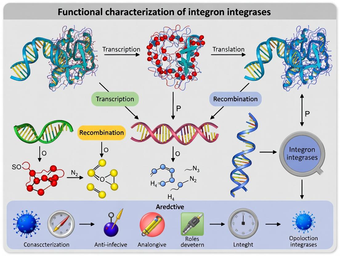
Decoding Integron Integrases: Core Biology, Structure, and Role in Antibiotic Resistance
Integrons are genetic assembly platforms found in bacteria that facilitate the capture, expression, and dissemination of gene cassettes, primarily those encoding antibiotic resistance. They are central to the adaptive evolution of bacteria, particularly in clinical settings. This content is framed within a thesis on the Functional characterization of integron integrases, focusing on experimental approaches to understand their recombination activity, regulation, and role in horizontal gene transfer.
The core component is the integron integrase (IntI), a tyrosine recombinase encoded by the intI gene. It catalyzes site-specific recombination between a proximal primary recombination site (attI) and a recombination site (attC) found within mobile gene cassettes. The captured cassettes are then expressed from a common promoter (Pc).
Table 1: Major Classes of Mobile Integrons and Their Key Features
| Integron Class | Typical Host(s) | attI Site Sequence (5'-3') | Common Cassette Array Length | Key Associated Phenotype |
|---|---|---|---|---|
| Class 1 | Plasmids, Transposons, Chromosomes | GTTGGCATCAATGC | 1-8 cassettes | Multi-drug resistance (e.g., β-lactams, aminoglycosides) |
| Class 2 | Tn7 transposon family | GTTAAGCACAATGC | Usually 2-3 cassettes (often truncated) | Trimethoprim, streptothricin resistance |
| Class 3 | Plasmids (e.g., pMET1) | GTTAGCGCAATGC | Variable | Metallo-β-lactamase (IMP-1) resistance |
| Chromosomal | Bacterial chromosomes (e.g., Vibrio spp.) | Varies by species | Up to 200+ cassettes | Diverse adaptive functions |
Table 2: Common attC Site Features Across Cassette Families
| attC Variant (Example) | Core Site (RYYYAAC) | Inverse Core (GTRRRY) | Length Range (bp) | Associated Gene Cassette |
|---|---|---|---|---|
| aadA1-type | GTTAGAC | GTCTAA | 68-72 | Aminoglycoside adenyltransferase |
| dfr-type | GTTAGGC | GCCTAA | ~60 | Dihydrofolate reductase |
| blaIMP-type | GTTAGAT | ATCTAA | 64-68 | Metallo-β-lactamase |
| Vibrio cholerae VCR | GTTAGTC | GACTAA | 123-126 | Diverse, often unknown |
Experimental Protocols for Integrase Characterization
Protocol 3.1:In VitroRecombination Assay for IntI Activity
Purpose: To directly assess the recombinase activity of a purified integron integrase (IntI) protein on DNA substrates containing attI and attC sites. Materials: Purified IntI protein, Supercoiled plasmid donor (containing attC-flanked cassette), Linearized plasmid acceptor (containing attI site), Reaction buffer (40 mM Tris-Cl pH 7.5, 50 mM NaCl, 5 mM EDTA, 10% glycerol), Stop solution (1% SDS, 50 mM EDTA), Proteinase K, Agarose gel electrophoresis system. Procedure:
- Setup: In a 20 µL reaction, mix 50 nM acceptor DNA, 25 nM donor DNA, and 1-2 µM IntI protein in reaction buffer.
- Incubation: Incubate at 30°C for 60 minutes. Include a no-enzyme control.
- Termination: Add 2 µL of Stop solution and 1 µL of Proteinase K (20 mg/mL). Incubate at 37°C for 15 min.
- Analysis: Resolve products by 1% agarose gel electrophoresis. Successful recombination integrates the cassette into the acceptor, increasing its size.
Protocol 3.2: PCR & Sequencing for Cassette Array Profiling
Purpose: To identify and sequence the repertoire of gene cassettes within an integron's variable region. Materials: Bacterial DNA template, Primers (5'-CS: GGCATCCAAGCAGCAAGC [intI proximal]; 3'-CS: AAGCAGACTTGACCTGA [qacEΔ1/sul1 proximal]), High-fidelity PCR mix, Sequencing reagents. Procedure:
- PCR Amplification: Using primers 5'-CS and 3'-CS, amplify the variable region. Cycling: 95°C 5 min; 30 cycles of (95°C 30s, 55°C 30s, 72°C 1-3 min/kb); 72°C 7 min.
- Product Analysis: Run PCR products on a 1.5% agarose gel. A smear suggests diverse cassette arrays.
- Cloning & Sequencing: Clone amplicons into a sequencing vector. Sequence multiple clones to assess population diversity.
- Bioinformatics: Use BLAST to identify cassette gene functions and analyze attC site structures.
Protocol 3.3: Reporter Assay for Integrase Expression Regulation
Purpose: To measure the activity of the intI promoter under different stress conditions (e.g., antibiotic exposure). Materials: Reporter strain (e.g., E. coli with PintI-lacZ fusion), LB broth, Substrate (X-gal, ONPG, or Luciferin), Test antibiotics, Microplate reader. Procedure:
- Culture & Induction: Grow reporter strain to mid-log phase. Split into cultures and expose to sub-inhibitory concentrations of antibiotics (e.g., ciprofloxacin, trimethoprim).
- Assay: After 2 hours, harvest cells. For β-galactosidase (lacZ), perform ONPG assay: measure absorbance at 420nm. Normalize to cell density (OD600).
- Analysis: Compare reporter activity in treated vs. untreated cells. Increased activity indicates SOS-response-mediated upregulation of intI.
Visualizations
Integron Cassette Mobility Pathways
SOS Regulation of Integrase Expression
The Scientist's Toolkit: Research Reagent Solutions
Table 3: Essential Reagents for Integron/Integrase Research
| Reagent/Material | Function/Application | Example/Notes |
|---|---|---|
| IntI Expression Vectors | Overproduction of His-tagged or GST-tagged integrase for purification. | pET-28a-intI1, pGEX-6P-intI3. |
| Defined attI/attC Substrate Plasmids | Provides standardized DNA targets for in vitro recombination assays. | pSUH-attI1, pKIL-aadA2 cassette. |
| SOS-Inducing Antibiotics | To study the regulatory link between stress and integrase expression. | Ciprofloxacin, Mitomycin C. |
| IHF Protein | Critical host factor for efficient IntI-mediated recombination in vitro. | Purified E. coli Integration Host Factor. |
| Cassette Primer Sets | Amplification and profiling of unknown integron cassette arrays. | 5'-CS / 3'-CS (Class 1), hep58 / hep59 (broad). |
| Integron-Positive Control Strains | Positive controls for PCR and phenotypic resistance assays. | E. coli J53/pMG267 (Class 1, multi-R), Acinetobacter BAJ (Class 3). |
| β-Galactosidase Reporter Plasmids | Measuring promoter activity of intI or cassette-associated promoters. | pRS550-based PintI-lacZ fusions. |
| Tyrosine Recombinase Inhibitors | Potential lead compounds for novel anti-resistance drugs. | SSB protein (non-specific), synthetic small molecules under research. |
Application Notes
Integron integrases are site-specific tyrosine recombinases that catalyze the insertion and excision of mobile gene cassettes, primarily driving the spread of antibiotic resistance. Within the broader thesis on the functional characterization of integron integrases, this section details the phylogeny, distinct features, and experimental protocols for studying the major classes associated with mobile genetic elements.
Phylogenetic Classification and Key Features
Integron integrases are phylogenetically grouped into three main classes (1, 2, 3) associated with mobile integrons, plus several other classes in sedentary chromosomal integrons. Mobile integron integrases share a common catalytic mechanism but differ in their genetic context, target site specificity, and prevalence.
Table 1: Key Features of Mobile Integron Integrase Classes
| Feature | Class 1 | Class 2 | Class 3 |
|---|---|---|---|
| Primary Host Platform | Tn402-like transposon | Tn7-like transposon | Uncharacterized transposon |
| Typical attI Site | attI1 (~130 bp) | attI2 (truncated, ~47 bp) | attI3 (~131 bp) |
| Common attC Sites | attC (59-be) variants: aadA, dfr, etc. | attC variants specific to In2- | attC variants specific to In3 |
| Catalytic Tyrosine | Y312 (IntI1 numbering) | Y302 (IntI2 numbering) | Y308 (IntI3 numbering) |
| Prevalence in Clinical Isolates | ~70-80% of resistant isolates | ~10-15% of resistant isolates | <5% (rare, sporadic) |
| Cofactor Requirement | Divalent cations (Mg²⁺/Ca²⁺) | Divalent cations (Mg²⁺/Ca²⁺) | Divalent cations (Mg²⁺/Ca²⁺) |
Table 2: Quantitative Recombination Efficiency Comparison (Representative Data)
| Integrase | attI x attC Recombination Frequency (Relative Units)* | Excision vs. Insertion Bias | Optimal Temperature | Optimal pH |
|---|---|---|---|---|
| IntI1 | 1.00 (reference) | Excision favored ~3:1 | 37°C | 7.5-8.0 |
| IntI2 | 0.15 - 0.30 | Excision favored ~1.5:1 | 30°C | 7.0-7.5 |
| IntI3 | 0.50 - 0.70 | Data limited | 37°C | 7.5-8.0 |
Frequency measured via plasmid-based *in vivo assay in E. coli; subject to assay conditions.
Experimental Protocols
Protocol 1:In VivoRecombination Assay for Integrase Activity
This protocol assesses the recombination activity between attI and attC sites catalyzed by a specific integrase in vivo.
Materials: See "The Scientist's Toolkit" (Table 3). Procedure:
- Construct Preparation: Clone the integrase gene (e.g., intI1) under a controllable promoter (e.g., PBAD) into plasmid pBAD24 (Reporter Plasmid). Clone a test attC cassette (e.g., aadA7 attC) containing a promoterless antibiotic resistance gene (e.g., aac(6')-Ib) into a compatible plasmid (Donor Plasmid). Clone the corresponding attI site upstream of a promoterless reporter gene (e.g., lacZ) in a third plasmid (Target Plasmid).
- Transformation: Co-transform the three plasmids into an E. coli ΔlacZ strain (Assay Strain).
- Induction and Selection: Grow cells to mid-log phase, induce integrase expression with 0.2% L-arabinose for 2 hours, and then plate on LB agar containing Amp, Cm, Km (for plasmid maintenance) + Gentamicin (for selection of recombinants where aac(6')-Ib is activated) + X-Gal.
- Analysis: Calculate recombination frequency as (CFU on Gent+X-Gal plates / CFU on control plates lacking Gent). Blue/white screening on X-Gal confirms correct attI-attC fusion.
Protocol 2: Purification of Histidine-Tagged Integrase forIn VitroStudies
This protocol describes the expression and purification of recombinant integrase for biochemical characterization.
Materials: See "The Scientist's Toolkit" (Table 3). Procedure:
- Expression: Transform expression plasmid (e.g., pET28a-intI1) into E. coli BL21(DE3). Grow culture in LB+KAN at 37°C to OD600 ~0.6. Induce with 0.5 mM IPTG at 16°C for 16-18 hours.
- Cell Lysis: Harvest cells by centrifugation. Resuspend pellet in Lysis Buffer. Lyse cells by sonication on ice. Clarify lysate by centrifugation at 20,000 x g for 30 min at 4°C.
- Immobilized Metal Affinity Chromatography (IMAC): Load supernatant onto a Ni-NTA column pre-equilibrated with Lysis Buffer. Wash with 10 column volumes of Wash Buffer. Elute protein with Elution Buffer in 1 mL fractions.
- Dialysis and Storage: Pool fractions containing the integrase (analyzed by SDS-PAGE). Dialyze against Storage Buffer overnight at 4°C. Concentrate, aliquot, flash-freeze in liquid N2, and store at -80°C. Determine concentration using a Bradford assay.
Protocol 3: Electrophoretic Mobility Shift Assay (EMSA) forattSite Binding
This protocol is used to study the direct binding of purified integrase to attI or attC DNA substrates.
Materials: See "The Scientist's Toolkit" (Table 3). Procedure:
- Probe Preparation: PCR-amplify or anneal oligonucleotides to generate a ~200 bp DNA fragment containing the attI1 site. Label the fragment using [γ-³²P]ATP and T4 Polynucleotide Kinase. Purify labeled probe using a spin column.
- Binding Reaction: In a 20 µL reaction, combine EMSA Binding Buffer, 1 µg poly(dI-dC), 1-10 fmol labeled probe, and purified IntI1 (0-500 nM). Incubate at 30°C for 20 min.
- Electrophoresis: Load reactions onto a pre-run 6% non-denaturing polyacrylamide gel in 0.5x TBE buffer. Run at 100 V at 4°C until the dye front migrates appropriately.
- Detection: Dry gel and expose to a phosphorimager screen overnight. Analyze shifts indicative of protein-DNA complex formation.
Visualizations
Diagram 1: IntI1 Integrase Catalytic Pathway
Diagram 2: Workflow for Measuring Integrase Activity
The Scientist's Toolkit
Table 3: Essential Research Reagents and Materials
| Item | Function/Description | Example Product/Catalog |
|---|---|---|
| Cloning & Expression | ||
| pBAD24 or pET28a vector | Tunable expression (araBAD) or strong T7-based expression of integrase genes. | pBAD24 (NCBI), pET-28a(+) (Novagen) |
| E. coli Assay Strain | Reporter strain lacking background activity (e.g., ΔlacZ). | E. coli MG1655 Δ*lacZ |
| E. coli Expression Strain | Protein expression host with T7 RNA polymerase. | E. coli BL21(DE3) |
| Biochemical Analysis | ||
| Ni-NTA Agarose Resin | Purification of polyhistidine-tagged recombinant integrase. | Qiagen Ni-NTA Superflow |
| Radiolabeled [γ-³²P]ATP | For end-labeling DNA probes in EMSA experiments. | PerkinElmer BLU002Z |
| Poly(dI-dC) | Non-specific competitor DNA to reduce background in EMSA. | Sigma-Aldrich P4929 |
| Culture & Selection | ||
| L-Arabinose | Inducer for PBAD promoter in in vivo assays. | Sigma-Aldrich A3256 |
| X-Gal (5-Bromo-4-chloro-3-indolyl-β-D-galactopyranoside) | Chromogenic substrate for LacZ in recombination screens. | GoldBio B4282 |
| Antibiotic Stock Solutions | For selection of plasmids and recombinant products (Amp, Cm, Km, Gm). | Various suppliers |
| Buffers | ||
| EMSA Binding Buffer (10X) | Provides optimal ionic conditions for protein-DNA interactions. | 200 mM Tris pH 7.5, 1M NaCl, 50 mM MgCl₂, 10 mM DTT, 50% Glycerol. |
| IMAC Lysis/Wash/Elution Buffers | For His-tag protein purification (with imidazole gradient). | Standard protocols with protease inhibitors. |
Integron integrases (IntIs) are tyrosine recombinases central to the capture and dissemination of antibiotic resistance genes within mobile integrons. Functional characterization of these enzymes requires a detailed understanding of their molecular architecture. This application note details the structural domains, their functions, and experimental protocols for probing IntI mechanics, providing a framework for research aimed at inhibiting multidrug resistance spread.
Domain Organization & Quantitative Characteristics
IntIs possess a conserved modular architecture. Key domains and their quantitative biochemical properties are summarized below.
Table 1: Core Domains of Integron Integrases
| Domain | Approximate Amino Acid Residues (in IntI1) | Key Motifs/Elements | Primary Function | Known Variants/Notes |
|---|---|---|---|---|
| Catalytic Domain | 1-180 | RHRY, KILGER, Box I, Box II | Tyrosine recombination chemistry; binds to core-type attC site bottom strand. | Contains the catalytic tyrosine (e.g., Y312 in IntI1). |
| DNA-Binding Domain (DBD) | ~180-280 | C-terminal Helix-Turn-Helix (HTH) | Sequence-specific recognition of attI and attC sites. | DBD swap experiments show specificity determinant. |
| Variable Region (VR) | Varies (e.g., 281-337 in IntI1) | Low sequence conservation | Proposed role in protein-protein interactions, oligomerization, and attC site recognition flexibility. | Length and sequence highly variable among IntI types. |
| Oligomerization Interface | Distributed | Hydrophobic patches & salt bridges | Mediates dimer/tetramer formation essential for synaptic complex assembly. | Often overlaps with catalytic core and VR. |
Table 2: Biochemical & Biophysical Parameters for IntI1
| Parameter | Value / Observation | Experimental Method |
|---|---|---|
| Molecular Weight | ~37 kDa (monomer) | SDS-PAGE / Mass Spectrometry |
| Active Oligomeric State | Dimer Tetramer equilibrium | Analytical Ultracentrifugation, SEC-MALS |
| DNA Binding Affinity (Kd) | attI site: ~20-50 nM; attC site: ~100-200 nM | EMSA, Fluorescence Anisotropy |
| Catalytic Rate (kcat) | ~0.1 - 1.0 min-1 (recombination) | In vitro recombination assay |
| Optimal Activity pH | 7.5 - 8.0 | Buffered activity assays |
| Divalent Cation Requirement | Mg2+ or Mn2+ (1-5 mM) | Chelation experiments |
Experimental Protocols
Protocol 3.1: Purification of Recombinant His-Tagged IntI
Objective: Obtain purified, active IntI protein from E. coli.
- Expression: Transform E. coli BL21(DE3) with pET28a-intI1. Grow culture in LB+KAN at 37°C to OD600 ~0.6. Induce with 0.5 mM IPTG. Shift to 18°C and incubate for 16h.
- Lysis: Pellet cells. Resuspend in Lysis Buffer (50 mM Tris-HCl pH 7.5, 500 mM NaCl, 10 mM imidazole, 10% glycerol, 1 mM PMSF). Lyse by sonication.
- Immobilized Metal Affinity Chromatography (IMAC): Clarify lysate. Load supernatant onto Ni-NTA column. Wash with 10 column volumes (CV) of Wash Buffer (Lysis Buffer with 25 mM imidazole). Elute with Elution Buffer (Lysis Buffer with 250 mM imidazole).
- Size Exclusion Chromatography (SEC): Pool elution fractions. Concentrate and inject onto HiLoad 16/600 Superdex 200 pg column equilibrated in SEC Buffer (20 mM HEPES pH 7.5, 300 mM NaCl, 1 mM DTT, 5% glycerol). Collect peak fractions corresponding to dimer/tetramer.
- Analysis: Verify purity by SDS-PAGE. Determine concentration (ε280). Aliquot, flash-freeze, and store at -80°C.
Protocol 3.2: Electrophoretic Mobility Shift Assay (EMSA) for DNA Binding
Objective: Quantify IntI binding affinity (Kd) for attI and attC sites.
- Probe Preparation: Generate 5'-Cy5-labeled double-stranded DNA probes (~200 bp containing attI or attC site) by PCR.
- Binding Reactions: Set up 20 µL reactions in Binding Buffer (20 mM Tris-HCl pH 7.5, 50 mM NaCl, 5 mM MgCl2, 1 mM DTT, 5% glycerol, 0.1 mg/mL BSA). Use a titration of purified IntI (0, 10, 25, 50, 100, 250, 500, 1000 nM). Include 2 nM DNA probe. Incubate at 30°C for 20 min.
- Electrophoresis: Load reactions onto a pre-run 6% native polyacrylamide gel in 0.5X TBE at 4°C. Run at 80 V for 90 min.
- Detection & Analysis: Image gel using a Cy5 channel. Quantify free and bound probe bands. Plot fraction bound vs. [IntI]. Fit data to a quadratic binding equation to determine Kd.
Protocol 3.3:In VitroSite-Specific Recombination Assay
Objective: Measure IntI catalytic activity.
- Substrate Preparation: Purify supercoiled plasmid (pSUMO-attI) and PCR-amplified linear attC cassette (with flanking attC sites). Use at least 200 ng of each.
- Recombination Reaction: Assemble in 25 µL Recombination Buffer (25 mM Tris-HCl pH 7.5, 100 mM NaCl, 5 mM MgCl2, 1 mM DTT). Add substrates and 200 nM IntI. Incubate at 37°C for 60 min.
- Reaction Stop: Add 2 µL of 10% SDS and 1 µL Proteinase K (20 mg/mL). Incubate at 55°C for 30 min.
- Analysis: Analyze products by agarose gel electrophoresis (1%). Successful recombination yields a distinct larger plasmid product. Quantify using band intensity.
Visualization of IntI Architecture and Function
Diagram Title: IntI Domain Function in Recombination
Diagram Title: IntI Protein Purification Workflow
The Scientist's Toolkit: Research Reagent Solutions
Table 3: Essential Reagents for IntI Characterization
| Reagent / Material | Function & Role in IntI Research | Example Product / Note |
|---|---|---|
| pET Expression Vectors | High-yield recombinant protein production in E. coli. | pET28a(+) for N-terminal His6-tag. |
| Ni-NTA Resin | Immobilized metal affinity chromatography for His-tagged IntI purification. | Commercially available from Qiagen, Thermo Fisher. |
| Superdex 200 Increase | SEC media for resolving IntI oligomeric states (dimers, tetramers). | GE Healthcare/Cytiva. |
| Fluorescent DNA Dyes | For labeling att site probes in EMSA and anisotropy. | Cy5, FAM, or TAMRA phosphoramidites. |
| Precision Proteases | For tag removal after purification if required. | TEV or 3C protease sites can be engineered. |
| Native attI/attC DNA Fragments | Critical substrates for binding and activity assays. | Must be cloned or synthesized as double-stranded. |
| Tyrosine Recombinase Inhibitors | Potential lead compounds for functional studies. | Novobiocin analogues; used as experimental controls. |
| SEC-MALS Instrumentation | Determines absolute molecular weight and oligomerization in solution. | Wyatt Technology systems coupled to HPLC. |
| Surface Plasmon Resonance (SPR) Chips | For real-time, label-free kinetics of IntI-DNA interactions. | Streptavidin chips for capturing biotinylated att sites. |
Within the broader functional characterization of integron integrases, understanding the attC x attI recombination mechanism is paramount. This site-specific recombination system governs the integration and excision of mobile gene cassettes, which are the primary drivers of antibiotic resistance dissemination in clinical pathogens. This document provides detailed application notes and protocols for studying this precise molecular mechanism, enabling researchers to dissect integrase activity, specificity, and kinetics.
Integron integrase (IntI) catalyzes recombination between the attI site (within the integron platform) and the attC site (within a mobile gene cassette). The reaction is reversible, facilitating both cassette acquisition (attI x attC) and excision (attC x attC). Key characteristics are summarized below.
Table 1: Key Characteristics of attC and attI Sites
| Feature | attI Site | attC Site (59-be) |
|---|---|---|
| Location | In the integron platform, 5' to inserted cassettes. | Within each mobile gene cassette. |
| Structure | Relatively conserved, simple core site (GTTRRRY). | Imperfect inverted repeats forming a secondary hairpin structure. |
| Size | ~65 bp core recombination region. | Variable length, typically 59-141 bp (hence "59-base element"). |
| Strand Bias | Recombined as double-stranded DNA. | Recombined as a single-stranded, folded substrate. |
| IntI Binding | Two direct binding sites (strong & weak). | Multiple binding sites within the hairpin arms. |
| Recombination Efficiency (Relative) | High in attI x attC reactions. | Low in attC x attC excision reactions. |
Table 2: Experimental Recombination Efficiency Metrics (Example Data)
| Recombination Substrate Pair | Relative Efficiency (%) | Key Influencing Factor |
|---|---|---|
| attI1 x attC (ss) | 100.0 ± 5.2 (Reference) | Standard condition, supercoiled donor. |
| attI1 x attC (ds) | 15.3 ± 3.1 | Double-stranded attC is a poor substrate. |
| attC x attC (ss) | 1.8 ± 0.7 | Excisive recombination is intrinsically less efficient. |
| attI1 x attI1 | < 0.1 | Integrase shows strong site specificity. |
| attI1 x attC (Mut. RY) | 8.5 ± 2.4 | Mutation in attC core sequence (R=purine, Y=pyrimidine). |
Experimental Protocols
Protocol 1:In VitroRecombination Assay (Gel-Based)
Purpose: To qualitatively and quantitatively assess IntI-mediated recombination between attI and attC substrates. Key Reagents: Purified IntI integrase, supercoiled plasmid donor DNA (containing attC cassette), linear recipient DNA fragment (containing attI site), recombination buffer.
- Reaction Setup: In a 20 µL final volume, combine:
- 2 µL 10X Recombination Buffer (250 mM Tris-Cl pH 7.5, 1 M NaCl, 100 mM MgCl2, 50% glycerol).
- 10-100 nM purified IntI protein.
- 5 nM supercoiled donor plasmid.
- 5 nM linear recipient fragment.
- Nuclease-free water to volume.
- Incubation: Incubate at 30°C for 60-120 minutes.
- Termination: Stop the reaction by adding 2 µL of 10% SDS and heating at 65°C for 10 min.
- Proteinase K Digestion: Add 1 µL of proteinase K (20 mg/mL), incubate at 37°C for 30 min.
- Analysis: Resolve products on a 1% agarose gel. A successful recombination event between a supercoiled donor and linear recipient produces a single, larger linear product.
Protocol 2:In VivoCassette Excision/Integration Assay (PCR-Based)
Purpose: To detect recombination events within a bacterial cell. Key Reagents: Bacterial strains (with chromosomal integron and donor cassette plasmid), specific PCR primers.
- Strain Construction: Introduce a plasmid containing an attC-flanked cassette (e.g., antibiotic resistance) into a reporter strain carrying an integron with an attI site.
- Induction: Induce expression of the integrase gene (e.g., from a regulated promoter like Ptac or PBAD) for 2-4 hours.
- DNA Extraction: Perform a quick plasmid and genomic DNA prep from induced cultures.
- Diagnostic PCR:
- Integration: Use a primer upstream of attI and a primer within the cassette. A product indicates site-specific integration.
- Excision: Use primers flanking a pre-integrated cassette. A reduction in amplicon size indicates precise excision.
- Quantification: Analyze PCR products by gel electrophoresis. For quantitative data, use qPCR with SYBR Green.
Visualizations
Diagram 1: attC x attI Recombination Mechanism
Diagram 2: Workflow for In Vitro Recombination Assay
The Scientist's Toolkit: Research Reagent Solutions
Table 3: Essential Research Reagents & Materials
| Reagent/Material | Function & Explanation |
|---|---|
| Purified IntI Integrase (Wild-type & Mutants) | Catalytic driver of recombination. Essential for in vitro assays and structure-function studies. |
| Supercoiled Plasmid Donor (attC+) | Provides the mobile gene cassette substrate. Supercoiling enhances attC activity. |
| Linear DNA Fragment (attI+) | Acts as the recipient target for integration in in vitro assays. |
| attC site Oligonucleotides (Single-stranded) | For studying the canonical single-stranded attC recombination pathway. |
| Recombination Buffer (10X) | Provides optimal ionic conditions (Mg2+, Na+, Tris) for integrase activity and synapse formation. |
| Clinical Integron Strain Collection | Source of natural attC and attI variants for studying sequence diversity impact on efficiency. |
| Site-Directed Mutagenesis Kit | For generating point mutations in attI, attC core sites, or integrase active site residues. |
| Electrophoretic Mobility Shift Assay (EMSA) Kit | To study and quantify integrase binding affinity to different att site variants. |
| High-Sensitivity DNA Gel Stain (e.g., SYBR Safe) | For visualizing low-abundance recombination products in gels. |
| qPCR System with SYBR Green | For quantifying in vivo recombination frequencies (excision/integration) with high sensitivity. |
Integrons as Major Drivers of Multidrug Resistance in Clinical and Environmental Pathogens
This document provides detailed application notes and protocols to support the functional characterization of integron integrases, a critical research area within the broader thesis on understanding the mobilization and spread of multidrug resistance. Integrons are genetic platforms that allow bacteria to capture, express, and exchange antibiotic resistance gene cassettes via site-specific recombination mediated by the integron integrase (IntI). Their role in driving resistance in both clinical (e.g., Pseudomonas aeruginosa, Acinetobacter baumannii) and environmental pathogens is paramount.
Current Epidemiological Data
Recent surveillance studies (2022-2024) highlight the prevalence of class 1 integrons across various pathogens and reservoirs.
Table 1: Prevalence of Class 1 Integrons in Key Pathogens (2022-2024 Meta-Analysis Data)
| Pathogen / Source | Sample Type | Prevalence (%) | Most Common Cassette Arrays (Examples) | Reference Region |
|---|---|---|---|---|
| Acinetobacter baumannii | Clinical isolates | 45-68% | aacA4, blaOXA-, dfrA1 | Global |
| Pseudomonas aeruginosa | Hospital wastewater | 52-60% | aadB, blaVIM-2, aac(6')-Ib | Europe, Asia |
| Escherichia coli | Poultry farm | 38-55% | dfrA17-aadA5, blaCTX-M-1 | North America |
| Klebsiella pneumoniae | Clinical (ICU) | 31-49% | aac(6')-Ib-cr, qnrB | South Asia |
| Riverine Biofilms | Environmental | 25-40% | sat, aadA, qac variants | South America |
Table 2: Common Integron Classes and Features
| Integron Class | Typical Integrase (IntI) | Primary attI Site | Common Mobilization Link | Primary Habitat |
|---|---|---|---|---|
| Class 1 | IntI1 | attI1 | Tn402-like transposon | Clinical, Environmental |
| Class 2 | IntI2 | attI2 | Tn7 transposon | Clinical |
| Class 3 | IntI3 | attI3 | Tn402-like transposon | Clinical |
| Mobile/Super | IntI* variants | Variable | Chromosomal | Environmental |
Protocols for Functional Characterization of Integron Integrases
Protocol 1: Detection and Typing of Integrons from Bacterial Isolates
Objective: To identify the presence, class, and cassette content of integrons from genomic DNA.
Materials (Research Reagent Solutions):
- Lysis Buffer (SDS-Proteinase K): For cell wall degradation and DNA release.
- IntI Gene-Specific Primers (IntI1-F/R, IntI2-F/R, IntI3-F/R): For PCR amplification of integrase genes.
- 5'-CS and 3'-CS Conserved Segment Primers: For amplification of the variable cassette region in class 1 integrons.
- High-Fidelity DNA Polymerase: For accurate amplification of GC-rich cassette arrays.
- Gel Extraction Kit: For purification of PCR amplicons.
- Sanger Sequencing Mix (BigDye Terminator v3.1): For sequencing cassette arrays.
- Bioinformatics Database (INTEGRALL, ResFinder): For cassette array annotation.
Procedure:
- Extract genomic DNA from bacterial culture using a standard phenol-chloroform method or commercial kit.
- Perform PCR for intI gene detection. Use primer sets specific for intI1, intI2, and intI3. Cycling: 95°C 5 min; 30 cycles of (95°C 30s, 55-60°C 30s, 72°C 1 min/kb); 72°C 5 min.
- For positive class 1 integron isolates, perform a second PCR using the 5'-CS (5'-GGCATCCAAGCAGCAAGC-3') and 3'-CS (5'-AAGCAGACTTGACCTGA-3') primers to amplify the variable region. Use a longer extension time (72°C for 3-5 min).
- Resolve amplicons on a 1.5% agarose gel. A smear or multiple bands indicate a variable cassette array.
- Purify the variable region amplicon and sequence using the 5'-CS primer as the sequencing primer.
- Analyze sequences using BLAST against the INTEGRALL database to identify gene cassette types and order.
Protocol 2:In VitroRecombination Assay for Integrase Activity
Objective: To functionally validate the recombination activity of a purified integron integrase (IntI) between attI and attC sites.
Materials (Research Reagent Solutions):
- Purified His-tagged IntI Protein: Recombinase of interest, purified via Ni-NTA chromatography.
- Supercoiled Plasmid DNA containing attI site: The recombination acceptor plasmid (e.g., pSUH series).
- PCR-amplified Linear attC Gene Cassette: The recombination donor fragment, containing an attC site and a resistance marker (e.g., aadA2).
- 10x Recombination Buffer: 250 mM Tris-HCl (pH 7.5), 1 M NaCl, 100 mM MgCl2, 10 mM DTT, 500 µg/mL BSA.
- Proteinase K Stop Solution: 1% SDS, 1 mg/mL Proteinase K.
- Electrocompetent E. coli cells: For transformation of recombination products.
- Selective Agar Plates: Containing appropriate antibiotics to select for recombinant plasmids.
Procedure:
- Set up a 20 µL recombination reaction: 50 ng acceptor plasmid, 100 ng donor attC cassette, 1x Recombination Buffer, and 200-500 ng purified IntI protein. Include a no-enzyme control.
- Incubate at 30-37°C for 2-4 hours.
- Stop the reaction by adding 2 µL of Proteinase K Stop Solution and incubating at 37°C for 15 min.
- Dialyze or dilute the reaction 10-fold in sterile water. Transform 2 µL into 50 µL of electrocompetent E. coli cells via electroporation.
- Plate cells onto agar plates selecting for the antibiotic resistance conferred by the inserted cassette (e.g., streptomycin for aadA2) AND the vector backbone.
- Count colonies. Recombination frequency is calculated as (CFU on double selection / CFU on vector selection) for the test reaction, normalized to the no-enzyme control.
- Confirm cassette insertion by colony PCR and sequencing across the attI-attC junction.
The Scientist's Toolkit: Key Research Reagents
Table 3: Essential Reagents for Integron/Integrase Research
| Reagent / Material | Function & Application |
|---|---|
| IntI1/2/3 Specific Primers | PCR-based detection and classification of integron classes. |
| 5'-CS / 3'-CS Primers | Amplification of the variable cassette array in class 1 integrons for profiling. |
| pSUH or pSW Rec. Plasmid Series | Standardized attI-containing supercoiled plasmids for in vitro recombination assays. |
| His-tagged IntI Expression Vectors (e.g., pET28a-intI1) | Recombinant production and purification of integrase proteins. |
| INTEGRALL / ResFinder Databases | Bioinformatics resources for annotating identified gene cassettes and integron structures. |
| High-Fidelity Polymerase | Accurate amplification of long, repetitive, or GC-rich cassette arrays. |
| Electrocompetent E. coli (recA-) | High-efficiency transformation for recovery of recombination assay products. |
Experimental Workflow & Pathway Visualizations
Diagram Title: Integron Detection & Integrase Assay Workflow
Diagram Title: Integrase-Mediated attI x attC Recombination
Application Notes
Role of Integron Integrases in Adaptive Evolution
Integron integrases (IntIs) are site-specific recombinases essential for the capture, rearrangement, and expression of gene cassettes within integron platforms. This activity drives bacterial genome plasticity, facilitating rapid adaptation to environmental stressors, including antibiotics. Within the broader thesis on the functional characterization of integron integrases, understanding their kinetic parameters, recombination efficiency, and regulatory mechanisms is paramount for developing novel strategies to curb the spread of antimicrobial resistance (AMR).
Quantitative Analysis of IntI Activity
Recent studies have characterized the activity of different IntI types. The data below summarize key enzymatic parameters critical for understanding their evolutionary impact.
Table 1: Comparative Kinetic Parameters of Major Integron Integrases
| Integrase Type | ( K_m ) (nM) | ( k_{cat} ) (min⁻¹) | Recombination Efficiency (%)* | Primary AttC Site Target |
|---|---|---|---|---|
| IntI1 | 15.2 ± 2.1 | 8.7 ± 0.9 | 95.5 ± 3.2 | attC1 |
| IntI2 | 22.5 ± 3.3 | 5.2 ± 0.7 | 82.1 ± 4.5 | attC2 |
| IntI3 | 18.7 ± 2.8 | 7.1 ± 0.8 | 88.7 ± 3.9 | attC3 |
| IntI9 | 30.1 ± 4.5 | 3.5 ± 0.5 | 75.4 ± 5.1 | attC9 |
Efficiency measured via *in vitro recombination assay between attI and attC sites.
Table 2: Impact of IntI1 Activity on Antibiotic Resistance Acquisition in E. coli
| Stress Condition | Cassette Acquisition Rate (events/cell/generation) | Mean Resistance Increase (Fold Change) | Most Frequently Captured Cassette |
|---|---|---|---|
| Ciprofloxacin | ( 4.2 \times 10^{-6} ) | 12.5 | aac(6')-Ib-cr |
| Ceftazidime | ( 3.8 \times 10^{-6} ) | 8.7 | blaVEB-1 |
| Meropenem | ( 1.5 \times 10^{-6} ) | 5.2 | blaIMP-1 |
| No Antibiotic | ( 0.7 \times 10^{-6} ) | 1.0 (baseline) | N/A |
Experimental Protocols
Protocol:In VitroIntegrase Recombination Assay
Purpose: To quantify the recombination efficiency of a purified IntI protein between attI and attC sites.
Materials: See "Research Reagent Solutions" table.
Procedure:
- Substrate Preparation: Generate DNA fragments containing the attI and attC sites (~200-300 bp) via PCR. Purify fragments using a PCR clean-up kit.
- Reaction Setup: In a 20 µL reaction, combine:
- 2 µL 10X Recombination Buffer (500 mM Tris-HCl pH 7.5, 1 M NaCl, 100 mM MgCl₂, 10 mM DTT).
- 1 µL purified IntI protein (100 nM final concentration).
- 1 µL attI substrate (10 nM final).
- 1 µL attC substrate (10 nM final).
- 15 µL nuclease-free water.
- Incubation: Incubate at 37°C for 60 minutes.
- Reaction Termination: Add 2 µL of 10% SDS and heat at 65°C for 10 minutes.
- Analysis: Resolve products on a 2% agarose gel. Recombination efficiency is calculated as (Intensity of Recombinant Product Band / Total Intensity of All DNA Bands) × 100%.
Protocol: MeasuringIn VivoCassette Acquisition Rates
Purpose: To determine the frequency of novel gene cassette integration mediated by IntI in bacterial populations under selective pressure.
Procedure:
- Strain Construction: Introduce a reporter plasmid containing an attI site and a promoterless antibiotic resistance gene into a strain harboring a chromosomal integron with a cognate IntI gene and a donor attC-flanked cassette.
- Culture Growth: Grow biological triplicates to mid-exponential phase (OD₆₀₀ ~0.6) in Lennox Broth (LB).
- Selection: Plate appropriate dilutions onto LB agar containing the antibiotic for which the donor cassette confers resistance.
- Calculation: The acquisition rate is calculated using the Ma-Sandri-Sarkar Maximum Likelihood Estimator (MSS-MLE) method, comparing counts on selective vs. non-selective plates after 24-48 hours incubation at 37°C.
- Validation: Confirm cassette structure and integration site via colony PCR and Sanger sequencing.
Diagrams
IntI-Mediated Cassette Recombination Pathway
Title: IntI-Mediated Cassette Recombination
Workflow for Functional Characterization of IntI Mutants
Title: IntI Mutant Characterization Workflow
Research Reagent Solutions
Table 3: Essential Reagents for Integrase Functional Studies
| Reagent/Material | Function/Benefit | Example Supplier/Catalog |
|---|---|---|
| Purified IntI Proteins (Wild-type & Mutants) | Substrate for in vitro assays; allows kinetic characterization without cellular background. | Recombinant expression in-house or from repositories like Addgene. |
| Synthetic attI and attC DNA Substrates (Fluorophore-labeled) | High-specificity substrates for gel-based or FRET-based recombination assays. | IDT DNA, Eurofins Genomics. |
| pSWITCH or pMASTER Reporter Plasmids | Modular plasmids for in vivo measurement of cassette acquisition frequency. | Available from research consortia (e.g., INTEGRALL). |
| Tunable Expression Vectors (e.g., pBAD, pET) | For controlled, high-yield expression of IntI proteins in E. coli for purification. | Thermo Fisher, Merck. |
| Electrophoretic Mobility Shift Assay (EMSA) Kit | To study protein-DNA binding affinity between IntI and att sites. | Thermo Fisher Scientific (EMSA Kit 20148). |
| MSS-MLE Calculation Software (e.g., FALCOR web tool) | Accurately calculates mutation or acquisition rates from fluctuation analysis data. | Open-access web tool (University of Michigan). |
| High-Fidelity DNA Polymerase (e.g., Q5) | For error-free amplification of integron and cassette sequences for cloning. | New England Biolabs (M0491). |
| His-tag Protein Purification Resin (Ni-NTA) | Standardized, high-purity isolation of recombinant His-tagged IntI proteins. | Qiagen (30210), Cytiva (17531801). |
Assaying Integrase Activity: From Standard Protocols to Advanced High-Throughput Screens
Within the broader thesis on the Functional Characterization of Integron Integrases, establishing robust, quantitative, and mechanistic assays is paramount. Integron integrases (IntIs) are site-specific recombinases responsible for the capture, excision, and rearrangement of antibiotic resistance gene cassettes. Their activity drives the adaptive evolution of multidrug-resistant bacterial pathogens. In vitro recombination assays constitute the gold standard for measuring IntI activity, enabling researchers to dissect the precise biochemical requirements, kinetics, and efficiencies of recombination events without the complexity of cellular systems. This document provides detailed application notes and protocols for these critical assays.
In vitro assays typically measure recombination between two DNA substrates: the attI site (on the integron platform) and an attC site (on a gene cassette). The reaction products are resolved by gel electrophoresis. Key quantitative outputs include recombination efficiency (%) and reaction kinetics.
Table 1: Standardized Reaction Conditions for IntI In Vitro Assays
| Component | Typical Concentration/Range | Function & Notes |
|---|---|---|
| Purified IntI Enzyme | 50-500 nM | Catalytic core of the reaction. Concentration is titrated based on specific activity. |
| Supercoiled attI Plasmid | 5-10 nM | Donor DNA molecule containing the attI recombination site. |
| Linear attC Substrate | 1-5 nM | Acceptor DNA fragment containing the attC site. Often PCR-generated and gel-purified. |
| Recombination Buffer | 1X | Typically: 25 mM Tris-Cl (pH 7.5), 1 mM DTT, 100 mM KCl, 5% (v/v) glycerol, 10 mM MgCl₂. |
| Divalent Cation (Mg²⁺) | 5-10 mM MgCl₂ | Essential cofactor for integrase activity. Some IntIs may use Mn²⁺. |
| Reaction Temperature | 30-37°C | Optimized for enzyme stability and activity. |
| Reaction Time | 60-180 min | Time-course experiments determine initial velocity and endpoint efficiency. |
| Stop Solution | 0.1% SDS, 10 mM EDTA, 0.1 mg/mL Proteinase K | Denatures IntI and stops the reaction for analysis. |
Table 2: Example Quantitative Data from an IntI1 Recombination Assay
| Experiment Variable | Condition | Recombination Efficiency (%) | Key Interpretation |
|---|---|---|---|
| Mg²⁺ Concentration | 0 mM | 0.0 ± 0.1 | Absolute requirement for divalent cation. |
| 5 mM | 45.2 ± 3.5 | Optimal concentration for IntI1. | |
| 20 mM | 32.1 ± 2.8 | Inhibition at high concentration. | |
| IntI1 Concentration | 50 nM | 12.5 ± 1.8 | Sub-saturating enzyme levels. |
| 200 nM | 48.7 ± 4.1 | Near-saturating, standard condition. | |
| 500 nM | 49.5 ± 4.3 | Plateau, substrate-limited. | |
| Inhibitor Screening | DMSO Control | 47.9 ± 2.9 | Baseline activity. |
| Compound A (100 µM) | 5.2 ± 0.9 | Potent inhibition of recombination. | |
| Compound B (100 µM) | 44.1 ± 3.7 | No significant effect. |
Detailed Experimental Protocols
Protocol 3.1: StandardattI x attCRecombination Assay
Purpose: To measure the efficiency of cassette integration.
Materials:
- Purified IntI protein (stored at -80°C in storage buffer).
- Supercoiled plasmid DNA containing the attI site (≥90% supercoiled, quantified by A₂₆₀).
- PCR-amplified, gel-purified linear DNA fragment containing the attC site.
- 5X Recombination Buffer: 125 mM Tris-Cl (pH 7.5), 5 mM DTT, 500 mM KCl, 25% glycerol.
- 100 mM MgCl₂ stock.
- 10% SDS, 0.5 M EDTA, Proteinase K (20 mg/mL).
- Gel loading dye (non-bromophenol blue, to avoid interference).
- Agarose, TAE buffer, DNA stain (e.g., SYBR Safe), DNA size ladder.
Procedure:
- Prepare Reaction Master Mix (for n reactions + 1 extra): For a single 20 µL reaction, combine:
- 4 µL 5X Recombination Buffer
- 1 µL 100 mM MgCl₂ (5 mM final)
- X µL Nuclease-free H₂O (to bring to final volume after adding DNA/enzyme)
- 1 µL attI plasmid (10 nM final)
- 1 µL attC fragment (2 nM final) Mix gently and centrifuge briefly.
- Aliquot 18 µL of Master Mix into thin-walled PCR tubes.
- Initiate Reaction: Add 2 µL of purified IntI enzyme (diluted in storage buffer to desired concentration) or storage buffer alone (No Enzyme Control) to each tube. Mix by gentle pipetting.
- Incubate at 30°C for 90 minutes in a thermal cycler or heating block.
- Stop Reaction: Add 2 µL of Stop Solution (0.1% SDS, 10 mM EDTA, 0.1 mg/mL Proteinase K). Mix and incubate at 37°C for 15 minutes to digest the integrase.
- Analyze Products: Add gel loading dye and load the entire reaction on a 1% agarose gel in 1X TAE. Run at 5-6 V/cm for 90 minutes. Stain, visualize, and image under UV/Gel Doc system.
- Quantification: Measure band intensities for substrate and product(s). Recombination Efficiency = [Intensity of Product(s)] / [Intensity of Product(s) + Intensity of remaining attI substrate)] × 100%.
Protocol 3.2: Cassette Excision Assay
Purpose: To measure the excision of a gene cassette (between attC and attI sites) from a supercoiled plasmid donor. Modification: The DNA substrate is a single supercoiled plasmid containing both attI and attC sites in direct orientation. The reaction produces a linear or nicked circular excision product and a relaxed plasmid. Follow Protocol 3.1, but use only the plasmid substrate. Resolve products on a 0.8% agarose gel for better separation of topologically distinct forms.
Visualization of Assay Workflows
Diagram Title: In Vitro IntI Recombination Assay Workflow
The Scientist's Toolkit: Research Reagent Solutions
Table 3: Essential Materials for IntI In Vitro Assays
| Item / Reagent | Supplier Examples | Function in Assay | Critical Notes |
|---|---|---|---|
| High-Fidelity DNA Polymerase | Thermo Fisher (Phusion), NEB (Q5) | Amplification of high-purity, error-free attC substrate fragments. | Essential for maintaining precise attC sequence and structure. |
| Gel Extraction Kit | Qiagen, Macherey-Nagel, Thermo Fisher | Purification of PCR-amplified attC fragments from agarose gels. | Removes primers, enzymes, and nonspecific products. |
| Plasmid Maxiprep Kit | Qiagen, Macherey-Nagel, IBI | Isolation of large quantities of pure, supercoiled attI plasmid DNA. | High supercoiled fraction is critical for consistent results. |
| Recombinant IntI Protein | In-house expression & purification | The active enzyme. Often expressed with a His-tag in E. coli. | Purity (>95%) and absence of nucleases must be verified. |
| Precision His-Tag Purification Resin | Cytiva (Ni Sepharose), Qiagen, Roche | Immobilized metal affinity chromatography (IMAC) for IntI purification. | Enables rapid, single-step purification of active integrase. |
| SYBR Safe DNA Gel Stain | Thermo Fisher | Safe, sensitive visualization of DNA bands on agarose gels. | Preferable to ethidium bromide; compatible with blue light transillumination. |
| Image Quantification Software | Image Lab (Bio-Rad), ImageJ (Fiji) | Densitometric analysis of gel images to calculate recombination efficiency. | Must be able to define lanes and subtract background. |
| MgCl₂ Stock Solution (Molecular Biology Grade) | Sigma-Aldrich, Thermo Fisher | Provides essential divalent cation cofactor for IntI activity. | Must be nuclease-free and prepared in high-purity water. |
Within the broader thesis on the Functional Characterization of Integron Integrases, the design and production of high-quality recombination site substrates—specifically the attC (59-be) and attI sites—is a foundational step. Integron integrases (IntIs) catalyze site-specific recombination between these sites, a key mechanism driving antimicrobial resistance gene capture and dissemination. Precise in vitro characterization of IntI activity, kinetics, and specificity demands rigorously defined, sequence-verified, and structurally consistent DNA fragments. These application notes detail protocols for the synthesis, validation, and application of optimal attC and attI substrates.
Recent research (2022-2024) emphasizes the structural nuances of att sites that govern recombination efficiency. Key parameters include attC site length, secondary structure stability (based on inverse core site RYYYAAC and extrahelical bases), and attI site sequence conservation.
Table 1: Key Parameters for Optimal attC/attI Substrate Design
| Parameter | attC (59-be) Site | attI Site | Optimal Value / Consensus | Impact on Recombination Efficiency |
|---|---|---|---|---|
| Length Range | 57-141 bp | ~65 bp (core region) | attC: ~60-80 bp; attI: 65 bp | attC length inversely correlates with efficiency beyond optimal stem-loop stability. |
| Core Sequence | RYYYAAC (Inverse Core) | GTTTRRY (Direct Core) | Strictly conserved | Absolute requirement for integrase binding and strand exchange. |
| Extrahelical Bases | Typically 2 (e.g., T, A) | 1 (usually A) | attC: T at position 2; attI: A | Critical for DNA distortion and cleavage; mutation abolishes activity. |
| Stem Stability (ΔG) | -5 to -15 kcal/mol | N/A (unstructured) | ~ -9 to -12 kcal/mol | Optimal stability required; too weak or too strong reduces efficiency by >80%. |
| Spacer (attC) | Variable (7-8 bp) | N/A | 8 bp | Influences attC site folding; 8 bp most common in natural arrays. |
| Flanking Sequences | 15-20 bp of native context | 15-20 bp of native context | Include 20 bp native flank | Context can alter local supercoiling and protein binding, affecting efficiency by up to 50%. |
Table 2: Recommended Recombination Assay Substrate Formats
| Substrate Format | Synthesis Method | Recommended Use | Pros | Cons |
|---|---|---|---|---|
| Linear dsDNA Fragment | PCR, Annealed Oligos | Standard in vitro assay, EMSA | Easy to produce, quantifiable | Lacks supercoiling, lower efficiency. |
| Supercoiled Plasmid | Cloning into high-copy vector | Topology-dependent studies | Mimics in vivo context, higher efficiency | More complex production, topology variability. |
| Biotin-/Fluor-labeled Oligo | Chemical synthesis | EMSA, FRET, Single-molecule assays | Enables detection & immobilization | Costly, may affect protein binding. |
| Donor/Acceptor Vectors | Molecular cloning (Gateway, etc.) | High-throughput screening | Functional readout (e.g., antibiotic resistance) | System-dependent, requires cloning. |
Detailed Protocols
Protocol 3.1:De NovoSynthesis and Annealing of attC and attI Oligonucleotides
Objective: Generate short, double-stranded DNA fragments containing a single att site for electrophoretic mobility shift assays (EMSAs) or initial cleavage assays.
Materials:
- Oligonucleotides: HPLC-purified single-stranded DNA (ssDNA) primers. For an attC site, design complementary strands with the attC sequence in the context of its imperfect inverted repeats.
- Annealing Buffer (10X): 100 mM Tris-HCl (pH 7.5), 1 M NaCl, 10 mM EDTA.
Method:
- Design: For a typical 60 bp attC fragment, design two 60-mer oligos with perfect complementarity except for the extrahelical bases and the central spacer region that forms the loop.
- Resuspension: Dilute each oligo to 100 µM in nuclease-free water.
- Mixing: Combine equal volumes of each complementary oligo (e.g., 10 µL each) with 5 µL of 10X Annealing Buffer and 25 µL nuclease-free water (total 50 µL).
- Annealing: Heat the mixture to 95°C for 5 min in a thermal cycler, then slowly cool to 25°C at a rate of -0.1°C/sec.
- Verification: Analyze 2 µL on a 10% native polyacrylamide gel. A single, sharp band lower than the ssDNA controls confirms correct duplex formation.
- Quantification: Measure dsDNA concentration via absorbance at 260 nm.
Protocol 3.2: Cloning att Sites into Plasmid Vectors for Supercoiled Substrates
Objective: Engineer plasmid-based substrates to study recombination in a supercoiled context, mimicking the physiological state.
Materials:
- Vector: pUC19 or similar high-copy-number cloning vector.
- Enzymes: Restriction enzymes (e.g., EcoRI, HindIII), T4 DNA Ligase.
- Competent Cells: High-efficiency E. coli DH5α.
Method:
- Insert Preparation: Amplify or synthesize attC or attI fragments with 20-25 bp of native flanking sequence. Add appropriate restriction sites to the 5' ends via PCR primers.
- Digestion: Digest both the purified PCR product (insert) and the pUC19 vector with the chosen restriction enzymes. Gel-purify the fragments.
- Ligation: Set up a ligation reaction with a 3:1 molar ratio of insert to vector. Incubate with T4 DNA Ligase at 16°C for 16 hours.
- Transformation: Transform the ligation mix into competent E. coli DH5α. Plate on LB-ampicillin plates.
- Screening: Pick colonies, perform colony PCR, and validate by Sanger sequencing across the cloned insert to ensure no mutations.
- Plasmid Preparation: Culture a positive clone and purify supercoiled plasmid using a maxi-prep kit. Verify topology by agarose gel electrophoresis with chloroquine for supercoiling analysis.
Protocol 3.3:In VitroRecombination Assay Using Purified Integrase
Objective: Functionally validate the synthesized att site substrates by measuring integrase-mediated recombination.
Materials:
- Purified IntI Protein: e.g., His-tagged IntI1, >95% purity.
- DNA Substrates: 50 nM each of supercoiled attI-plasmid (donor) and linear attC fragment (acceptor).
- Reaction Buffer (2X): 40 mM Tris-HCl (pH 7.5), 100 mM NaCl, 10 mM MgCl₂, 2 mM DTT, 0.2 mg/mL BSA, 10% (v/v) glycerol.
Method:
- Setup: Mix 100 ng of each DNA substrate in a 15 µL volume.
- Reaction: Add 15 µL of 2X Reaction Buffer. Initiate the reaction by adding purified IntI to a final concentration of 500 nM. Include a no-enzyme control.
- Incubation: Incubate at 37°C for 90 minutes.
- Termination: Add 3 µL of 10% SDS and 2 µL of Proteinase K (20 mg/mL). Incubate at 55°C for 30 min to digest the integrase.
- Analysis: Resolve the DNA products on a 1% agarose gel at 80V for 90 min. Visualize with ethidium bromide or SYBR Safe.
- Quantification: The appearance of a recombinant band (e.g., linearized or larger plasmid product) indicates successful recombination. Calculate efficiency as (% recombinant DNA / total DNA) using densitometry software.
Visualizations
Diagram 1: IntI-Mediated attC × attI Recombination Pathway
Diagram 2: Workflow for Cloning att Sites into Plasmid Vectors
The Scientist's Toolkit: Research Reagent Solutions
Table 3: Essential Materials for attC/attI Substrate Studies
| Reagent / Material | Function in Research | Key Considerations |
|---|---|---|
| High-Fidelity DNA Polymerase (e.g., Q5, Phusion) | Error-free amplification of att site inserts for cloning. | Critical for maintaining exact core sequences; low error rate (< 1 in 1 Mb). |
| HPLC-Purified Oligonucleotides | De novo synthesis of att site strands for annealing or probes. | Essential for consistent annealing and labeling; removes truncated sequences. |
| Covalently Supercoiled Plasmid Prep Kits | Production of topologically uniform plasmid substrates. | Avoids nicked or relaxed plasmids which give false-negative results. |
| Recombinant His-Tagged Integrase (IntI) | Catalytic protein for in vitro assays. | Requires high purity (>95%); activity varies by purification batch. |
| Electrophoresis Grade Agarose & Gels | Analysis of DNA substrates and recombination products. | Use high-resolution gels (e.g., 3-4%) for small fragment analysis. |
| Fluorescent DNA Dyes (e.g., SYBR Safe, Cy3/Cy5 labels) | Sensitive detection and quantification of DNA. | Safer than ethidium bromide; allows for real-time or gel-based quantification. |
| Mobility Shift Assay (EMSA) Kit | Validation of integrase binding to att sites. | Includes non-specific competitor DNA (poly dI-dC) and specialized buffers. |
| Thermocycler with Gradient Function | Optimization of annealing temperatures and PCR. | Crucial for handling attC fragments with varying GC content and stem stability. |
Application Notes
In the functional characterization of integron integrases, a trio of key readout technologies is indispensable. Integron integrases are site-specific recombinases that capture, excise, and rearrange mobile gene cassettes, driving bacterial adaptation and antibiotic resistance spread. These enzymes are central to understanding horizontal gene transfer and developing novel antimicrobial strategies. Gel electrophoresis provides foundational analysis of DNA substrate recombination and cleavage products. PCR-based detection, particularly qPCR, offers sensitive, quantitative measurement of cassette excision and integration events. Finally, reporter systems like GFP enable real-time, in vivo visualization of integrase activity and promoter switching within dynamic integron platforms. Together, these methods form a hierarchical validation pipeline from in vitro biochemical confirmation to complex cellular phenotyping.
Protocols
Protocol 1: Agarose Gel Electrophoresis for Integrase Recombination Assay
Objective: To visualize the products of integron integrase-mediated recombination between attI and attC sites.
Materials:
- Purified integron integrase (e.g., IntI1)
- DNA substrates: Supercoiled plasmid containing attI site and PCR-amplified linear attC cassette.
- Reaction Buffer (10X): 250 mM Tris-HCl (pH 7.5), 1 M NaCl, 100 mM MgCl2, 10 mM DTT.
- Stop Solution: 50 mM EDTA, 40% glycerol, 0.5% SDS, 0.1% bromophenol blue.
- 1% Agarose gel in 1X TAE, stained with SYBR Safe.
- Electrophoresis system and UV transilluminator.
Procedure:
- Set up a 20 µL recombination reaction: 2 µL 10X Buffer, 50 ng attI plasmid, 50 ng attC fragment, 100 ng integrase, nuclease-free water.
- Incubate at 30°C for 60 minutes.
- Stop the reaction by adding 5 µL of Stop Solution and heating at 65°C for 10 minutes.
- Load the entire sample onto a 1% agarose gel. Include controls (substrates without enzyme, enzyme without DNA).
- Run at 5 V/cm in 1X TAE until sufficient separation.
- Image using a UV transilluminator with a SYBR Safe filter. Successful recombination yields novel linear or relaxed circular products distinct from substrate bands.
Protocol 2: qPCR Detection of Cassette Excision Frequency
Objective: To quantify the excision of a gene cassette from a model integron platform upon integrase expression.
Materials:
- Bacterial strain with chromosomal model integron (e.g., attI1-aadB-attC).
- Inducible plasmid for integrase (IntI1) expression.
- qPCR reagents: SYBR Green Master Mix, primers specific for the empty attI site (post-excision) and a reference chromosomal locus.
- Real-time PCR instrument.
- DNA extraction kit.
Procedure:
- Induce integrase expression in test culture. Harvest cells at timed intervals (0, 30, 60, 120 min).
- Extract genomic DNA and quantify.
- Prepare qPCR reactions in triplicate: 10 µL SYBR Green Mix, 0.5 µM each primer, 20 ng gDNA, water to 20 µL.
- Use the following cycling protocol: 95°C for 5 min; 40 cycles of 95°C for 15 sec, 60°C for 30 sec, 72°C for 30 sec (with plate read).
- Analyze using the ΔΔCt method. Normalize the attI empty site signal to the reference gene. The fold-change relative to the uninduced control represents excision activity.
Protocol 3: GFP Reporter Assay for Integrase Promoter Activity
Objective: To monitor the activity of the integron integrase promoter (PintI) in response to stress or genetic perturbation.
Materials:
- Reporter plasmid: PintI driving GFPuv transcription.
- Competent E. coli strain.
- Microplate reader with fluorescence excitation/emission (395/509 nm).
- LB broth with appropriate antibiotics.
- Stress inducers (e.g., sub-inhibitory antibiotics, oxidative stress agents).
Procedure:
- Transform reporter plasmid into bacterial strain. Plate and incubate overnight.
- Inoculate 3-5 colonies into 5 mL LB. Grow to mid-log phase.
- Dilute culture to OD600 of 0.05 in fresh medium in a 96-well flat-bottom plate. Add test compounds. Include a vector-only control.
- Incubate in plate reader at 37°C with continuous shaking. Measure OD600 and GFP fluorescence every 15-30 minutes for 12-16 hours.
- For each time point, normalize fluorescence to OD600. Plot normalized fluorescence over time. Area under the curve provides a quantitative measure of promoter activity under each condition.
Data Tables
Table 1: Comparative Analysis of Key Readout Methods in Integrase Characterization
| Method | Key Parameter Measured | Typical Assay Time | Quantitative/Qualitative | Primary Application in Integrase Research |
|---|---|---|---|---|
| Agarose Gel Electrophoresis | DNA product size/shape | 2-4 hours | Qualitative/Semi-Quantitative | Initial validation of recombination activity in vitro. |
| qPCR Detection | Target DNA copy number | 2-3 hours | Quantitative | Measuring cassette excision/integration frequencies in vivo. |
| GFP Reporter System | Promoter activity/Protein expression | 12-24 hours | Quantitative | Real-time monitoring of integrase expression dynamics. |
Table 2: Example qPCR Data for IntI1-Mediated Cassette Excision
| Sample (Time post-induction) | Mean Ct (Target attI) | Mean Ct (Reference) | ΔCt | ΔΔCt | Fold-Change in Excision |
|---|---|---|---|---|---|
| Uninduced (0 min) | 24.5 | 20.1 | 4.4 | 0.0 | 1.0 |
| 60 min | 23.1 | 19.9 | 3.2 | -1.2 | 2.3 |
| 120 min | 21.8 | 20.0 | 1.8 | -2.6 | 6.1 |
Diagrams
Title: Hierarchical Experimental Workflow for Integrase Characterization
Title: Integrase Expression Pathway and GFP Reporter Readout
The Scientist's Toolkit
Table 3: Essential Research Reagents for Integrase Characterization Assays
| Reagent/Material | Function/Application | Example Product/Catalog Number (Hypothetical) |
|---|---|---|
| Purified Integrase Protein | Core enzyme for in vitro recombination and cleavage assays. | Recombinant His6-tagged IntI1, >95% pure. |
| attI & attC DNA Substrates | Defined recombination targets for activity assays. | Gel-extracted pBR322-attI plasmid; attC PCR fragment. |
| SYBR Safe DNA Gel Stain | Safer, sensitive alternative to ethidium bromide for visualizing DNA on gels. | Thermo Fisher Scientific S33102. |
| SYBR Green qPCR Master Mix | For quantitative detection of cassette excision/integration events. | Bio-Rad 1725274. |
| GFPuv Reporter Plasmid | Optimized GFP variant for transcriptional fusion to P_intI. | Addgene #123456 (PintI-GFPuv). |
| SOS-Inducing Antibiotic | To stimulate the natural promoter of integron integrases. | Ciprofloxacin hydrochloride. |
| Microplate Reader | For kinetic measurement of GFP fluorescence and cell density (OD600). | Multi-mode reader with temperature control. |
High-Throughput Screening (HTS) Platforms for Integrase Inhibitor Discovery
Application Notes
Within the broader thesis research on the functional characterization of integron integrases, HTS platforms are indispensable for the rapid discovery of novel integrase inhibitors. These platforms enable the testing of hundreds of thousands of chemical or biological compounds against specific integrase activities, primarily recombination or binding. The primary goal is to identify "hits" that modulate integrase function, which can serve as lead compounds for antimicrobial development, particularly against multi-drug resistant bacteria where integrons play a key role in disseminating antibiotic resistance genes.
Core Assay Principles: Modern HTS for integrase inhibitors predominantly employs fluorescence-based or luminescence-based assays in microtiter plate formats (96-, 384-, or 1536-well). These assays are designed to measure either the decrease in recombination efficiency (for inhibitors of catalytic activity) or the disruption of protein-DNA/protein-protein interactions (for binding inhibitors). Key considerations include assay robustness, quantified by the Z'-factor (>0.5 is excellent), and suitability for automation.
Integration with Functional Characterization: Hits identified through primary HTS must undergo rigorous secondary validation within the functional characterization pipeline. This includes dose-response analysis (IC50 determination), counter-screens to rule out non-specific inhibition (e.g., against generic DNA-binding proteins), and mechanistic studies using tools like surface plasmon resonance (SPR) and electrophoretic mobility shift assays (EMSAs) to confirm direct target engagement.
Protocols
Protocol 1: Primary HTS Using a Fluorescent Recombination Assay
Objective: To screen a compound library for inhibitors of integron integrase catalytic recombination activity.
Principle: A plasmid-based assay where recombination between two specific attC and attI sites separates a quenched fluorophore from its quencher, resulting in increased fluorescence upon successful recombination. Inhibitors reduce the fluorescence signal.
Materials:
- Purified integron integrase (IntI) protein.
- Donor plasmid containing attC site with fluorescent reporter (e.g., FAM) and quencher (e.g., BHQ1).
- Recipient plasmid containing attI site.
- Assay Buffer: 25 mM Tris-HCl (pH 7.5), 100 mM NaCl, 5 mM MgCl2, 1 mM DTT, 0.1 mg/mL BSA.
- Compound library (in DMSO), pre-dispensed in 384-well assay plates.
- 384-well, black-walled, clear-bottom microtiter plates.
- Plate reader capable of fluorescence measurement (e.g., excitation/emission: 485/535 nm).
- Liquid handling robotics for reagent dispensing.
Procedure:
- Plate Preparation: Using a non-contact dispenser, add 20 nL of each compound (or DMSO control) to assigned wells of the 384-well assay plate. Final DMSO concentration should not exceed 1%.
- Reaction Mixture: Prepare a master mix on ice containing:
- Assay Buffer.
- Donor plasmid (10 nM final).
- Recipient plasmid (20 nM final).
- Initiation: Add integrase enzyme to the master mix (final concentration 50 nM). Immediately dispense 20 µL of the master mix into each well of the compound plate using a multidrop dispenser. Centrifuge briefly (1000 rpm, 1 min).
- Incubation: Seal plate and incubate at 30°C for 60 minutes.
- Detection: Read fluorescence on a plate reader.
- Data Analysis: Calculate percent inhibition for each well:
% Inhibition = [1 - (Fluor_compound - Fluor_negative_control) / (Fluor_positive_control - Fluor_negative_control)] * 100. A hit is typically defined as a compound showing >50% inhibition at the screening concentration.
Protocol 2: Secondary Validation – Dose-Response (IC50) Determination
Objective: To determine the half-maximal inhibitory concentration (IC50) of primary HTS hits.
Procedure:
- Prepare a 10-point, 1:2 serial dilution of each confirmed hit compound in DMSO.
- Transfer diluted compounds to a 384-well plate as in Protocol 1, in triplicate.
- Perform the fluorescent recombination assay as described in Protocol 1.
- Fit the dose-response data to a four-parameter logistic equation using software (e.g., GraphPad Prism) to calculate the IC50 value.
Data Tables
Table 1: Performance Metrics of Common HTS Assays for Integrase Inhibition
| Assay Type | Readout | Z'-Factor Typical Range | Throughput (Compounds/Day) | Primary Cost Driver |
|---|---|---|---|---|
| Fluorescent Recombination | Fluorescence Intensity | 0.6 – 0.8 | 50,000 – 100,000 | Fluorescently-labeled DNA substrates |
| AlphaScreen (Protein-DNA) | Luminescence | 0.5 – 0.7 | 30,000 – 70,000 | Donor/Acceptor beads |
| Fluorescence Polarization (FP) | Polarization (mP) | 0.7 – 0.9 | 100,000+ | Tracer DNA/peptide |
| Reporter Gene (Cell-based) | Luminescence/Fluorescence | 0.4 – 0.6 | 20,000 – 50,000 | Cell culture & reagents |
Table 2: Example IC50 Data from a Hypothetical HTS Campaign
| Compound ID | Primary HTS % Inhibition (at 10 µM) | IC50 (µM) [Mean ± SD] | Counter-Screen (DNA-binding) Result | Classification |
|---|---|---|---|---|
| INT-001 | 95% | 0.12 ± 0.03 | Inactive | Confirmed Hit |
| INT-002 | 88% | 15.6 ± 2.1 | Active | Non-specific |
| INT-003 | 78% | 0.87 ± 0.11 | Inactive | Confirmed Hit |
| INT-004 | 92% | 2.3 ± 0.4 | Borderline | Requires further study |
Diagrams
Title: Integrase Inhibitor HTS & Validation Workflow
Title: Fluorescent Recombination Assay Principle
The Scientist's Toolkit
Table 3: Key Research Reagent Solutions for Integrase HTS
| Reagent/Material | Function/Benefit | Example/Specification |
|---|---|---|
| Purified Recombinant Integrase | Essential enzyme for in vitro assays. Requires high purity (>95%) and confirmed catalytic activity. | His6-tagged IntI1 from E. coli, stored in glycerol at -80°C. |
| Fluorescent DNA Substrates | Report on recombination activity via de-quenching or FRET. Must contain specific att sites. | Oligos/plasmids with FAM (donor) and TAMRA (acceptor) for FRET-based assays. |
| HTS-Optimized Assay Buffer | Maintains enzyme stability and activity while minimizing non-specific compound aggregation. | Contains Tris buffer, salts (Mg2+), DTT, and stabilizing agents like BSA or CHAPS. |
| Positive Control Inhibitor | Validates assay performance in each run. Usually a known weak binder or metal chelator. | EDTA (chelates Mg2+), or a known DNA-binding dye like Suramin. |
| Low-Volume Microtiter Plates | Enables miniaturization to reduce reagent costs. Essential for 384/1536-well formats. | 384-well, black-walled, clear-bottom, non-binding surface plates. |
| DMSO-Tolerant Liquid Handler | Precisely dispenses nanoliter volumes of compound libraries in DMSO without clogging. | Non-contact acoustic or piezoelectric dispensers. |
| Multimode Plate Reader | Detects fluorescence/luminescence signals with high sensitivity and speed. | Equipped with appropriate filters/lasers for chosen fluorophores (e.g., 485/535 nm for FAM). |
Electrophoretic Mobility Shift Assays (EMSAs) for Protein-DNA Interaction Analysis
Within the functional characterization of integron integrases—key recombinase enzymes that drive antimicrobial resistance gene capture and dissemination—quantifying DNA-binding affinity and specificity is fundamental. Electrophoretic Mobility Shift Assays (EMSAs, or gel shift assays) serve as a cornerstone technique for this purpose. These applications are critical for:
- Verifying Integrase-DNA Complex Formation: Confirming binding of integrase (IntI) to attC and attI recombination sites.
- Determining Binding Affinity (Kd): Quantifying the strength of interaction between integrase and target DNA sequences.
- Assessing Binding Specificity: Using competitor DNA to distinguish specific from non-specific interactions.
- Mapping Binding Sites: Employing truncated or mutated DNA probes to identify minimal binding sequences.
- Evaluating Drug Candidate Effects: Screening for small molecules that inhibit integrase-DNA complex formation, a potential anti-resistance strategy.
Detailed Protocol: EMSA for Integron Integrase Binding toattCDNA
Objective: To detect and characterize the binding of purified integron integrase (IntI) to a fluorescently labeled attC site DNA probe.
Materials & Reagents:
- Purified Integrase Protein (IntI): Recombinantly expressed and purified.
- DNA Probe: A 40-50 bp double-stranded DNA containing the attC site, labeled at the 5' end with Cy5 or FAM.
- Non-specific Competitor DNA: Poly(dI-dC) or sheared salmon sperm DNA.
- Specific Competitor DNA: Unlabeled identical attC probe.
- Binding Buffer: 20 mM HEPES (pH 7.5), 50 mM KCl, 5 mM MgCl2, 1 mM DTT, 0.1 mg/mL BSA, 5% glycerol.
- Non-denaturing Polyacrylamide Gel: 6-8% acrylamide:bis-acrylamide (29:1) in 0.5X TBE.
- Electrophoresis Equipment: Gel system, power supply, and fluorescent gel imager.
Procedure:
- Prepare Binding Reactions (20 µL final volume):
- Combine in a nuclease-free tube:
- Binding Buffer (as per master mix).
- 1 µg non-specific competitor DNA (e.g., poly(dI-dC)).
- 10-50 fmol fluorescently labeled attC DNA probe.
- Purified IntI protein (0, 10, 25, 50, 100, 200 nM final concentration).
- Include control reactions: probe alone; probe + protein + 100x molar excess unlabeled specific competitor.
- Mix gently and incubate at 30°C for 30 minutes.
- Combine in a nuclease-free tube:
Load and Run the Gel:
- Pre-run the 6% non-denaturing polyacrylamide gel in 0.5X TBE buffer at 100 V for 60 minutes at 4°C.
- After incubation, add 2 µL of 10X gel loading dye (non-denaturing, e.g., 30% glycerol, 0.25% bromophenol blue) to each reaction.
- Load samples onto the pre-run gel.
- Run the gel at 100 V, 4°C, for 60-90 minutes, until the dye front is near the bottom.
Visualization & Analysis:
- Carefully disassemble the gel apparatus.
- Scan the gel directly using a fluorescence imager (Cy5 or FAM channel).
- Quantify the bands corresponding to free probe and protein-DNA complex using image analysis software (e.g., ImageJ).
- Calculate the fraction of DNA bound at each protein concentration.
- Plot fraction bound vs. protein concentration and fit the data to a hyperbolic binding equation to estimate the apparent dissociation constant (Kd).
Data Presentation
Table 1: Representative EMSA Data for IntI1 Binding to attC Probe
| IntI1 Concentration (nM) | Free DNA (%) | Bound Complex (%) | Fraction Bound |
|---|---|---|---|
| 0 | 100.0 ± 2.1 | 0.0 | 0.00 |
| 10 | 85.4 ± 3.5 | 14.6 ± 3.5 | 0.15 |
| 25 | 62.1 ± 4.2 | 37.9 ± 4.2 | 0.38 |
| 50 | 38.8 ± 3.8 | 61.2 ± 3.8 | 0.61 |
| 100 | 19.5 ± 2.9 | 80.5 ± 2.9 | 0.81 |
| 200 | 8.2 ± 1.7 | 91.8 ± 1.7 | 0.92 |
| 200 + 100x Specific Competitor | 96.3 ± 2.8 | 3.7 ± 2.8 | 0.04 |
| Apparent Kd | 32.5 ± 4.7 nM |
Table 2: The Scientist's Toolkit - Key Reagents for Integrase EMSA
| Reagent | Function & Rationale |
|---|---|
| Cy5/FAM-labeled attC Probe | High-sensitivity detection; allows quantification without radioactive materials. |
| Poly(dI-dC) | Non-specific competitor; sequesters non-specific DNA-binding proteins. |
| HEPES-based Binding Buffer | Maintains physiological pH; Mg2+ is often a critical cofactor for integrase activity. |
| Glycerol (in Buffer) | Adds density for sample loading; stabilizes protein-DNA interactions. |
| Non-denaturing PAGE Gel | Separates complex from free probe based on size and charge shift in native state. |
| BSA (in Buffer) | Stabilizes dilute protein solutions and reduces non-specific adhesion to tubes. |
Visualized Workflows & Pathways
Title: EMSA Experimental Workflow for Integrase-DNA Binding
Title: EMSA's Role in Integrase Functional Characterization
This document provides Application Notes and Protocols within the broader thesis on the Functional characterization of integron integrases (IntIs). IntIs are site-specific tyrosine recombinases that catalyze the integration and excision of gene cassettes within mobile integrons. Their ability to recombine specific DNA attachment sites (attC and attI) with high efficiency and directionality makes them powerful tools for synthetic biology. This work details methods to exploit IntIs for programmable DNA rearrangements in engineered biological systems, with direct relevance to researchers, scientists, and drug development professionals aiming to build genetic circuits, pathways, and adaptive therapies.
Integron integrases catalyze recombination between a primary attI site and a target attC site (excision also occurs between two attC sites). Key parameters for application include recombination efficiency, directionality, and specificity under various conditions.
Table 1: Key Quantitative Parameters of Characterized Integron Integases
| Integrase (Source) | Optimal Temp. (°C) | Cofactor Requirement | attI x attC Efficiency (%)* | attC x attC Efficiency (%)* | Primary Reference |
|---|---|---|---|---|---|
| IntI1 (Tn21) | 30-37 | Mg²⁺ / Ca²⁺ | 85 ± 7 | 12 ± 4 | Bikard et al., 2010 |
| IntI2 (pVS1) | 30 | Mg²⁺ | 45 ± 5 | <5 | Loot et al., 2012 |
| IntI5 (pRASS) | 37 | Mg²⁺ | 92 ± 3 | 20 ± 6 | Nivina et al., 2016 |
| IntI3 (pLMA550) | 25-30 | Mg²⁺ / Mn²⁺ | 78 ± 8 | 15 ± 3 | Escudero et al., 2015 |
Efficiency measured via plasmid resolution assay in *E. coli after 24h induction.
Table 2: Characteristics of Common att Site Pairs
| Site Pair | Core Site (bp) | Recombinant Products | Relative Recombination Rate (IntI1) |
|---|---|---|---|
| attI1 x attC1 | 7 | attI1-attC1 fusion, excised cassette | 1.00 (Reference) |
| attI1 x attC2 | 7 | Fusion product | 0.65 ± 0.08 |
| attI1 x attC4 | 7 | Fusion product | 0.41 ± 0.05 |
| attC1 x attC1 | Variable | Excised cassette (circle), donor backbone | 0.14 ± 0.03 |
Research Reagent Solutions Toolkit
Table 3: Essential Reagents for IntI-Based DNA Rearrangement Experiments
| Reagent / Material | Function / Description | Example Product/Catalog |
|---|---|---|
| Purified IntI Enzyme | Catalyzes the recombination reaction between att sites. | Recombinant His-tagged IntI1, purified via Ni-NTA. |
| att-Site Donor Plasmids | Plasmid vectors containing attI and/or attC sites flanking cargo genes. | pATTO-I (AmpR, attI1), pATTO-C (KanR, attC1). |
| Reporter Strain | Engineered cell line with chromosomal recombination reporter (e.g., split antibiotic resistance). | E. coli DH10B attI1::lacZ. |
| Recombination Buffer | Optimized buffer providing optimal pH, salt, and divalent cations (Mg²⁺). | 40 mM Tris-HCl (pH 7.5), 50 mM NaCl, 10 mM MgCl₂, 1 mM DTT. |
| Stop Solution | Halts recombination reaction for analysis (contains EDTA, SDS). | 50 mM EDTA, 1% SDS. |
| Cassette Recovery Primers | Oligonucleotides for PCR amplification of excised circular cassettes. | Primers binding to conserved segments of attC sites. |
| In-Gel FRET Substrate | Double-stranded DNA oligonucleotide with att sites and fluorophore/quencher pairs for real-time activity assay. | FAM-attI1-BHQ1 / Cy5-attC1-BHQ2 duplex. |
Detailed Experimental Protocols
Protocol 4.1:In VitroRecombination Assay for IntI Activity Characterization
Objective: To quantitatively measure the recombination efficiency of a purified IntI enzyme between defined att site substrates.
Materials:
- Purified IntI protein (stock at 1 µM in storage buffer).
- Supercoiled donor plasmid (pATTO-I, 100 ng/µL) and acceptor plasmid (pATTO-C, 100 ng/µL).
- 5X Recombination Buffer: 200 mM Tris-HCl (pH 7.5), 250 mM NaCl, 50 mM MgCl₂.
- Stop Solution: 50 mM EDTA, 1% SDS.
- Proteinase K (20 mg/mL).
- Agarose gel electrophoresis supplies.
Method:
- Prepare a 50 µL reaction mix on ice: 10 µL 5X Recombination Buffer, 200 ng donor plasmid, 200 ng acceptor plasmid, X µL IntI protein (final conc. typically 10-100 nM), nuclease-free water to 50 µL.
- Incubate at optimal temperature (e.g., 30°C for IntI1) for 60 minutes.
- Stop the reaction by adding 5 µL Stop Solution and 2 µL Proteinase K. Incubate at 37°C for 15 min to digest proteins.
- Analyze products by agarose gel electrophoresis (0.8% gel). Key bands: donor plasmid (~4 kb), acceptor plasmid (~3 kb), recombinant co-integrate plasmid (~7 kb).
- Quantify band intensities using gel analysis software. Calculate efficiency as: (Intensity of Co-integrate) / (Intensity of Co-integrate + Intensity of Donor Plasmid) x 100%.
Protocol 4.2:In VivoCassette Exchange and Selection in EngineeredE. coli
Objective: To programmably swap a gene cassette flanked by attC sites into a chromosomal attI landing pad.
Materials:
- Engineered E. coli strain with chromosomal attI1 site upstream of a promoter-less essential gene or reporter.
- Donor plasmid pDONOR-Cassette (SpecR, attC1-flanked GOI, R6Kγ origin).
- Helper plasmid pHELP-IntI1 (AmpR, arabinose-inducible intI1, expresses Pi protein for R6Kγ replication).
- LB media with appropriate antibiotics.
Method:
- Transform the pHELP-IntI1 helper plasmid into the engineered E. coli strain. Select on LB+Amp plates.
- Transform the pDONOR-Cassette plasmid into the strain from step 1. Plate on LB+Amp+Spec. (Note: pDONOR can only replicate in presence of pHELP).
- Inoculate a single colony into LB+Amp+Spec + 0.2% L-arabinose to induce IntI1 expression. Grow overnight at 30°C.
- Plate dilutions on LB+Spec only. Colonies growing on Spec without Amp have potentially integrated the GOI cassette into the chromosome via attI1 x attC1 recombination and lost the helper plasmid.
- Verify integration by colony PCR using primers annealing to the chromosomal region outside the attI site and within the integrated GOI.
Visualization of Pathways and Workflows
IntI-Mediated Cassette Integration Pathway
In Vivo Cassette Exchange Workflow
Overcoming Experimental Hurdles: Optimization Strategies for Reliable Integrase Characterization
Common Pitfalls in Recombinant IntI Expression and Purification
Application Note for Functional Characterization of Integron Integrases
Successful functional characterization of integron integrases (IntI) hinges on obtaining high-purity, active, soluble protein. This note details common obstacles encountered during recombinant IntI production in E. coli and provides optimized protocols to mitigate them, framed within a thesis on IntI structure-function analysis.
Table 1: Impact and Resolution of Key Expression & Purification Pitfalls
| Pitfall | Typical Yield/Outcome | Optimal Yield/Outcome | Key Mitigation Strategy |
|---|---|---|---|
| Inclusion Body Formation | >80% insoluble, <0.5 mg/L soluble | Up to 8-10 mg/L soluble | Lower growth temperature (16-18°C), inducer titration |
| Proteolytic Degradation | Multiple lower MW bands on SDS-PAGE | Single band at predicted MW | Use of protease-deficient strains, addition of cocktail inhibitors |
| Non-Specific DNA Binding | Co-purification of genomic DNA, >40% A260/A280 | A260/A280 ~0.6, >90% purity | Benzonase/lysozyme treatment, high-salt wash (500 mM NaCl) |
| Low Affinity Purification | ≥50% flow-through of target protein | ≥95% binding efficiency | Tag optimization (His-SUMO vs. His-only), extended binding time |
| Loss of Activity Post-Purification | >70% activity loss after 24h at 4°C | <20% activity loss | Inclusion of 10% glycerol, 150 mM NaCl, store at -80°C |
Detailed Experimental Protocols
Protocol 1: Optimized Expression for Soluble IntI1
- Expression Vector & Strain: pET-SUMO-IntI1 in E. coli BL21(DE3) pLysS.
- Culture: Inoculate 50 mL LB with antibiotics, grow overnight at 37°C. Dilute 1:100 into 1 L TB auto-induction media (containing 0.5% glycerol, 0.05% glucose, 0.2% lactose).
- Induction: Grow at 37°C to OD600 ~0.6. Shift temperature to 16°C for 30 min. Continue incubation at 16°C for 20-24 hours with shaking (220 rpm).
- Harvest: Centrifuge at 4,500 x g for 20 min at 4°C. Pellet can be processed immediately or stored at -80°C.
Protocol 2: Purification with Benzonase Treatment
- Lysis: Resuspend cell pellet in 40 mL Lysis Buffer (50 mM Tris-HCl pH 7.5, 500 mM NaCl, 10% glycerol, 20 mM imidazole, 1 mM PMSF). Add lysozyme (1 mg/mL final) and Benzonase (25 units/mL). Incubate on ice for 30 min.
- Clarification: Sonicate on ice (5 cycles of 30 sec pulse, 59 sec rest). Centrifuge at 18,000 x g for 45 min at 4°C. Filter supernatant through a 0.45 μm membrane.
- IMAC Purification: Load supernatant onto a 5 mL HisTrap HP column pre-equilibrated with Lysis Buffer. Wash with 10 column volumes (CV) of Wash Buffer (Lysis Buffer with 50 mM imidazole). Elute with 5 CV of Elution Buffer (Lysis Buffer with 300 mM imidazole).
- Tag Cleavage & Final Purification: Add SUMO protease (1:100 w/w) to pooled eluate. Dialyze overnight at 4°C into Dialysis Buffer (20 mM Tris-HCl pH 7.5, 150 mM NaCl, 10% glycerol). Pass cleaved sample over a second HisTrap column; collect flow-through containing pure IntI.
Visualization of Workflows and Relationships
Diagram 1: IntI Expression & Purification Pitfall Pathways
Diagram 2: Optimized IntI Purification Workflow
The Scientist's Toolkit: Key Research Reagent Solutions
Table 2: Essential Materials for Recombinant IntI Production
| Reagent/Material | Function & Rationale |
|---|---|
| pET-SUMO Vector | Enhances solubility of fused IntI; provides protease site for tag removal. |
| E. coli BL21(DE3) pLysS | T7 expression host; pLysS lowers basal expression, enhancing control. |
| Terrific Broth (TB) Auto-induction Media | High-density growth; lactose auto-induction minimizes handling at low temps. |
| Benzonase Nuclease | Degrades genomic DNA, reducing viscosity and non-specific binding. |
| HisTrap HP Column (Ni²⁺ Sepharose) | Robust, high-capacity immobilized metal affinity chromatography (IMAC). |
| SUMO Protease (Ulp1) | Highly specific cleavage after SUMO tag, leaving no artifactual residues. |
| Protease Inhibitor Cocktail (EDTA-free) | Prevents degradation by cellular proteases during lysis and purification. |
| Glycerol & NaCl | Critical stabilizers in storage buffer to maintain IntI activity post-purification. |
Application Notes
This document provides detailed protocols and application notes for the optimization of in vitro recombination assays for integron integrases (IntI), within the broader thesis context of "Functional characterization of integron integrases." Integron integrases catalyze site-specific recombination between attC (cassette) and attI (integrase) sites, a key mechanism in the acquisition of antibiotic resistance genes. Precise optimization of reaction conditions is critical for robust, reproducible activity assays used in mechanistic studies and inhibitor screening for novel antimicrobial drug development.
The Critical Role of Buffer and Ionic Strength
The buffer system maintains pH and ionic conditions that preserve integrase structure and facilitate DNA binding and strand exchange. Tris-based buffers are commonly used. Optimal ionic strength balances electrostatic interactions: low salt promotes non-specific DNA binding, while moderate salt supports specific att site recognition.
Table 1: Buffer and Salt Optimization for IntI Activity
| Condition | Typical Range | Optimal Point (IntI1 Example) | Effect on Recombination Efficiency |
|---|---|---|---|
| Buffer (pH) | Tris-HCl, pH 7.0 - 8.5 | 25 mM Tris-HCl, pH 7.6 | Stabilizes protein fold; pH 7.6 maximizes catalytic residue activity. |
| NaCl (mM) | 0 - 200 mM | 50 - 100 mM | Reduces non-specific DNA binding; >150 mM often inhibits complex formation. |
| KCl (mM) | 0 - 150 mM | 50 mM | Can substitute for NaCl; may influence DNA supercoiling state. |
Divalent Cation Requirements
Integron integrases are metal-dependent enzymes. Mg²⁺ is the primary physiological cofactor, essential for catalysis. Mn²⁺ can often substitute and may enhance activity under suboptimal conditions. Ca²⁺ can support binding but not catalysis.
Table 2: Divalent Cation Effects on IntI Activity
| Cation | Typical Conc. | Role in Mechanism | Notes for Optimization |
|---|---|---|---|
| Mg²⁺ | 5 - 15 mM | Catalytic cofactor; stabilizes DNA transition state. | Absolute requirement for strand cleavage. Titrate from 1-20 mM. |
| Mn²⁺ | 1 - 10 mM | Alternative cofactor; may increase relaxation of specificity. | Can lead to increased non-productive recombination products. |
| Ca²⁺ | 5 - 10 mM | Supports synapsis of att sites but inhibits cleavage. | Useful for trapping pre-cleavage complexes for study. |
Temperature and Incubation Time
Recombination efficiency is highly temperature-sensitive. The optimal temperature reflects the host's physiological conditions and the enzyme's stability.
Table 3: Temperature and Time Optimization
| Parameter | Tested Range | Optimal Condition (IntI1) | Rationale |
|---|---|---|---|
| Incubation Temp. | 4°C - 45°C | 30°C - 37°C | Matches host enterobacterial physiology; balances activity & stability. |
| Reaction Time | 15 min - 16 hrs | 1 - 4 hours | Longer incubations increase yield but risk product degradation or precipitation. |
| Enzyme Pre-incubation | 5 min on ice | With DNA, without Mg²⁺ | Allows specific complex formation before initiating catalysis with Mg²⁺. |
Co-factors and Additives
Various additives can stabilize the enzyme or modulate activity, crucial for high-throughput screening assays.
Table 4: Effects of Key Additives and Co-factors
| Additive | Typical Concentration | Purpose | Impact on Recombination |
|---|---|---|---|
| DTT / β-mercaptoethanol | 1 - 5 mM | Reducing agent; maintains cysteine residues. | Essential for activity; prevents oxidation-induced inactivation. |
| BSA or Gelatin | 0.1 - 1 mg/mL | Protein stabilizer; reduces surface adsorption. | Increases signal in low-protein-concentration assays. |
| Glycerol | 5 - 10% (v/v) | Stabilizes protein storage; affects solution viscosity. | >10% can inhibit DNA-protein interactions. |
| Polyethylene Glycol (PEG-8000) | 5 - 10% (w/v) | Molecular crowding agent. | Dramatically increases reaction rate by promoting synapsis. |
Detailed Protocols
Protocol 1: StandardIn VitroRecombination Assay (AttI x AttC)
Objective: To assess integron integrase activity under optimized conditions by measuring recombination between a supercoiled plasmid carrying attI and a linear PCR fragment carrying an attC site.
Materials:
- Purified IntI protein (store in 20 mM Tris-HCl pH 7.6, 200 mM NaCl, 1 mM DTT, 50% glycerol at -80°C).
- Supercoiled donor plasmid (e.g., pSU18-attI).
- Linear attC-containing DNA fragment (100-200 bp, gel-purified).
- Reaction Buffer (10X stock): 250 mM Tris-HCl (pH 7.6), 500 mM NaCl, 50 mM DTT, 10 mg/mL BSA.
- 100 mM MgCl₂ stock.
- Sterile nuclease-free water.
- Stop Solution: 2% SDS, 50 mM EDTA.
- Phenol:Chloroform:Isoamyl Alcohol (25:24:1).
- Equipment: Thermostatic water bath or heating block, agarose gel electrophoresis system.
Procedure:
- Prepare Master Mix (on ice): For a 20 μL reaction, combine:
- 2 μL 10X Reaction Buffer
- ~20 fmol supercoiled attI plasmid
- ~40 fmol linear attC fragment (2:1 molar ratio attC:attI is typical)
- Nuclease-free water to 18 μL.
- Add Enzyme: Add 1-2 μL of purified IntI (final 50-200 nM). Include a no-enzyme negative control.
- Initiate Reaction: Pre-incubate mixture for 5 minutes at 30°C. Start the reaction by adding 2 μL of 100 mM MgCl₂ (final 10 mM). Mix gently.
- Incubate: Continue incubation at 30°C for 1-4 hours.
- Stop Reaction: Add 5 μL of Stop Solution and incubate at 65°C for 10 minutes to denature proteins.
- DNA Extraction: Add 25 μL of phenol:chloroform:isoamyl alcohol. Vortex for 30 seconds. Centrifuge at 16,000 x g for 5 minutes. Carefully transfer the upper aqueous phase to a new tube.
- Analysis: Precipitate DNA with ethanol/sodium acetate or load directly onto a 0.8-1.2% agarose gel. Recombination is indicated by the formation of new linear or relaxed circular products from the supercoiled donor plasmid.
Protocol 2: Cation Substitution and Optimization Assay
Objective: To determine the optimal type and concentration of divalent cation for a specific IntI variant or inhibitor screening condition.
Procedure:
- Prepare the Master Mix from Protocol 1, steps 1-2, omitting MgCl₂.
- Aliquot the master mix into separate tubes.
- To each tube, add a different divalent cation stock (MgCl₂, MnCl₂, CaCl₂) to achieve a final concentration series (e.g., 0, 1, 2, 5, 10, 20 mM). Use a no-cation control.
- Initiate and incubate reactions as in Protocol 1.
- Stop, process, and analyze reactions. Quantify product bands via gel densitometry to generate a profile of activity vs. cation concentration/type.
Protocol 3: High-Throughput Screening (HTS) Compatible Assay
Objective: A scaled-down, quenched assay suitable for 96-well or 384-well plates to screen for integrase inhibitors. Modifications:
- Volume: Scale reaction to 50 μL.
- Detection: Use fluorescently labeled (e.g., FAM) attC oligonucleotide substrate and a quencher-labeled complementary strand. Successful recombination separates fluor from quencher, increasing fluorescence.
- Stop & Read: EDTA (final 25 mM) is added to chelate Mg²⁺ and stop the reaction. Fluorescence is measured in a plate reader (excitation/emission appropriate for the fluorophore).
- Controls: Include no-enzyme (background) and no-inhibitor (100% activity) controls on every plate. Z'-factor should be >0.5 for robust screening.
Visualization
Diagram Title: Integron Integrase Reaction Optimization Workflow
Diagram Title: Integrase Catalytic Mechanism & Key Conditions
The Scientist's Toolkit: Research Reagent Solutions
Table 5: Essential Materials for Integron Integrase Studies
| Reagent / Solution | Function & Rationale | Example Supplier / Catalog Consideration |
|---|---|---|
| Recombinant IntI Protein | Purified enzyme (His-tag or native). Activity varies by purification method and storage buffer. | Express in E. coli BL21(DE3); purify via Ni-NTA and size-exclusion chromatography. |
| attI & attC DNA Substrates | Supercoiled plasmid and PCR fragments with validated recombination sites. Critical for specific activity measurement. | Clone attI into pSU18; synthesize attC oligonucleotides for PCR amplification. |
| 10X Optimized Reaction Buffer | Provides consistent pH, ionic strength, reducing environment, and protein stability. | 250 mM Tris-HCl pH 7.6, 500 mM NaCl, 50 mM DTT, 10 mg/mL BSA. Aliquot and store at -20°C. |
| 100 mM MgCl₂ Stock | Essential catalytic cofactor. Must be prepared in nuclease-free water and filter-sterilized. | Molecular biology grade, DNase/RNase free. |
| Molecular Crowding Agent (PEG-8000) | Increases effective macromolecular concentration, dramatically improving recombination synapsis kinetics. | Prepare 40% (w/v) stock in reaction buffer (without DTT/BSA). |
| FRET-based attC Oligonucleotide | Fluorescently-labeled substrate for rapid, quantitative HTS assays. FAM donor/TAMRA quencher common. | HPLC-purified oligonucleotides from IDT or Sigma. |
| Stop Solution (2% SDS, 50 mM EDTA) | Denatures integrase and chelates Mg²⁺, instantly halting the reaction for accurate endpoint measurement. | EDTA is critical for complete cessation of catalytic activity. |
| Phenol:Chloroform:IAA (25:24:1) | For clean extraction of DNA products from stopped reactions prior to gel analysis. Removes denatured protein. | Use molecular biology grade, pH-balanced phenol. |
Addressing Substrate Specificity and Low Recombination Efficiency
Within the broader thesis on the Functional Characterization of Integron Integrases, a central challenge is the inherent substrate specificity and frequently low recombination efficiency exhibited by these enzymes in vitro. These limitations hinder robust experimental analysis and potential biotechnological applications. This application note provides updated protocols and strategies to address these issues, leveraging current molecular and structural insights.
Table 1: Reported Recombination Efficiencies of Selected Integrase Variants
| Integrase (IntI) Type | Natural attC Site | Recombination Efficiency in vitro (%) | Key Limiting Factor |
|---|---|---|---|
| IntI1 (Wild Type) | attCaadA7 | 0.5 - 2.0 | Stringent sequence/structure recognition |
| IntI1 (R28K/R29A variant) | attCaadA7 | ~15.0 | Reduced specificity, increased promiscuity |
| IntI3 | attCGES | < 1.0 | Requirement for specific host factors |
| Engineered Tn4430 Int | Synthetic att sites | 25.0 - 40.0 | Optimized buffer/system |
Table 2: Strategies to Modulate Specificity and Efficiency
| Strategy | Principle | Typical Fold-Change in Yield |
|---|---|---|
| Buffer Optimization (Mg²⁺, K⁺, pH) | Stabilizes synaptic complex | 2x - 5x |
| Addition of Molecular Crowders (PEG-8000) | Mimics cellular crowding, promotes synapsis | 3x - 10x |
| Directed Evolution (Site-Saturation at R28/R29) | Alters att site recognition cleft | Up to 30x (for non-cognate sites) |
| Fusion to DNA-Binding Domains (e.g., LacI) | Targets integrase to specific plasmid regions | 5x - 15x (local concentration) |
| att Site Codon Optimization & Engineering | Reduces secondary structure conflict for in vitro systems | 4x - 8x |
Detailed Experimental Protocols
Protocol 1: High-Efficiencyin vitroRecombination Assay
Objective: To measure and improve integrase-mediated recombination between attI and attC sites. Materials: See "The Scientist's Toolkit" below. Procedure:
- Substrate Preparation: Generate linear DNA fragments (150-300 bp) containing attI and attC sites via PCR. Purify using silica columns. Quantify via spectrophotometry.
- Reaction Assembly (50 µL):
- 10 µL 5X Recombination Buffer (250 mM Tris-OAc pH 7.5, 1 M K-OAc, 50 mM Mg-OAc, 5 mM DTT).
- 1 µL Molecular Crowder (40% PEG-8000).
- 50 ng each attI and attC substrate DNA.
- 100-200 nM purified Integrase (IntI1 variant).
- Nuclease-free water to 49 µL.
- Incubation: Mix gently, centrifuge briefly. Incubate at 30°C for 2 hours.
- Termination: Add 2 µL Proteinase K (20 mg/mL) and incubate at 55°C for 15 min.
- Analysis: Resolve products on a 2% agarose gel. Quantify band intensities using image analysis software (e.g., ImageJ). Calculate efficiency as (Product/(Product + Substrate)) x 100%.
Protocol 2: Screening Integrase Variants for Broadened Specificity
Objective: To identify integrase mutants with activity on non-cognate attC sites. Procedure:
- Library Construction: Perform site-saturation mutagenesis on residues IntI1-R28 and R29 using NNK primers. Clone into an expression vector.
- Screening:
- Express variant libraries in E. coli BL21(DE3) in 96-well format.
- Induce with 0.5 mM IPTG at OD600 ~0.6 for 4 hours at 30°C.
- Lyse cells via freeze-thaw and benzonase treatment.
- Use crude lysate in Protocol 1, substituting with a panel of divergent attC sites.
- Detect recombination via qPCR using primers spanning the recombinant junction.
- Validation: Isolate high-activity clones, sequence, purify protein, and re-assay using Protocol 1 for quantitative efficiency.
Visualizations
Integrase Recombination Workflow
Strategies to Overcome Low Efficiency
The Scientist's Toolkit
Table 3: Essential Research Reagent Solutions
| Item | Function & Rationale | Example/Supplier |
|---|---|---|
| 5X Recombination Buffer | Provides optimal ionic conditions (K⁺ for DNA annealing, Mg²⁺ for catalysis) and pH stability for integrase activity. | In-house formulation: 250 mM Tris-OAc pH 7.5, 1 M K-OAc, 50 mM Mg-OAc, 5 mM DTT. |
| PEG-8000 (40%) | Molecular crowding agent. Mimics intracellular environment, increases effective reactant concentration, promotes synapsis. | Sigma-Aldrich, P2139. |
| IntI1 (R28K/R29A) Purified Protein | Engineered integrase variant with broadened attC site specificity and higher baseline activity. | Purified from expression vector pET28a-IntI1R28KR29A. |
| att Site DNA Fragments | High-purity, linear PCR fragments containing canonical or variant attI and attC sites. Substrates for in vitro assays. | Generated via PCR from clinical integron templates. |
| Heteroduplex-Specific Nuclease (e.g., T7 Endonuclease I) | Detects recombination products by cleaving mismatches at att site junctions in early screening. | NEB, M0302. |
| Mobility Shift Assay Gel Buffer | For EMSAs to visualize integrase-DNA complex formation. Contains low ionic strength and glycerol to stabilize complexes. | 0.5X TBE, 10% glycerol, 2.5 mM MgCl₂. |
Troubleshooting High Background in Reporter or HTS Assays
Application Note: Minimizing Background in Integron Integrase Activity Assays
Within the functional characterization of integron integrases, reporter gene assays (e.g., luciferase, β-galactosidase) and High-Throughput Screening (HTS) assays are pivotal for measuring recombination activity and identifying inhibitors. High background signal is a critical obstacle, masking true activity and compromising assay robustness (Z-score < 0.5). This note details common causes and protocols for mitigation, framed within integrase research.
Primary Sources of High Background & Quantitative Impact
| Source of Background | Typical Signal Increase (vs. Baseline) | Impact on Z' / S:B |
|---|---|---|
| Non-specific attC site recombination | 2 to 5-fold | Reduces Z' from >0.5 to <0.3 |
| Spontaneous promoter activity (e.g., Pc promoter leak) | 3 to 8-fold | Signal-to-Background (S:B) < 3:1 |
| Auto-luminescence of library compounds | 1.5 to 10-fold (compound-dependent) | Major source of false positives in HTS |
| Non-optimal cell lysis / substrate kinetics | 2 to 4-fold | Increases CV > 20% |
| Endogenous cellular enzyme interference | 1.5 to 3-fold | Alters dose-response curve shape |
Detailed Protocols for Background Reduction
Protocol 1: Validating attC Site Specificity in a Bacterial Two-Hybrid Reporter Assay Objective: To confirm that reporter activation is due to integrase-mediated attI x attC recombination, not non-specific recombination. Materials: E. coli reporter strain (e.g., DH5α), plasmid with integrase gene under inducible promoter, reporter plasmid with attI site upstream of promoter-less reporter gene and a separate, promoter-driven attC donor cassette, control plasmids with mutated att sites, LB media, antibiotic selection, inducer (e.g., IPTG, arabinose), luciferase assay kit. Procedure:
- Co-transform the reporter strain with the integrase plasmid and one of the following reporter constructs: a) wild-type attI-attC, b) mutated attI, c) mutated attC, d) empty vector control.
- For each combination, inoculate 3-5 biological replicate colonies into selective media.
- Grow to mid-log phase (OD600 ~0.5-0.6) and induce integrase expression with optimal inducer concentration (determined empirically).
- Incubate for a precise window (e.g., 2 hours) to limit non-specific effects.
- Measure reporter activity (luminescence) and normalize to cell density (OD600).
- Data Analysis: True integrase activity is defined as signal from wild-type combination significantly exceeding all mutated controls (>10 SD). High background in mutated controls indicates non-specific recombination or promoter leak, necessitating vector redesign.
Protocol 2: Optimizing HTS Conditions for an Integrase Inhibitor Screen Objective: To establish a robust, low-background HTS protocol using a cell-based recombinase reporter assay. Materials: 384-well white, solid-bottom plates, stable integrase reporter cell line (bacterial or mammalian), positive control (known inhibitor or siRNA), negative control (vehicle/DMSO), compound library, luciferase assay reagent with "add-and-read" lysis, multipipettor, plate reader. Procedure:
- Day 1: Cell Seeding. Harvest and resuspend reporter cells in assay-complete medium. Using a multidispenser, seed 50 μL/well at an optimized density (e.g., 10,000 cells/well for mammalian) to ensure confluency at assay end.
- Day 2: Compound Addition. Pin-transfer or dilute compounds to a final concentration (e.g., 10 μM) in 0.5% DMSO. Include high (no integrase, e.g., with inhibitor) and low (active integrase, vehicle only) controls on each plate. Incubate for predetermined time (e.g., 24h).
- Day 3: Reporter Measurement. Equilibrate "add-and-read" luciferase substrate to room temperature. Add 25 μL directly to each well. Shake plates briefly and measure luminescence after a fixed delay (e.g., 5-10 minutes).
- Quality Control Analysis:
- Calculate plate-wise Z' factor: Z' = 1 - [ (3σhigh + 3σlow) / |μhigh - μlow| ]
- Calculate Signal-to-Background: S:B = μlow / μhigh
- Accept plates where Z' > 0.5 and S:B > 5.
The Scientist's Toolkit: Research Reagent Solutions
| Item | Function in Integrase Assays |
|---|---|
| Low-Autofluorescence 384-Well Plates | Minimizes background signal in luminescence/fluorescence reads, crucial for HTS. |
| "Add-and-Read" Luciferase Assays | Provides uniform, rapid cell lysis and stable glow-type signal, reducing kinetic variability. |
| att Site Mutant Control Plasmids | Essential controls to define baseline background from non-specific recombination. |
| Tightly Regulated Inducible Promoters (araBAD, T7) | Minimizes leaky integrase expression before induction, lowering basal recombination. |
| DMSO-Tolerant Assay Reagents | Maintains performance in compound screening where DMSO is the standard solvent. |
Visualization of Experimental Workflow and Pathway
Diagram Title: HTS Workflow & Cellular Reporter Pathway
Diagram Title: High Background Signal Decision Tree
Strategies for Enhancing Assay Sensitivity and Reproducibility
Within the context of a thesis on the functional characterization of integron integrases, robust assays are paramount. Integron integrases, such as IntI1, are recombinases that catalyze site-specific recombination, enabling bacteria to capture and rearrange antibiotic resistance gene cassettes. Accurately measuring their recombination activity, binding affinity, and kinetics is critical for understanding integron dynamics and identifying potential inhibitors. The following application notes and protocols detail strategies to enhance the sensitivity and reproducibility of key assays in this field.
Application Notes
Quantitative Recombination Assay (qRA) for Integrase Activity
Traditional gel-based recombination assays are semi-quantitative and low-throughput. The qRA utilizes real-time PCR to monitor the formation of recombinant DNA products with high sensitivity and a broad dynamic range. This is essential for characterizing mutant integrase variants or screening small molecule effectors where activity differences may be subtle.
Surface Plasmon Resonance (SPR) for Binding Kinetics
SPR provides label-free, real-time data on integrase binding to its DNA substrates (attI and attC sites). Reproducibility hinges on meticulous surface preparation (e.g., biotinylated DNA capture on streptavidin chips) and standardized regeneration conditions to maintain ligand integrity across multiple analyte cycles.
Fluorescence Polarization (FP) for High-Throughput Binding
FP assays are ideal for competitive binding studies to identify compounds that disrupt integrase-DNA interactions. Sensitivity depends on the fluorophore choice and the molecular weight difference between bound and free DNA. Using a high-quantum-yield dye (e.g., FAM) and a purified, stable integrase protein preparation is critical.
Protocols
Protocol 1: Sensitive qRA for IntI1 Activity
Objective: Quantify recombination efficiency between attI and attC substrates.
Materials:
- Purified IntI1 integrase.
- Linear attI donor DNA fragment (200 bp) and supercoiled attC plasmid acceptor.
- Recombination buffer: 25 mM Tris-HCl (pH 7.5), 60 mM KCl, 1 mM DTT, 5% glycerol.
- Proteinase K and SDS stop solution.
- SYBR Green qPCR Master Mix.
- Primers specific for the recombinant product (not amplifying parental substrates).
Method:
- Set up 20 µL recombination reactions containing 10 nM attI donor, 5 nM attC acceptor, and IntI1 (0-500 nM) in recombination buffer. Include no-enzyme controls.
- Incubate at 30°C for 60 minutes.
- Terminate reactions by adding 1 µL of 10% SDS and 1 µL of proteinase K (20 mg/mL). Incubate at 55°C for 30 min.
- Dilute reaction products 1:10 in nuclease-free water.
- Perform qPCR in triplicate: 5 µL diluted product, 10 µL SYBR Green mix, 0.5 µM primers in a 20 µL reaction.
- Use the cycle threshold (Ct) values. Generate a standard curve using a known quantity of recombinant plasmid to determine product copy number. Plot integrase concentration vs. recombinant product formed.
Data Analysis Table: Table 1: qRA Data for Wild-Type vs. Mutant IntI1 (KD R391A).
| Integrase Variant | Concentration (nM) | Mean Ct Value (±SD) | Calculated Product (Molecules/µL) | % Activity vs. WT |
|---|---|---|---|---|
| Wild-Type IntI1 | 50 | 24.1 ± 0.3 | 1.2 x 10⁸ | 100% |
| 100 | 22.8 ± 0.2 | 3.5 x 10⁸ | 100% | |
| 200 | 21.5 ± 0.4 | 8.9 x 10⁸ | 100% | |
| Mutant (R391A) | 50 | 32.5 ± 0.5 | 5.1 x 10⁵ | 0.4% |
| 100 | 31.8 ± 0.6 | 7.2 x 10⁵ | 0.2% | |
| 200 | 31.0 ± 0.4 | 1.1 x 10⁶ | 0.1% |
Protocol 2: Reproducible SPR Analysis of IntI1-DNA Binding
Objective: Measure kinetic constants (ka, kd, KD) for integrase binding to immobilized attC DNA.
Materials:
- Biotinylated double-stranded attC DNA (35 bp).
- Streptavidin (SA) sensor chip.
- HBS-EP+ running buffer: 10 mM HEPES, 150 mM NaCl, 3 mM EDTA, 0.05% v/v Surfactant P20, pH 7.4.
- Purified IntI1 in running buffer (0-200 nM series).
- Regeneration solution: 1 M NaCl, 50 mM NaOH.
Method:
- Dock a fresh SA chip and prime with HBS-EP+.
- Immobilize biotinylated attC DNA to a target density of 50-100 Response Units (RU) in one flow cell. Use a second flow cell as a reference.
- Set flow rate to 30 µL/min. Inject IntI1 analyte concentrations in random order for 120s (association), followed by a 300s dissociation phase in running buffer.
- Regenerate the surface with a 30s pulse of regeneration solution.
- Double-reference the data (reference flow cell and blank buffer injections).
- Fit the corrected sensorgrams to a 1:1 binding model using the SPR evaluation software.
Data Analysis Table: Table 2: SPR Kinetic Parameters for IntI1 Binding to attC Site.
| DNA Substrate | ka (1/Ms) | kd (1/s) | KD (nM) [kd/ka] | χ² (RU²) |
|---|---|---|---|---|
| attC (WT) | 2.8 x 10⁵ ± 3.1 x 10³ | 4.7 x 10⁻³ ± 1.2 x 10⁻⁴ | 16.8 ± 0.5 | 0.12 |
| attC (Mut) | 1.1 x 10⁵ ± 2.5 x 10³ | 4.5 x 10⁻³ ± 1.0 x 10⁻⁴ | 40.9 ± 1.2 | 0.15 |
The Scientist's Toolkit
Table 3: Key Research Reagent Solutions for Integron Integrase Characterization.
| Reagent/Material | Function & Importance for Sensitivity/Reproducibility |
|---|---|
| High-Purity, Tag-Free Integrase | Affinity tags can interfere with activity; purity minimizes non-specific background in binding assays. |
| Biotinylated DNA Oligonucleotides | For consistent, oriented immobilization on SPR or microscopy surfaces, enabling reproducible ligand presentation. |
| Fluorescein (FAM)-Labeled DNA Probes | High-quantum-yield fluorophore for sensitive FP and EMSA detection, enabling low nM concentration measurements. |
| Magnetic Streptavidin Beads | For rapid pulldown assays to isolate protein-DNA complexes, reducing handling time and non-specific loss. |
| Single-Use, Low-Binding Microplates & Tubes | Minimizes adsorption of proteins and DNA to plastic surfaces, ensuring accurate concentration delivery. |
| Precision Digital Dilutors | Critical for creating accurate, reproducible serial dilutions of proteins and compounds for dose-response assays. |
Visualizations
Title: qRA Experimental Workflow for Integrase Activity
Title: SPR Binding Analysis Cycle
Title: Integrase Function & Assay Mapping
Best Practices for Data Quantification and Statistical Analysis in Integrase Studies
Within the functional characterization of integron integrases, robust quantification and statistical analysis are paramount. These enzymes catalyze site-specific recombination, integrating gene cassettes into attI sites, a mechanism central to horizontal gene transfer and antibiotic resistance dissemination. This document outlines application notes and standardized protocols for generating statistically defensible data in integrase activity assays, binding studies, and in vivo recombination analyses.
Quantitative Assays for Integrase Activity & Kinetics
Application Note 1.1:In VitroRecombination Efficiency Assay
This protocol quantifies the strand transfer activity of purified integrase.
Experimental Protocol:
- Substrate Preparation: Prepare linear DNA fragments containing the attC site (or variant) and supercoiled plasmid containing the attI site. Fluorescently label (e.g., Cy5) the 5' end of the attC substrate.
- Reaction Setup: In a 20 µL reaction, combine:
- 10 nM fluorescent attC substrate
- 5 nM supercoiled attI plasmid
- 50 mM Tris-HCl (pH 7.5)
- 100 mM NaCl
- 5 mM MgCl₂
- 1 mM DTT
- 0.1 mg/mL BSA
- Purified integrase (titrate from 0-500 nM).
- Incubation: Incubate at 37°C for 60 minutes.
- Termination & Separation: Stop reactions with 1% SDS/25 mM EDTA. Separate products using 1% agarose gel electrophoresis.
- Quantification: Image gels using a fluorescence scanner. Quantify band intensities for substrate and recombinant product using software (e.g., ImageQuant, ImageJ).
- Data Analysis: Calculate recombination efficiency (%) as (Product Intensity / (Product + Substrate Intensity)) × 100. Fit data to the Michaelis-Menten equation to derive
KmandVmax.
Table 1: Representative Data from In Vitro Recombination Assay
| Integrase Variant | [Enzyme] (nM) | Mean Efficiency (%) ± SD | Calculated Vmax (nM/min) |
Calculated Km (nM) |
|---|---|---|---|---|
| IntI1-WT | 50 | 25.3 ± 2.1 | 12.5 ± 0.8 | 18.4 ± 2.2 |
| IntI1-WT | 200 | 68.7 ± 3.5 | ||
| IntI1-WT | 500 | 85.2 ± 1.9 | ||
| IntI1-H₁ (Mutant) | 50 | 2.1 ± 0.5 | 1.8 ± 0.3 | 15.2 ± 4.1 |
| IntI1-H₁ (Mutant) | 200 | 7.9 ± 1.2 | ||
| IntI1-H₁ (Mutant) | 500 | 14.3 ± 2.0 |
Application Note 1.2: Electrophoretic Mobility Shift Assay (EMSA) for Binding Affinity
Quantifies integrase binding affinity (Kd) to att site DNA.
Experimental Protocol:
- Probe Labeling: End-label a 40-bp duplex DNA containing the attI or attC site with [γ-³²P] ATP or a fluorescent dye.
- Binding Reaction: In 10 µL, combine:
- 1 nM labeled DNA probe.
- Titrated integrase (0-1000 nM).
- 10 mM HEPES (pH 7.9), 50 mM KCl, 1 mM DTT, 2.5 mM MgCl₂, 0.1% NP-40, 10% glycerol, 1 µg poly(dI-dC).
- Electrophoresis: Incubate 30 min at 25°C, then load onto a pre-run 6% native polyacrylamide gel in 0.5x TBE. Run at 4°C.
- Analysis: Detect bands (phosphorimager or fluorescence). Quantify free vs. bound probe. Fit fraction bound vs. [integrase] to a quadratic binding equation to determine Kd.
Table 2: EMSA-Derived Binding Affinities (Kd)
| DNA Substrate | Integrase Variant | Mean Kd (nM) ± 95% CI | Hill Coefficient (n) |
|---|---|---|---|
| attI Site (Consensus) | IntI1-WT | 15.3 ± 3.1 | 1.1 ± 0.2 |
| attC (aadA7 cassette) | IntI1-WT | 102.5 ± 15.6 | 1.8 ± 0.3 |
| attI Site (Consensus) | IntI1-ΔC₇ (Mutant) | 210.4 ± 45.2 | 1.0 ± 0.3 |
In VivoRecombination Frequency Analysis
Application Note 2.1: Plasmid-to-Chromosome Capture Assay
Measures recombination frequency in a bacterial cell model.
Experimental Protocol:
- Strain Construction: Engineer an E. coli reporter strain with a chromosomally integrated
attIsite upstream of a promoter-less antibiotic resistance gene (e.g., cat). Provide integrase in trans from an inducible plasmid. - Donor Plasmid: Transform a non-replicative donor plasmid carrying an attC cassette with a promoter driving the cat gene.
- Assay: Induce integrase expression. Plate on media with antibiotic selecting for recombination events (chloramphenicol) and media for total viable count. Incubate 24-48 hours.
- Statistical Analysis: Calculate recombination frequency as (CFU/mL on selective media) / (CFU/mL on non-selective media). Perform ≥3 biological replicates. Compare genotypes using a non-parametric test (Mann-Whitney U) due to log-normal distribution of frequencies.
Table 3: In Vivo Recombination Frequency Data
| Experimental Condition | Median Recombination Frequency | IQR (25%-75%) | p-value (vs. IntI1-WT) |
|---|---|---|---|
| IntI1-WT (Induced) | 4.7 x 10⁻⁵ | 2.1-8.9 x 10⁻⁵ | - |
| IntI1-DD₁ (Catalytic Mutant, Induced) | <1.0 x 10⁻⁸ | - | <0.0001 |
| Vector Only (Induced) | <1.0 x 10⁻⁸ | - | <0.0001 |
| IntI1-WT (Uninduced) | 5.2 x 10⁻⁸ | 1.3-12.1 x 10⁻⁸ | 0.0002 |
The Scientist's Toolkit: Research Reagent Solutions
Table 4: Essential Reagents for Integrase Studies
| Reagent/Material | Function & Rationale |
|---|---|
| Purified Integrase (WT & Mutants) | Essential substrate for in vitro assays. Requires >95% purity (SDS-PAGE). |
| attI & attC DNA Substrates | Defined, sequence-verified oligonucleotides or PCR fragments for binding/activity assays. |
| Fluorescent/Radioactive Nucleotides | (e.g., Cy5-dCTP, [γ-³²P] ATP) Enable sensitive detection in EMSA and activity gels. |
| Native Gel Electrophoresis System | For EMSA and native analysis of protein-DNA complexes. |
| Phosphorimager / Fluorescence Scanner | For accurate quantification of gel-based assays. |
| Statistical Software (e.g., R, Prism) | For nonlinear regression (Kd, kinetics), descriptive stats, and hypothesis testing. |
| attI Chromosomal Reporter Strain | Validated bacterial strain for in vivo recombination frequency measurement. |
Visualization of Protocols and Pathways
Diagram Title: In Vitro Recombination Assay Workflow
Diagram Title: Integrase att Site Recombination Pathway
Benchmarking Integrase Function: Validation Frameworks and Cross-Family Comparisons
Within the functional characterization of integron integrases, establishing rigorous validation criteria for catalytic function and specificity is paramount. Integron integrases are site-specific recombinases that catalyze the excision and integration of mobile gene cassettes, driving bacterial adaptation and antibiotic resistance spread. This document provides application notes and protocols to definitively prove an integron integrase's catalytic activity and its specificity for target attC and attI recombination sites.
Core Validation Criteria: Catalytic Function
Proof of catalytic function requires demonstration of in vitro recombination activity using purified components.
Table 1: Quantitative Benchmarks for Catalytic Activity
| Parameter | Target Benchmark | Typical Method of Measurement | Significance for Integrase IntI1 |
|---|---|---|---|
| Specific Activity | >50% substrate conversion in 60 min | Gel-based assay quantification | Confirms enzyme is functional post-purification. |
| Reaction Velocity (Vmax) | 0.1 - 1.0 nM product/min/nM enzyme | Kinetic assay with real-time PCR or fluorescence | Measures turnover rate. |
| Michaelis Constant (Km) | 5-50 nM for attC or attI DNA substrate | Steady-state kinetics | Affinity for target recombination sites. |
| Metal Ion Dependency | Complete loss of activity in 1 mM EDTA | Activity assay +/- Mg²⁺/Mn²⁺ | Integrases are metal-dependent recombinases. |
| Optimal pH | 7.5 - 8.5 (Tris-HCl buffer) | Activity across pH gradient | Reflects physiological conditions. |
| Optimal Temperature | 30-37°C | Activity across temperature gradient | Physiological relevance. |
Protocol 1.1:In VitroRecombination Assay
Objective: To demonstrate site-specific recombination between attI and attC DNA substrates.
Reagents:
- Purified Integrase (e.g., IntI1).
- Supercoiled plasmid donor (containing attI site).
- PCR-amplified linear acceptor DNA (containing an attC cassette).
- Reaction Buffer: 40 mM Tris-HCl (pH 7.5), 50 mM NaCl, 10% glycerol, 1 mM DTT, 5 mM MgCl₂.
- Stop Solution: 50 mM EDTA, 0.5% SDS, 20% glycerol.
- Proteinase K.
- Agarose gel electrophoresis supplies.
Procedure:
- Assemble a 20 µL reaction on ice: 10 nM donor plasmid, 20 nM acceptor DNA, 100-500 nM Integrase in Reaction Buffer.
- Incubate at 37°C for 60 minutes.
- Stop the reaction by adding 2 µL Stop Solution and 1 µL Proteinase K (10 mg/mL). Incubate at 55°C for 15 min.
- Analyze products by 1% agarose gel electrophoresis. A successful recombination event produces a linear recombinant plasmid (from integration) or a free cassette (from excision), visible as a distinct band shift.
- Include controls: No enzyme, heat-inactivated enzyme, and substrate-only.
Core Validation Criteria: Specificity
Specificity is demonstrated through controlled comparison against non-cognate sites and mutant variants.
Table 2: Specificity Validation Experiments
| Experiment | Control/Target Comparison | Expected Outcome for Specific Integrase | Measurement Method |
|---|---|---|---|
| Non-cognate Site Competition | Reaction with attI/attC vs. random DNA sequence of equal length. | >90% reduction in product with non-cognate competitor. | Gel quantification. |
| attC Site Mutagenesis | Wild-type attC vs. attC with mutations in conserved RYYYAAC consensus. | >95% loss of recombination activity with mutant. | Gel assay & qPCR. |
| Cross-Integrase Testing | IntI1 vs. IntI2 with attI1/attC and attI2/attC pairs. | Minimal activity with non-cognate integrase/site pairs. | Gel assay. |
| Electrophoretic Mobility Shift Assay (EMSA) | Binding to attI/attC vs. non-specific DNA. | High-affinity shift with cognate sites; no shift with non-specific DNA. | Radiolabeled or fluorescent gel shift. |
Protocol 2.1: Specificity via EMSA
Objective: To validate protein-DNA binding specificity for att sites.
Reagents:
- Purified Integrase.
- Cy5-labeled attI and attC DNA probes (30-50 bp).
- Unlabeled specific (cold att) and non-specific (random sequence) competitor DNA.
- Binding Buffer: 20 mM HEPES (pH 7.6), 50 mM KCl, 5 mM MgCl₂, 1 mM DTT, 0.1 mg/mL BSA, 5% glycerol.
- 6% native polyacrylamide gel, pre-run in 0.5x TBE at 100V for 60 min at 4°C.
Procedure:
- Prepare binding reactions (20 µL): 1 nM labeled probe, 0-200 nM Integrase, 1 µg poly(dI-dC) in Binding Buffer. For competition, add 100x molar excess of unlabeled competitor DNA.
- Incubate 20 min at room temperature.
- Load samples onto the pre-run gel. Run at 100V, 4°C for 60-90 min in 0.5x TBE.
- Visualize using a fluorescence gel imager (Cy5 channel). Specific binding is indicated by a retarded band, outcompeted by cold att DNA but not by non-specific DNA.
The Scientist's Toolkit: Research Reagent Solutions
| Item | Function in Integrase Characterization |
|---|---|
| His-tagged Integrase Clone | Enables one-step purification via immobilized metal affinity chromatography (IMAC). |
| attI & attC Plasmid Vectors | Standardized, sequence-validated sources of recombination substrates. |
| Fluorescently-labeled att Probes (Cy5, FAM) | Essential for EMSA and real-time binding assays (e.g., FRET). |
| Precision Protease (e.g., TEV, 3C) | For cleaving off purification tags that may interfere with activity. |
| Gel Filtration Standards | To determine the oligomeric state (monomer/dimer/tetramer) of purified integrase. |
| Surface Plasmon Resonance (SPR) Chip (SA) | For immobilizing biotinylated att site DNA to measure binding kinetics (KA, KD). |
| Hot-Start High-Fidelity DNA Polymerase | For error-free PCR amplification of att site substrates and mutant constructs. |
| Mobility Shift 6% DNA Retardation Gels | Pre-cast gels for consistent, high-resolution EMSA. |
Visualizations
Title: Workflow for Validating Integrase Catalysis and Specificity
Title: Integrase Site-Specific Recombination Pathway
Comparative Analysis of Recombination Efficiency Across IntI Types
This Application Note is framed within a broader thesis on the Functional Characterization of Integron Integrases. Integron integrases are site-specific recombinases responsible for the capture, excision, and rearrangement of gene cassettes, primarily within mobile genetic elements. These systems are pivotal in the rapid dissemination of antibiotic resistance among bacterial pathogens. A critical functional metric is recombination efficiency, which varies significantly among the different integrase types (IntI1, IntI2, IntI3, etc.). This document provides a standardized protocol for the comparative quantification of attC x attI recombination efficiency and details key reagents and methodologies essential for researchers and drug development professionals aiming to identify integrase-specific inhibitors or understand mobilization dynamics.
Table 1: Reported Recombination Efficiencies of Major IntI Types
| Integrase Type | Common Source | Standard attI site | Model attC site | Relative Recombination Efficiency (%)* | Key Influencing Factor |
|---|---|---|---|---|---|
| IntI1 | Tn402, E. coli | attI1 | attCaadA7 | 100.0 ± 5.0 (Reference) | Divalent cations (Mg²⁺/Ca²⁺) |
| IntI2 | Tn7-like | attI2 | attCaadB | 42.3 ± 3.5 | Lower pH optimum (~6.8) |
| IntI3 | pMG220 | attI3 | attCblaIMP-1 | 18.7 ± 2.1 | Stringent supercoiling requirement |
| IntI4 | Vibrio spp. | attI4 | attCdfrA1 | 65.5 ± 4.8 | Temperature sensitivity (>30°C) |
| IntI9 | pWW0 | attI9 | attCqacH | 10.2 ± 1.5 | Specific host factor dependence |
*Efficiency normalized to the IntI1 reaction under optimal conditions (37°C, pH 7.5, 5 mM MgCl₂). Data compiled from recent literature.
Core Protocol:In VitroRecombination Assay
Principle
This protocol measures the formation of recombinant plasmid products (cointegrates) from separate donor (attC-containing) and recipient (attI-containing) plasmid substrates in the presence of purified integrase.
Materials & Reagents (The Scientist's Toolkit)
Table 2: Essential Research Reagent Solutions
| Item | Function/Description | Example/Catalog Consideration |
|---|---|---|
| Purified His-tagged IntI proteins | Catalytic driver of recombination. Purification via Ni-NTA chromatography is standard. | Prepare aliquots of IntI1, IntI2, IntI3 in storage buffer (20 mM Tris-HCl, pH 7.5, 500 mM NaCl, 50% glycerol). |
| Supercoiled Plasmid Substrates | Donor (pDonor-attC) and Recipient (pRecipient-attI). Must be highly supercoiled. | pMS050 (attI1), pMS051 (attCaadA7); alternatively, clone specific att sites into pUC19. |
| 10x Recombination Buffer | Provides optimal ionic and pH conditions. | 250 mM Tris-HCl (pH 7.5 for IntI1/2, adjust for others), 100 mM NaCl, 50 mM MgCl₂, 1 mM DTT. |
| Stop Buffer | Halts reaction and prepares for electrophoresis. | 2% SDS, 50 mM EDTA, 20% Ficoll-400, 0.05% Bromophenol Blue. |
| Proteinase K Solution | Digests integrase to prevent DNA binding interference during analysis. | 10 mg/mL stock in 10 mM Tris-HCl, pH 7.5. |
| Agarose Gel Electrophoresis System | For separation and visualization of substrate and product DNA. | Use 0.8% agarose gels in 1x TAE buffer. SYBR Safe DNA stain. |
| DpnI Restriction Enzyme | Digests methylated substrate DNA (plasmid prep from Dam⁺ E. coli) but not de novo synthesized cointegrate. | Confirms recombinant product identity in control experiments. |
Detailed Methodology
Step 1: Reaction Setup
- Prepare a 20 µL reaction mixture on ice:
- 2 µL 10x Recombination Buffer (optimized for the specific IntI type).
- 100 ng pDonor-attC supercoiled plasmid.
- 100 ng pRecipient-attI supercoiled plasmid.
- 200 ng purified IntI protein (titrate for linear range, typically 150-250 ng).
- Nuclease-free water to 19 µL.
- Include negative controls: (a) No enzyme, (b) No Mg²⁺ (replace with EDTA).
- Initiate reaction by transferring tubes to a 37°C (or type-optimal temperature) heat block. Incubate for 4 hours.
Step 2: Reaction Termination & Analysis
- Stop reactions by adding 2 µL of Proteinase K solution and incubating at 55°C for 30 minutes.
- Add 5 µL of Stop Buffer and mix thoroughly.
- Load entire reaction (~25 µL) onto a 0.8% agarose gel. Run electrophoresis at 5 V/cm in 1x TAE buffer until adequate separation.
- Stain gel with SYBR Safe and image using a gel documentation system.
Step 3: Quantification
- Measure band intensities for substrate (donor, recipient) and product (cointegrate) using ImageJ or similar software.
- Calculate recombination efficiency:
- Efficiency (%) = [Intensity(Cointegrate) / (Intensity(Cointegrate) + Intensity(Recipient))] × 100
- Note: The recipient plasmid is limiting and directly reports on successful recombination events.
Visualization of Experimental Workflow and Logic
Diagram 1: Recombination Assay Workflow
Diagram 2: Integrase Efficiency Determinants
Structural and Functional Insights from Mutational Studies and Crystal Structures
Application Notes
This document provides detailed protocols and resources for the structural and functional characterization of integron integrases (IntIs), critical recombinases in bacterial adaptation and antibiotic resistance gene dissemination. The insights derived from these methods are foundational for a thesis on the Functional characterization of integron integrases, aiming to elucidate recombination mechanisms and identify potential targets for interference.
1. Key Structural Insights: Crystal structures of IntI catalytic domains (e.g., IntI1) reveal a conserved RNase H-like fold shared with tyrosine recombinases. A pivotal difference is the presence of a flexible "L-loop" region, implicated in partner DNA selection and synapsis. Structures in complex with DNA substrates (e.g., PDB IDs: 7P3M, 7P3N) show direct interactions between this loop and the bottom strand of the recombination site (attC), explaining its role in discriminating the structurally uneven attC from the symmetric attI site.
2. Functional Correlates from Mutational Studies: Systematic alanine-scanning mutagenesis has quantified the contribution of specific residues to recombination activity, measured via plasmid-based recombination assays in E. coli. Data highlights critical functional clusters.
Table 1: Impact of Key IntI1 Mutations on Recombination Efficiency
| Residue / Region | Structural Context | attI x attC Recombination (% of WT) | Proposed Functional Role |
|---|---|---|---|
| R146 | Catalytic pentad (R-H-R-H-R) | <1% | Transition state stabilization, DNA bending |
| H208 | Catalytic pentad | ~2% | Acid-base catalysis |
| Y312 | Active site | <1% | Nucleophile attacking DNA backbone |
| L-loop (G142) | Flexible loop near active site | ~15% | attC bottom strand recognition, synapsis |
| K249 | DNA-binding helix | ~8% | Electrostatic interaction with DNA backbone |
| Wild-Type (WT) | - | 100% | Baseline activity |
Protocols
Protocol 1: Site-Directed Mutagenesis of Integron Integrase Plasmid Objective: Generate point mutations in the intI gene for functional testing.
- Design: Create complementary primers (25-35 bp) with the desired mutation in the center.
- PCR: Set up a 50 µL reaction using a high-fidelity polymerase (e.g., Phusion).
- Template DNA: 10-50 ng of plasmid encoding intI (e.g., pSU18-intI1).
- Primers: 0.5 µM each.
- Cycling: 98°C/30s; 18 cycles of [98°C/10s, 55-72°C/30s, 72°C/2-5 min/kb]; 72°C/5 min.
- DpnI Digestion: Add 1 µL of DpnI enzyme directly to PCR product. Incubate at 37°C for 1 hour to digest methylated parental template.
- Transformation: Transform 5 µL of digested product into competent E. coli DH5α. Plate on selective agar.
- Verification: Pick colonies, sequence the entire intI gene to confirm mutation and exclude PCR errors.
Protocol 2: In Vivo Recombination Assay (Liquid Culture Method) Objective: Quantify integrase-mediated recombination between attI and attC sites.
- Strains: Co-transform E. coli with two plasmids:
- pSU-Integrase: Carries wild-type or mutant intI under inducible (e.g., Ptac) control.
- pAC-attI-attC-Reporter: Carries attI and attC sites flanking a transcription terminator upstream of a promoterless cat (chloramphenicol resistance) gene.
- Induction & Outgrowth: Grow triplicate cultures in LB with appropriate antibiotics to mid-log phase. Induce integrase expression with 0.5 mM IPTG for 2 hours.
- Plasmid Harvest & Retransformation: Isolate total plasmid DNA. Electroporate into a recombination-deficient E. coli strain (e.g., DH5α) to separate recombined products.
- Selection & Analysis: Plate on media with chloramphenicol (selects for successful recombination event) and on non-selective media for total plasmid count. Calculate recombination frequency as (CFU on chloramphenicol / total CFU) x 100%. Normalize mutant activity to wild-type control.
Protocol 3: Crystallization of IntI-DNA Complexes Objective: Obtain crystals for X-ray diffraction studies.
- Protein Purification: Express His-tagged IntI catalytic core in E. coli BL21(DE3). Purify via Ni-NTA affinity, followed by size-exclusion chromatography in buffer (20 mM Tris pH 7.5, 150 mM NaCl, 2 mM DTT).
- DNA Substrate Preparation: Anneal complementary oligonucleotides containing the attC bottom strand or attI site. Purify via HPLC.
- Complex Formation: Mix protein and DNA at 1:1.2 molar ratio. Incubate on ice for 30 min.
- Crystallization Screening: Use sitting-drop vapor diffusion at 18°C. Mix 0.2 µL of complex (10 mg/mL) with 0.2 µL of reservoir solution from commercial screens (e.g., Hampton Index, Morpheus).
- Optimization: Optimize initial hits. A typical condition: 0.1 M Tris pH 8.5, 20% PEG 8000, 0.2 M MgCl₂. Flash-cool crystals in liquid N₂ with 20% glycerol as cryoprotectant.
Visualizations
Title: Integrase Mutant Creation & In Vivo Assay Workflow
Title: Key Structural Features & Functions of IntI
The Scientist's Toolkit: Research Reagent Solutions
Table 2: Essential Reagents for Integrase Characterization
| Item | Function & Application |
|---|---|
| Phusion High-Fidelity DNA Polymerase | Ensures accurate amplification during site-directed mutagenesis with low error rate. |
| DpnI Restriction Enzyme | Selectively digests methylated parental plasmid DNA post-PCR, enriching for mutant plasmids. |
| pSU18 or pET-based Expression Vectors | Modular plasmids for controlled (IPTG-inducible) expression of intI genes in E. coli. |
| Hampton Research Crystal Screen Kits | Sparse-matrix screens for initial identification of crystallization conditions for IntI-DNA complexes. |
| Ni-NTA Superflow Resin | Immobilized metal affinity chromatography for rapid purification of His-tagged IntI proteins. |
| Superdex 200 Increase GL Size-Exclusion Column | Final polishing step to obtain monodisperse, high-purity protein for crystallization and biochemical assays. |
| Chloramphenicol (CAT) Antibiotic | Selective agent in recombination reporter assays; resistance is conferred only upon successful attI x attC recombination. |
Correlating In Vitro Activity with In Vivo Resistance Phenotypes
Within the broader thesis on the Functional Characterization of Integron Integrases, this application note addresses a critical challenge: translating the enzymatic activity of integrases measured in vitro to clinically relevant antibiotic resistance phenotypes observed in vivo. Integrons, particularly class 1 integrons, are key genetic elements driving the acquisition and expression of antibiotic resistance genes in pathogens. Their integrase enzymes (IntI) catalyze the site-specific recombination of mobile gene cassettes, creating diverse resistance arrays. Accurately correlating in vitro integrase recombination efficiency with the in vivo resistance levels of bacterial hosts is essential for understanding resistance evolution and assessing the threat of novel integrase variants.
Table 1: Correlation of IntI1 Variant Activity with Resistance Phenotypes
| IntI1 Variant | In Vitro Recombination Efficiency (% ± SD) | Most Impacted Antibiotic(s) | In Vivo MIC Fold-Change (Mean ± SEM) | Clinical Isolate Source |
|---|---|---|---|---|
| Wild-Type (WT) | 100.0 ± 5.2 | Trimethoprim, Aminoglycosides | 1.0 (Baseline) | NA |
| G128D | 12.3 ± 1.8 | Chloramphenicol | 2.5 ± 0.3 | Urinary |
| E257K | 165.4 ± 15.7 | Fluoroquinolones, β-lactams | 8.2 ± 1.1 | Bloodstream |
| S252A (Catalytic Mutant) | 0.5 ± 0.1 | None | 1.0 ± 0.1 | Engineered |
| ΔIntI1 (Knock-out) | 0.0 ± 0.0 | N/A | 1.0 ± 0.0 | Engineered |
Table 2: Experimental Conditions for Correlation Studies
| Parameter | In Vitro Assay Condition | In Vivo / Ex Vivo Assay Condition | Rationale for Alignment |
|---|---|---|---|
| Temperature | 37°C | 37°C (host infection model) | Physiological relevance |
| Cofactor (Divalent Cations) | 5 mM Mg²⁺ | Host cytosolic [Mg²⁺] ~1-2 mM | Mimics intracellular environment |
| Supercoiling State | Supercoiled plasmid substrate | Chromosomal attI site in native context | Accounts for DNA topology effect |
| Reaction Time | 60 min | Measured over 16-24h growth | Captures cumulative cassette integration |
Experimental Protocols
Protocol 1:In VitroIntegrase Activity Assay (Fluorescent Reporter)
Objective: Quantify integrase-mediated recombination efficiency. Materials: See "Research Reagent Solutions" below. Procedure:
- Substrate Preparation: Prepare donor plasmid (pDonor) containing attC site flanking a transcription terminator and acceptor plasmid (pAcceptor) with attI site upstream of a promoterless GFP gene. Purify plasmids via midiprep.
- Integrase Purification: Express His-tagged IntI variant in E. coli BL21(DE3). Purify using Ni-NTA affinity chromatography under native conditions. Confirm purity via SDS-PAGE.
- Recombination Reaction: In a 50 µL reaction mix: 25 mM Tris-HCl (pH 7.5), 5 mM MgCl₂, 70 mM NaCl, 10 nM supercoiled pDonor, 10 nM supercoiled pAcceptor, 200 nM purified IntI. Incubate at 37°C for 60 minutes. Include a no-integrase control.
- Termination & Quantification: Stop reaction with 1 µL Proteinase K (20 mg/mL) and incubate at 55°C for 15 min. Transform 5 µL into competent E. coli DH5α. Plate on LB+Amp. Count GFP-positive (fluorescent) and total colonies under a blue light transilluminator.
- Calculation: % Recombination Efficiency = (Number of GFP+ colonies / Total colonies) x 100. Normalize to wild-type IntI1 activity.
Protocol 2:Ex VivoPhenotypic Correlation in Isogenic Strains
Objective: Measure antibiotic MIC changes directly linked to specific integrase activity. Procedure:
- Strain Engineering: Using λ-Red recombineering, construct isogenic E. coli K-12 strains where the chromosomal attI site of a mini-integron is preceded by a constitutive promoter. Introduce a "landing pad" cassette with a promoterless aadA2 (streptomycin resistance) gene.
- Donor Cassette Introduction: Transform each strain with a pBAD33-derived plasmid carrying a catB3 (chloramphenicol resistance) gene cassette with a native attC site. The plasmid is induced with 0.2% arabinose.
- Integration Event Selection: Plate serial dilutions of cultures on LB agar containing Streptomycin (50 µg/mL) + Chloramphenicol (25 µg/mL). Only cells where the integrase has recombined the catB3 cassette into the chromosomal attI, placing it under the constitutive promoter, will grow.
- Phenotypic Confirmation: For each IntI variant, pick 10 double-resistant colonies. Perform colony PCR to verify correct cassette insertion. Grow verified isolates in Mueller-Hinton broth for standard broth microdilution MIC testing against a panel of antibiotics.
- Correlation Analysis: Plot the in vitro recombination efficiency (%) for each IntI variant against the fold-change in MIC for chloramphenicol. Perform linear regression analysis.
Diagrams & Workflows
Title: Integrase Activity to Phenotype Correlation Workflow
Title: Molecular Pathway from Integrase to Resistance
The Scientist's Toolkit: Research Reagent Solutions
Table 3: Essential Materials for Correlation Studies
| Item | Function in Protocol | Key Provider/Example | Critical Specification |
|---|---|---|---|
| HisTrap HP Column | Affinity purification of His-tagged integrase variants. | Cytiva | High-flow for native protein purification. |
| pSWITCH/T or pKMA | Validated plasmid systems for in vitro attI x attC recombination assays. | Available via Addgene (#123456) | Contains promoterless GFP reporter for easy quantification. |
| E. coli BW25141 ΔintI | Isogenic, integrase knock-out strain for clean background studies. | Keio Collection | Allows for precise complementation with variant IntI genes. |
| Mueller-Hinton II Broth | Standardized medium for in vivo MIC determination. | BD Biosciences | Compliance with CLSI guidelines for reproducibility. |
| Phusion High-Fidelity DNA Polymerase | Error-free amplification of integron cassettes and variant genes. | Thermo Fisher Scientific | Essential for constructing precise clinical variant mutants. |
| Clinical Isolate Genomic DNA | Source of intI genes from resistant pathogens. | ATCC, BEI Resources | Ensure metadata includes antibiogram for correlation. |
| Automated Microplate Reader (Fluorescence) | High-throughput quantification of GFP in in vitro assays. | BioTek Synergy | Enables kinetic measurements of recombination. |
Evaluating Novel IntI Variants and Their Potential Clinical Impact
Within the broader thesis on Functional Characterization of Integron Integrases, this document details the application notes and protocols for evaluating novel IntI (Integrase Integron) variants. Integrons are genetic platforms that enable bacteria to capture, excise, and rearrange mobile gene cassettes, primarily via the activity of the integron integrase (IntI). Novel variants, often identified through metagenomic surveillance, may exhibit altered recombination activity, substrate specificity, or regulatory properties, directly impacting the acquisition and spread of antimicrobial resistance (AMR). Their functional characterization is critical for assessing clinical impact, particularly in predicting and mitigating multidrug-resistant infections.
Table 1: Reported Novel IntI Variant Features and Frequencies
| Variant Designation | Closest Canonical Type (% Identity) | Primary Host Environment | Clinical Association | Estimated Prevalence in Metagenomic Surveys (%) |
|---|---|---|---|---|
| IntI-1C | IntI-1 (92%) | Wastewater | K. pneumoniae ST258 | 0.15 |
| IntI-2B | IntI-2 (88%) | Agricultural soil | None confirmed | 0.07 |
| IntI-3-like | IntI-3 (76%) | Aquaculture | V. cholerae O139 | 0.04 |
| IntI-1M | IntI-1 (95%) | Hospital effluent | MDR P. aeruginosa | 0.22 |
Table 2: In Vitro Recombination Efficiency of Selected Variants
| IntI Variant | attI x attC Recombination Rate (rel. to IntI-1) | Error Rate (Non-canonical recombination) | Minimum Divergent attC Site Tolerated (%) |
|---|---|---|---|
| IntI-1 (WT) | 1.00 | < 0.1% | 15 |
| IntI-1C | 1.45 ± 0.12 | 0.5% | 12 |
| IntI-1M | 0.33 ± 0.05 | < 0.1% | 25 |
| IntI-2B | 0.78 ± 0.08 (with attI2) | 1.2% | 18 |
Detailed Experimental Protocols
Protocol: Cloning and Purification of Novel IntI Variants
Objective: To express and purify His-tagged IntI variants for biochemical assays. Materials: See Scientist's Toolkit, Table 3. Procedure:
- Gene Synthesis & Cloning: Codon-optimize the novel intI gene sequence for E. coli and synthesize. Clone into pET-28a(+) vector using NdeI and XhoI restriction sites, ensuring an N-terminal 6xHis tag.
- Transformation: Transform the construct into E. coli BL21(DE3) chemically competent cells. Select on LB agar plates containing 50 µg/mL kanamycin.
- Expression: Inoculate a single colony into 5 mL LB+Kan, grow overnight at 37°C. Dilute 1:100 into 1 L of auto-induction medium (ZYP-5052). Grow at 37°C until OD600 ~0.6, then incubate at 18°C for 18-20 hours with shaking.
- Purification (IMAC):
- Harvest cells by centrifugation (4,000 x g, 20 min, 4°C).
- Resuspend pellet in 30 mL Lysis Buffer (50 mM Tris-HCl pH 8.0, 300 mM NaCl, 10 mM imidazole, 1 mM PMSF, 1 mg/mL lysozyme). Incubate on ice for 30 min.
- Sonicate on ice (10 cycles of 30 sec pulse, 30 sec rest).
- Clarify lysate by centrifugation (20,000 x g, 45 min, 4°C).
- Filter supernatant (0.45 µm) and apply to a 5 mL Ni-NTA column pre-equilibrated with Wash Buffer (50 mM Tris-HCl pH 8.0, 300 mM NaCl, 20 mM imidazole).
- Wash with 10 column volumes of Wash Buffer.
- Elute with 5 column volumes of Elution Buffer (50 mM Tris-HCl pH 8.0, 300 mM NaCl, 250 mM imidazole).
- Buffer Exchange & Storage: Dialyze pooled elution fractions into Storage Buffer (40 mM HEPES-KOH pH 7.5, 200 mM KCl, 10% glycerol, 1 mM DTT). Concentrate, aliquot, flash-freeze in LN2, and store at -80°C. Determine concentration via Bradford assay.
Protocol:In VitroSite-Specific Recombination Assay
Objective: To quantify the recombination activity and fidelity of novel IntI variants. Procedure:
- Substrate Preparation: Generate fluorescently labeled (e.g., FAM) PCR products containing the attI site and a candidate attC site (e.g., from aadA2 cassette). Purify using a PCR cleanup kit.
- Reaction Setup: For a 20 µL reaction, combine:
- 2 µL 10X Reaction Buffer (400 mM Tris-HCl pH 7.5, 1 M NaCl, 100 mM MgCl2, 10 mM DTT, 1 mg/mL BSA)
- 10 nM each DNA substrate
- 50-200 nM purified IntI variant
- Nuclease-free water to 20 µL.
- Incubation: Run parallel reactions. Incubate at 30°C for 60 minutes.
- Reaction Termination: Add 2 µL of 10% SDS and heat at 65°C for 10 min to inactivate integrase.
- Analysis: Analyze products by 2% agarose gel electrophoresis or capillary electrophoresis. Quantify band intensities using ImageJ. Calculate recombination efficiency as: (Product / (Product + Substrate)) * 100%.
Diagrams and Visualizations
Diagram Title: Functional Characterization Workflow for Novel IntI Variants
Diagram Title: IntI-Mediated attI x attC Recombination Pathway
The Scientist's Toolkit
Table 3: Essential Research Reagents and Materials
| Item Name | Supplier/Example (Catalog #) | Function in Experiments |
|---|---|---|
| pET-28a(+) Vector | Novagen/MilliporeSigma (69864-3) | High-level expression vector with N-terminal His-tag for protein purification. |
| Ni-NTA Superflow Resin | Qiagen (30410) | Immobilized metal affinity chromatography resin for purifying 6xHis-tagged IntI proteins. |
| Auto-induction Medium, ZYP-5052 | Custom formulation or commercial kits (e.g., MBP0510, Sigma) | Medium for high-yield, auto-induced recombinant protein expression in E. coli. |
| DTT (Dithiothreitol) | Thermo Fisher Scientific (R0861) | Reducing agent to maintain integrase activity by preventing disulfide bond formation. |
| FAM-labeled dNTPs | PerkinElmer (NEL544001EA) | Fluorescent nucleotides for labeling PCR-generated att site substrates in recombination assays. |
| attI and attC Oligonucleotides | IDT DNA | Custom primers and synthetic DNA for generating recombination substrates and cloning. |
| Pre-cast Polyacrylamide Gels | Bio-Rad (4561094) | For analyzing protein purity (SDS-PAGE) and DNA recombination products (native PAGE). |
| Gel Doc XR+ System with Image Lab | Bio-Rad | Imaging and quantification software for analyzing gel-based assay results. |
| Site-Directed Mutagenesis Kit | NEB (E0554S) | For generating point mutants in novel IntI variants to test structure-function hypotheses. |
Within the broader thesis on the Functional characterization of integron integrases, this application note details protocols for validating the mechanism of action (MoA) of integrase inhibitors and assessing their therapeutic potential. Integrase inhibitors, particularly those targeting viral integrases like HIV-1 IN, are established antiviral agents. This work extends the concept to targeting bacterial integron integrases, key drivers of multi-drug resistance acquisition, to develop novel antimicrobials.
Table 1: Representative Integrase Inhibitors and Key Biochemical Parameters
| Inhibitor Name (Class) | Target Integrase | IC₅₀ (nM) Biochemical | EC₅₀ (nM) Cellular | Selectivity Index (CC₅₀/EC₅₀) | Key Resistance Mutations |
|---|---|---|---|---|---|
| Raltegravir (RAL) | HIV-1 IN | 2 - 10 | ~10 | >1000 | Y143R/C, Q148H/K/R, N155H |
| Elvitegravir (EVG) | HIV-1 IN | 0.7 - 2.5 | 0.5 - 1.5 | >5000 | T66I/A/K, E92Q, Q148R, N155H |
| Dolutegravir (DTG) | HIV-1 IN | 2.7 | 0.5 | >7000 | G118R, R263K |
| Candidate BCI-1 | IntI1 (Class 1) | 150 | 1200 (in E. coli) | ~25 | Not Determined |
Table 2: Comparison of Integrase Functional Assay Formats
| Assay Type | Readout | Throughput | Z'-factor | Key Advantage | Key Limitation |
|---|---|---|---|---|---|
| Strand Transfer (FRET) | Fluorescence Quenching | High | 0.7 - 0.8 | Homogeneous, real-time kinetic data | Susceptible to fluorescent compound interference |
| LEDGF/p75-Dependent | Time-Resolved FRET | Medium | 0.6 - 0.75 | Measures inhibitor effect on cofactor binding | Specific to lentiviral IN |
| 3'-Processing (Gel-Based) | Radiolabeled DNA Fragment Analysis | Low | N/A | Direct, quantitative visual confirmation | Low throughput, radioactive material |
| AlphaScreen | Luminescent Proximity | High | >0.7 | High sensitivity, low background | Bead and buffer optimization critical |
Experimental Protocols
Protocol 3.1:In VitroStrand Transfer Assay (FRET-based)
Purpose: To quantify inhibitor potency against integrase catalytic activity. Reagents:
- Purified integrase (HIV-1 IN or bacterial IntI1).
- Double-stranded oligonucleotide donor substrate with 5' fluorescein (FAM) and 3' Iowa Black FQ quencher.
- Target DNA (supercoiled pUC19 or pre-cleaved target duplex).
- Reaction Buffer: 20 mM HEPES (pH 7.5), 10 mM MgCl₂, 10 mM DTT, 0.1 mg/mL BSA, 5% PEG-8000.
- Test compounds in DMSO (final DMSO ≤1%).
Procedure:
- Prepare a 2x reaction mix containing buffer, MgCl₂, and 50 nM donor substrate.
- In a black 384-well plate, add 10 μL of 2x reaction mix to 5 μL of serially diluted inhibitor or DMSO control.
- Initiate the reaction by adding 5 μL of purified integrase (final conc. 100 nM). Centrifuge briefly.
- Incubate at 37°C for 90 minutes.
- Measure fluorescence (excitation 485 nm, emission 528 nm) on a plate reader.
- Data Analysis: Calculate % inhibition relative to no-enzyme (100% inhibition) and DMSO-only (0% inhibition) controls. Fit dose-response curve to determine IC₅₀.
Protocol 3.2: Time-Resolved FRET (TR-FRET) Integrase-LEDGF/p75 Interaction Assay
Purpose: To identify allosteric inhibitors disrupting the integrase-cofactor protein-protein interaction. Reagents:
- Biotinylated Integrase Catalytic Core Domain (IN-CCD).
- His-tagged LEDGF/p75 IBD (integrase binding domain).
- Europium (Eu)-labeled Streptavidin.
- APC-labeled anti-His antibody.
- Assay Buffer: 50 mM Tris (pH 7.5), 150 mM NaCl, 0.5 mM TCEP, 0.1% BSA.
Procedure:
- In an assay plate, pre-mix IN-CCD (5 nM final) with Eu-Streptavidin (2 nM) in buffer for 30 min.
- Add serially diluted inhibitor or control.
- Add the LEDGF/p75 IBD-APC complex (10 nM IBD, 2 nM APC-antibody final).
- Incubate at room temperature for 60 min in the dark.
- Measure TR-FRET signal (excitation 340 nm, emissions at 615 nm (Eu) and 665 nm (APC)).
- Calculate the 665/615 nm emission ratio. Determine % disruption relative to controls.
Protocol 3.3: Cell-Based Recombination Inhibition Assay (for Bacterial Integrons)
Purpose: To assess inhibitor efficacy in a physiologically relevant bacterial context. Reagents:
- E. coli reporter strain harboring a plasmid with attI1 site and a promoterless aadA7 aminoglycoside resistance gene cassette.
- Donor plasmid containing attC site and a strong promoter.
- LB broth and agar with appropriate antibiotics (e.g., spectinomycin for selection of successful recombination).
- Test compounds.
Procedure:
- Co-transform the reporter and donor plasmids into competent E. coli ΔrecA cells.
- Grow transformed cells in LB with primary antibiotics (no spectinomycin) to mid-log phase.
- Dilute and culture in the presence of serially diluted integrase inhibitor for 16 hours at 37°C.
- Plate serial dilutions onto LB agar with and without spectinomycin.
- Count colonies after 24h. The frequency of recombination is calculated as (CFU on spec plates / CFU on non-spec plates).
- Normalize frequency to DMSO-treated control (100%) to determine % inhibition of recombination.
Diagrams
Diagram 1: Integrase Inhibitor Validation Workflow
Title: Integrase Inhibitor Validation Workflow
Diagram 2: Integrase Catalytic Cycle & Inhibitor Sites
Title: Integrase Catalytic Cycle and Inhibitor Sites
The Scientist's Toolkit: Key Research Reagent Solutions
Table 3: Essential Reagents for Integrase Inhibitor Research
| Reagent / Solution | Supplier Examples (Illustrative) | Function & Application Notes |
|---|---|---|
| Purified Recombinant Integrase (HIV-1, IntI1) | ProSpec, Sino Biological, in-house expression | Source of enzyme for all in vitro biochemical assays (FRET, gel-based). Catalytic activity and purity are critical. |
| FRET-based Strand Transfer Assay Kits | Reaction Biology, BPS Bioscience | Homogeneous, high-throughput kits for primary screening. Contains optimized donor/target DNA and buffer. |
| TR-FRET Integrase-LEDGF/p75 Interaction Kit | Cisbio, Thermo Fisher | For identifying allosteric inhibitors. Includes tagged proteins and detection beads/antibodies. |
| Pre-Processed Donor DNA Substrates (for 3'-Processing Assays) | Integrated DNA Technologies (IDT) | Custom, radiolabeled (³²P) or fluorescently labeled oligonucleotides mimicking the viral DNA end. |
| Integrase Inhibitor Resistance Test Plasmids | NIH AIDS Reagent Program | Plasmids encoding IN with common resistance mutations (Y143R, Q148H, N155H) for cross-resistance profiling. |
| Cell Lines for Viral Replication Assays (TZM-bl, MT-4) | ATCC, NIH ARP | Reporter cell lines expressing luciferase/β-gal upon HIV infection for cellular EC₅₀ determination. |
| Bacterial ΔrecA Reporter Strains (for integron studies) | Constructed in-house or via academic collaboration | Engineered to measure integron-mediated cassette recombination frequency in a controlled genetic background. |
| AlphaScreen His-Tag Detection Kit | PerkinElmer | Useful for developing alternative protein-protein interaction assays (e.g., IN dimerization). |
Conclusion
The functional characterization of integron integrases is a critical frontier in understanding and combating the AMR crisis. Foundational studies have elucidated their sophisticated recombination mechanisms and pivotal role as genetic engineers of resistance. Methodological advances now enable robust, high-throughput analysis of their activity, paving the way for targeted drug discovery. By systematically troubleshooting experimental variables and employing rigorous validation frameworks, researchers can generate reliable, comparative data on diverse IntI families. The synthesis of knowledge across these four intents underscores IntIs as high-value therapeutic targets. Future research must focus on translating in vitro findings into in vivo models, developing potent and specific integrase inhibitors, and exploring the ecological dynamics of integrons beyond clinical settings. Success in these areas promises not only new anti-resistance strategies but also novel tools for genetic engineering and a deeper comprehension of bacterial evolution.