INT Assay for Mycobacterial DST: A Comprehensive Guide to Principles, Protocols, and Clinical Applications
This article provides a detailed examination of the INT (2,3-diphenyl-5-thienyl-(2)-tetrazolium chloride) colorimetric assay for drug susceptibility testing (DST) of mycobacteria, including Mycobacterium tuberculosis complex (MTBC) and nontuberculous mycobacteria (NTM).
INT Assay for Mycobacterial DST: A Comprehensive Guide to Principles, Protocols, and Clinical Applications
Abstract
This article provides a detailed examination of the INT (2,3-diphenyl-5-thienyl-(2)-tetrazolium chloride) colorimetric assay for drug susceptibility testing (DST) of mycobacteria, including Mycobacterium tuberculosis complex (MTBC) and nontuberculous mycobacteria (NTM). Aimed at researchers and drug development professionals, it covers the biochemical foundation of the assay, step-by-step methodological protocols, troubleshooting strategies for common pitfalls, and validation data comparing INT assay performance against reference standards like MGIT and agar proportion methods. The review synthesizes current literature to evaluate the assay's role in accelerating drug discovery and supporting clinical decision-making in the fight against drug-resistant tuberculosis and NTM infections.
Understanding the INT Assay: Core Principles and Biochemical Mechanisms for Mycobacterial Viability
This whitepaper elucidates the chemistry and application of 2-(4-Iodophenyl)-3-(4-nitrophenyl)-5-phenyl-2H-tetrazolium chloride (INT) as a critical redox indicator in microbiological assays, with specific emphasis on its role in mycobacterial drug susceptibility testing (DST). The content is framed within a broader thesis aimed at optimizing phenotypic DST methods for Mycobacterium tuberculosis, a necessity for curbing antimicrobial resistance. INT serves as a vital tool for visualizing metabolic activity through a colorimetric reduction reaction, providing a quantifiable endpoint for assessing bacterial viability in the presence of antimicrobial agents.
Chemical Structure and Redox Chemistry
INT is a tetrazolium salt characterized by a heterocyclic core. The redox-sensitive tetrazolium ring is cleaved upon reduction by electron transfer from biological systems (e.g., NADH, NADPH via electron transport chain dehydrogenases), yielding an intensely colored, water-insoluble formazan product. The key structural features enabling this function are:
- Tetrazolium Ring: The site of reduction (C2-N3 bond cleavage).
- Aromatic Substituents (Phenyl groups): Provide stability and influence the solubility and extinction coefficient of the formazan.
- p-Iodophenyl and p-Nitrophenyl Groups: Electron-withdrawing groups that increase the positive redox potential of INT, making it more readily reducible than older tetrazolium salts like MTT.
The reduction reaction is summarized as follows: INT (Colorless) + 2e⁻ + H⁺ → INT-Formazan (Purple/Red, Insoluble)
Table 1: Key Physicochemical Properties of INT
| Property | Value / Description | Significance in Assays |
|---|---|---|
| Molecular Formula | C₁₉H₁₃ClIN₅O₂ | - |
| Molecular Weight | 505.69 g/mol | For solution preparation. |
| Redox Potential (E'₀) | ~ -0.1 V (Approx.) | More positive than NADH/NAD⁺, facilitating spontaneous reduction. |
| Formazan λmax | ~ 490 nm (in DMF) | Determines optimal spectrophotometric reading wavelength. |
| Solubility | Soluble in water, PBS, culture media. Formazan is insoluble in aqueous solutions. | Requires detergent (e.g., SDS) or organic solvent for solubilization for OD reading. |
INT in Mycobacterial Drug Susceptibility Testing: Thesis Context
The overarching thesis posits that INT-based colorimetric assays offer a rapid, cost-effective, and reliable alternative to conventional agar-based proportion methods for first- and second-line anti-tuberculosis drug DST. The core hypothesis is that the rate and extent of INT reduction correlate directly with viable mycobacterial load, enabling visual and spectrophotometric detection of growth inhibition.
Advantages in Mycobacteriology:
- Rapid Result: Color change can be observed in 7-14 days for M. tuberculosis, compared to 21-42 days for standard Löwenstein-Jensen (LJ) slopes.
- Objective Endpoint: Quantitative measurement via Optical Density (OD).
- Amenable to Automation: Suitable for microtiter plate formats.
- Safety: Avoids generation of radioactive waste (cf. BACTEC MGIT).
Detailed Experimental Protocol: INT Assay for M. tuberculosis DST
The following protocol is adapted from recent literature (e.g., Mokaddas et al., 2021; Shaan et al., 2023) and represents a core methodology within the thesis research.
A. Principle: Metabolically active mycobacteria reduce the yellow, water-soluble INT to a red-purple, insoluble INT-formazan. Inhibition of metabolism by an effective antimicrobial drug prevents this color change.
B. Reagents and Materials:
- Mycobacterial Suspension: M. tuberculosis isolate, adjusted to 1.0 McFarland standard in Middlebrook 7H9 broth.
- Drug Solutions: Critical concentrations of drugs (e.g., Isoniazid 0.2 μg/mL, Rifampicin 1.0 μg/mL) prepared in Middlebrook 7H9 broth.
- INT Solution: 0.2% (w/v) INT in sterile distilled water. Filter sterilize (0.22 μm). Store at 4°C in the dark for up to 1 month.
- Culture Medium: Middlebrook 7H9 broth supplemented with OADC (Oleic Acid, Albumin, Dextrose, Catalase).
- Solubilization Solution: 10% SDS in 50% Isopropanol (or DMSO).
- Equipment: Microtiter plates (96-well, U-bottom), Biosafety Cabinet Level III, incubator (37°C, 5% CO₂), plate reader (490 nm).
C. Procedure:
- Inoculum Preparation: Dilute the 1.0 McFarland suspension 1:20 in supplemented 7H9 broth.
- Plate Setup: In a sterile 96-well plate:
- Column 1: Growth control (100μL broth + 100μL diluted inoculum).
- Column 2: Sterility control (200μL broth).
- Test wells: Add 100μL of drug solution at critical concentration, then add 100μL of diluted inoculum. Perform in duplicate/triplicate.
- Incubation: Seal plates, incubate at 37°C with 5% CO₂ for 7 days.
- INT Addition: Under sterile conditions, add 20μL of 0.2% INT solution to all wells except the sterility control.
- Secondary Incubation: Re-incubate plates for 24-48 hours.
- Formazan Solubilization & Reading: Add 50μL of solubilization solution (10% SDS) to all wells. Incubate at 37°C for 30-60 minutes to dissolve formazan crystals. Read absorbance at 490 nm.
D. Interpretation:
- Visual: Red-purple pellet indicates growth (resistance). No color change or marked reduction in color intensity indicates inhibition (susceptibility).
- Spectrophotometric: Calculate percentage reduction using formula:
% Reduction = [(OD Drug Well) / (OD Growth Control)] * 100A cutoff value (e.g., <10% reduction) defines susceptibility, validated against a reference method.
Table 2: Example INT-DST Results for Key Anti-TB Drugs
| Drug | Critical Concentration (μg/mL) | OD₄₉₀ (Susceptible Strain) | OD₄₉₀ (Resistant Strain) | % Reduction (vs. Control) | Interpretation |
|---|---|---|---|---|---|
| Growth Control | - | 0.85 | 0.82 | 100% | - |
| Isoniazid | 0.2 | 0.09 | 0.78 | 10.6% | Susceptible |
| Rifampicin | 1.0 | 0.07 | 0.81 | 8.2% | Susceptible |
| Moxifloxacin | 0.5 | 0.10 | 0.12 | 11.8% | Susceptible |
| Sterility Control | - | 0.05 | 0.05 | - | - |
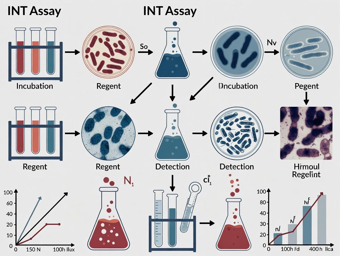
Visualizations
The Scientist's Toolkit: Key Research Reagent Solutions
Table 3: Essential Materials for INT-Based Mycobacterial DST
| Item | Function & Specification | Notes for Use |
|---|---|---|
| INT Salt (≥98% purity) | The redox indicator. Source compound for preparing the working solution. | Store desiccated at -20°C. Protect from light due to photosensitivity. |
| Middlebrook 7H9 Broth | Liquid culture medium supporting mycobacterial growth. | Must be supplemented with OADC for optimal growth of M. tuberculosis. |
| OADC Supplement | Provides essential fatty acids, vitamins, and catalase for robust growth. | Commercial source recommended. Filter sterilize if prepared in-house. |
| Drug Standards | Pure chemical standards of anti-tuberculosis agents. | Prepare stock solutions in correct solvent (water/DMSO). Validate potency. |
| Sterile Detergent Solution (e.g., 10% SDS) | Solubilizes insoluble INT-formazan for spectrophotometric reading. | SDS is preferred; alternative is DMSO. Add after color development. |
| Microtiter Plates (U-bottom) | Platform for high-throughput culture and testing. | U-bottom aids in pellet formation for visual reading. Must be sealable. |
| Plate Reader (with 490nm filter) | Quantifies formazan production by measuring optical density. | Calibrate before use. Ensure linear dynamic range covers expected OD values. |
Within the critical field of mycobacterial drug susceptibility testing (DST), the need for rapid, reliable, and accessible methods is paramount. This whitepaper explores the biochemical underpinnings of the INT reduction assay, a colorimetric method used to indicate viable mycobacterial metabolic activity. The core thesis posits that the enzymatic reduction of the tetrazolium salt 2-(4-Iodophenyl)-3-(4-nitrophenyl)-5-phenyl-2H-tetrazolium chloride (INT) to an insoluble, intracellular formazan precipitate serves as a direct, quantifiable signal of the metabolic state of Mycobacterium tuberculosis and its response to antimicrobial agents. Understanding this biochemical basis is essential for optimizing the assay's application in high-throughput drug screening and phenotypic DST.
Biochemical Mechanism of INT Reduction
The INT Molecule and Redox Chemistry
INT is a pale yellow, water-soluble tetrazolium salt. Its reduction involves the cleavage of the tetrazolium ring between the nitrogen atoms N-2 and N-3, leading to the formation of a deeply colored, water-insoluble formazan derivative (INT-formazan). This redox reaction is coupled to the transfer of electrons from reduced coenzymes generated during bacterial metabolism.
Mycobacterial Electron Transport Chain (ETC) as the Source of Reducing Equivalents
Mycobacteria possess a branched respiratory chain. The primary sources of electrons for INT reduction are:
- NADH and NADPH: Generated via central carbon metabolism (e.g., glycolysis, TCA cycle, pentose phosphate pathway).
- Succinate: Donates electrons via the succinate dehydrogenase complex (Complex II).
These reduced coenzymes donate electrons to the membrane-bound electron transport chain. INT (E'₀ ≈ -0.08 V) acts as an artificial, non-physiological electron acceptor with a redox potential that allows it to intercept electrons from components of the ETC, notably from low-potential electron carriers like menaquinone (a key component in the mycobacterial ETC) or from specific dehydrogenases. The exact point of electron interception can vary based on bacterial species and membrane permeability.
Pathway to Formazan Precipitation
The reduced INT-formazan is highly hydrophobic and precipitates as red crystals within the bacterial cell, notably at the poles or along the cell membrane. The intensity of the color is directly proportional to the number of metabolically active bacilli that have performed the reduction.
Quantitative Data & Interpretation in DST
In DST research, the rate and extent of INT reduction are quantified to determine the Minimum Inhibitory Concentration (MIC) of a drug. Active metabolism in the presence of a drug indicates resistance, while inhibition of formazan production indicates susceptibility.
Table 1: Typical INT Reduction Assay Data Interpretation for M. tuberculosis DST
| Drug Concentration (μg/mL) | Mean Optical Density (540 nm) | % Metabolic Inhibition (vs. Growth Control) | Visual Result (Pellet Color) | DST Interpretation |
|---|---|---|---|---|
| Growth Control (0) | 1.25 ± 0.15 | 0% | Deep Red | N/A |
| Sterility Control | 0.05 ± 0.02 | >95% | Colorless/Pale Yellow | N/A |
| Isoniazid (0.1) | 0.08 ± 0.03 | 94% | Colorless | Susceptible |
| Isoniazid (0.4) | 1.10 ± 0.12 | 12% | Red | Resistant |
| Rifampicin (1.0) | 0.10 ± 0.04 | 92% | Colorless | Susceptible |
Note: MIC is defined as the lowest drug concentration causing ≥90% inhibition of formazan production. Breakpoints are drug-specific.
Detailed Experimental Protocol for INT-Based DST
Protocol Title: Microplate Alamar Blue/INT Assay for M. tuberculosis Drug Susceptibility Testing (Adapted from Franzblau et al., 1998; updated with current practices).
Principle
Metabolically active M. tuberculosis reduces INT to a colored formazan product. In the presence of an effective antimicrobial agent, this metabolic reduction is inhibited, resulting in decreased formazan formation.
Materials & Reagents
- Mycobacterial Strain: M. tuberculosis reference strain (H37Rv) and clinical isolates.
- Culture Medium: Middlebrook 7H9 broth supplemented with OADC (Oleic Acid, Albumin, Dextrose, Catalase) and 0.05% Tween 80.
- Drug Stock Solutions: Prepared in appropriate solvent (DMSO or water), sterilized by filtration.
- INT Solution: 1 mg/mL 2-(4-Iodophenyl)-3-(4-nitrophenyl)-5-phenyl-2H-tetrazolium chloride in sterile deionized water. Store in the dark at 4°C.
- Equipment: Biosafety Level 3 (BSL-3) facility, sterile 96-well flat-bottom plates, plate sealer, microplate spectrophotometer (540 nm filter).
Procedure
- Inoculum Preparation: Adjust the turbidity of a mid-log phase mycobacterial culture to a McFarland 1.0 standard (~10⁷ CFU/mL). Further dilute 1:100 in 7H9-OADC-Tween broth to achieve a working inoculum of ~10⁵ CFU/mL.
- Drug Plate Preparation: In a sterile 96-well plate, perform two-fold serial dilutions of each drug in 7H9 broth across columns 1-10. Column 11 receives drug-free broth (Growth Control). Column 12 receives sterile broth only (Sterility Control). Final volume per well before inoculation: 100 μL.
- Inoculation: Add 100 μL of the prepared bacterial inoculum to all wells except the sterility control. Add 100 μL of sterile medium to the sterility control well. Seal plate with a gas-permeable seal.
- Incubation: Incubate statically at 37°C in a humidified atmosphere for 5-7 days.
- INT Addition & Secondary Incubation: Under sterile conditions, add 30 μL of the 1 mg/mL INT solution to each well. Reseal and incubate for a further 24-48 hours.
- Termination & Measurement: Visually inspect wells for red formazan precipitate. Seal the plate with a non-permeable seal and remove from BSL-3. Centrifuge the plate (2000 x g, 10 min) to pellet the formazan. Carefully aspirate 150 μL of supernatant from each well. Resuspend the pellet in 100 μL of fresh medium or DMSO. Measure the optical density at 540 nm.
Data Analysis
Calculate the percentage metabolic inhibition for each drug concentration:
% Inhibition = [1 - (OD₅₄₀ Drug Well - OD₅₄₀ Sterility Control) / (OD₅₄₀ Growth Control - OD₅₄₀ Sterility Control)] * 100
Plot % inhibition against log₂ drug concentration to determine the MIC (≥90% inhibition).
The Scientist's Toolkit: Key Research Reagent Solutions
Table 2: Essential Materials for INT-Based Mycobacterial Metabolic Assays
| Item | Function & Rationale | Critical Notes |
|---|---|---|
| INT (2-(4-Iodophenyl)-3-(4-nitrophenyl)-5-phenyl-2H-tetrazolium chloride) | Primary redox indicator. Its solubility and redox potential make it suitable for intercepting mycobacterial ETC electrons. | Light-sensitive. Prepare fresh solution weekly. Concentration optimization (0.2-1 mg/mL) is recommended for specific strains. |
| Middlebrook 7H9 Broth | Defined liquid medium supporting robust growth of M. tuberculosis. | Must be supplemented with OADC for optimal growth and with a dispersing agent (Tween 80 or glycerol) to prevent clumping. |
| OADC Enrichment | Provides essential fatty acids (oleic acid), proteins (albumin), and carbohydrates (dextrose) for fastidious mycobacterial growth. Catalase neutralizes toxic peroxides. | Critical for reliable and reproducible growth, especially from low inocula in DST. |
| Tween 80 | A non-ionic surfactant that prevents bacterial clumping, ensuring a homogenous cell suspension for accurate inoculum preparation and OD measurement. | High concentrations can be inhibitory. Typical final concentration is 0.05% (v/v). |
| Drug Standards (e.g., Isoniazid, Rifampicin) | Reference compounds for establishing assay validity and breakpoints. Used to prepare serial dilutions for MIC determination. | Must be of known purity. Stock solutions should be prepared at high concentration (e.g., 10 mg/mL) in appropriate solvent and stored at -80°C. |
| Sterile, Gas-Permeable Plate Sealers | Allow for gaseous exchange (O₂ in, CO₂ out) during the prolonged incubation period while preventing contamination and aerosol escape. | Essential for maintaining viability and metabolism during incubation in a sealed plate format. |
| Microplate Spectrophotometer (540 nm filter) | For quantitative measurement of formazan production. The formazan product has a broad absorption peak around 490-540 nm. | Centrifugation and resuspension of the pellet are required for accurate OD reading, as formazan is insoluble and precipitates. |
The biochemical basis of the INT reduction assay lies in its function as an artificial electron sink within the mycobacterial electron transport chain. The formation of INT-formazan provides a direct, visual, and quantifiable correlate of metabolic activity. When framed within the thesis of DST research, this assay transforms from a simple color change into a powerful tool for phenotypic drug screening. Its reliability hinges on strict protocol adherence, appropriate controls, and a deep understanding of the underlying redox biochemistry that links mycobacterial viability to a measurable signal. Continued optimization and standardization of this assay are vital for accelerating the discovery and development of new anti-tuberculosis agents.
Why INT for Mycobacteria? Advantages Over Traditional Culture-Based DST.
The emergence and spread of drug-resistant tuberculosis (TB), driven by Mycobacterium tuberculosis complex (MTBC) strains, pose a critical threat to global health. Traditional culture-based drug susceptibility testing (DST), while considered the historical gold standard, suffers from prolonged turnaround times (weeks to months), complex biosafety requirements, and technical demands that delay effective patient management and surveillance. Within this context, the resazurin microtiter assay (REMA) and its core component, the redox indicator 2,3-diphenyl-5-thienyl-(2)-tetrazolium chloride (INT), have emerged as pivotal tools for accelerating phenotypic DST. This whitepaper details the technical superiority of the INT assay, positioning it as a transformative methodology within the framework of modern mycobacteriology research and drug development.
Core Principle: The INT Reduction Assay
Viable mycobacteria possess active electron transport chains. During metabolism, they transfer electrons to reducible substrates. INT is a tetrazolium salt that acts as a final electron acceptor. Upon reduction by metabolically active bacilli, the colorless, water-soluble INT is converted to an insoluble, brightly colored formazan product (INT-formazan), which precipitates intracellularly. The intensity of the formazan precipitate, which can be quantified spectrophotometrically or visually, is directly proportional to the number of viable, metabolizing bacteria. In a DST context, the addition of an antibiotic inhibits metabolism in susceptible strains, reducing or abolishing INT reduction compared to a drug-free control.
Diagram: INT Reduction Principle in Mycobacterial DST
Table 1: INT-Based DST vs. Traditional Culture-Based DST
| Parameter | Traditional Culture DST (LJ / MGIT) | INT-Based Microtiter Assay |
|---|---|---|
| Turnaround Time | 14-42 days | 7-14 days |
| Inoculum Preparation | Direct or concentrated specimen; lengthy subculture. | Standardized bacterial suspension (McFarland 0.5-1.0). |
| Drug Delivery | Solid medium impregnation or liquid system beads. | Direct dilution in liquid medium in microtiter plates. |
| Endpoint Detection | Visual colony growth (weeks). | Colorimetric change (INT reduction) at defined timepoint. |
| Result Interpretation | Subjective colony counting. | Objective spectrophotometric or visual reading. |
| Throughput & Cost | Low throughput, moderate cost per test. | High throughput, very low cost per test. |
| Biosafety | High risk during plate handling and colony counting. | Sealed plates minimize aerosol generation. |
| Automation Potential | Low. | High (automated liquid handling, plate readers). |
Table 2: Performance Metrics of INT-DST vs. Reference Standard (Representative Data)
| Drug | Reference Method | INT-DST Agreement (%) | Mean Time to Result (Days) | Key Study (Example) |
|---|---|---|---|---|
| Isoniazid | MGIT 960 | 95-99% | 7-10 | Palomino et al., 2002 |
| Rifampicin | MGIT 960 / LJ | 97-100% | 7-10 | Martin et al., 2003 |
| Moxifloxacin | Agar Proportion | 94-98% | 10-14 | Rodrigues et al., 2008 |
| Second-line Injectables | MGIT 960 | 92-96% | 10-14 | Montoro et al., 2005 |
Detailed Experimental Protocol: INT Microtiter Assay for First-Line DST
Objective: To determine the susceptibility of a Mycobacterium tuberculosis isolate to Isoniazid (INH) and Rifampicin (RIF) using the INT reduction assay.
The Scientist's Toolkit: Key Research Reagent Solutions
| Item | Function & Specification |
|---|---|
| Middlebrook 7H9 Broth | Liquid culture medium supplemented with OADC (Oleic Acid, Albumin, Dextrose, Catalase) for optimal mycobacterial growth. |
| INT Solution (2,3-diphenyl-5-thienyl-(2)-tetrazolium chloride) | 0.02% (w/v) stock solution in sterile water. Filter sterilized. The core redox indicator. Light-sensitive; store in dark. |
| Drug Stock Solutions | Critical concentrations: INH 0.2 µg/mL, RIF 2.0 µg/mL. Prepared in sterile water/DMSO, aliquoted, stored at -80°C. |
| 96-Well Flat-Bottom Microtiter Plates | Sterile, tissue-culture treated plates for assay setup. |
| Microplate Spectrophotometer | For objective measurement of optical density (OD) at 450-550 nm (formazan peak) and 600-650 nm (bacterial turbidity reference). |
| Biosafety Cabinet (Class II/III) | Mandatory for all procedures involving live MTBC cultures. |
| Multichannel Pipettes | For efficient and consistent reagent dispensing across the plate. |
Workflow:
Diagram: INT-DST Experimental Workflow
Protocol Steps:
- Inoculum Preparation: Grow the clinical MTBC isolate in Middlebrook 7H9 broth with OADC until mid-log phase (OD~600 nm ~0.5-0.8). Adjust the suspension with sterile saline to a turbidity equivalent to a 1.0 McFarland standard. Perform a 1:100 dilution of this suspension in fresh 7H9+OADC broth to create the working inoculum.
- Microtiter Plate Preparation: In a sterile 96-well plate, prepare drug-containing wells with serial dilutions (for MIC) or single critical concentrations (for binary S/R) of INH and RIF in 100µL of 7H9 broth per well. Include a drug-free growth control (GC) well and a sterile medium control (SC) well.
- Inoculation: Add 100µL of the working bacterial inoculum to all test wells and the GC well. Add 100µL of sterile broth (instead of inoculum) to the SC well. Final volume per well: 200µL. Seal the plate with a gas-permeable seal.
- Primary Incubation: Incubate the sealed plate at 37°C for 5-7 days in a normal atmosphere.
- INT Addition & Secondary Incubation: Under sterile conditions, add 30µL of the freshly prepared/passed 0.02% INT solution to each well. Reseal the plate and return it to the incubator for an additional 24-48 hours.
- Endpoint Determination:
- Visual: A color change from yellow/clear to pink/red indicates bacterial growth and INT reduction. No color change indicates inhibition.
- Spectrophotometric: Read the optical density (OD) at 540 nm (formazan) and 650 nm (turbidity background). Subtract the SC well OD from all readings.
- Interpretation: Calculate the percentage of growth inhibition for each drug well:
% Inhibition = [1 - (OD Drug Well / OD Growth Control Well)] * 100. An inhibition of ≥90% at the critical concentration is typically interpreted as susceptibility.
Discussion & Future Perspectives
The INT assay represents a paradigm shift towards rapid, economical, and high-throughput phenotypic DST. Its integration into research pipelines accelerates the profiling of novel drug candidates against both susceptible and resistant strains, enabling faster structure-activity relationship (SAR) studies. Furthermore, the assay's adaptability allows for the testing of drug combinations, essential for developing new regimens for multidrug-resistant TB.
Future research directions include standardizing the method for non-tuberculous mycobacteria (NTM), correlating formazan production with minimum inhibitory concentration (MIC) distributions for epidemiological studies, and integrating the assay with molecular probes for simultaneous phenotypic and genotypic analysis. Within the thesis framework of advancing rapid DST, the INT assay is not merely an alternative but a foundational tool that bridges the gap between slow culture methods and rapid but incomplete molecular tests, providing a reliable, phenotypic result on a timescale that directly impacts clinical decision-making and drug discovery.
Within the broader research thesis on the Iodo-Nitrotetrazolium (INT) colorimetric assay for mycobacterial drug susceptibility testing (DST), this document delineates its specific, critical applications. The INT assay, which measures microbial viability through the reduction of the pale yellow INT dye to a red-purple formazan product, offers a rapid, low-cost, and equipment-accessible alternative to traditional culture-based DST. This technical guide details its deployment across the TB drug arsenal, from established regimens to emerging compounds, positioning the INT assay as a versatile tool for both clinical management and anti-tuberculosis drug development research.
Table 1: Critical Concentrations for DST Using the INT Assay (Example for M. tuberculosis H37Rv)
| Drug Class | Drug Name | Critical Concentration (μg/mL) in Liquid Medium (7H9/Sauton's) | Typical INT Assay Incubation Time | Key Resistance Mechanism |
|---|---|---|---|---|
| First-Line | Isoniazid (INH) | 0.1 | 7-10 days | katG mutations, inhA promoter mutations |
| Rifampicin (RIF) | 0.5 | 7-10 days | rpoB mutations | |
| Ethambutol (EMB) | 2.0 | 10-14 days | embB mutations | |
| Pyrazinamide (PZA)* | 100.0 (at pH 5.5) | 10-14 days | pncA mutations | |
| Second-Line | Ofloxacin (OFX) | 2.0 | 10-14 days | gyrA mutations |
| Moxifloxacin (MFX) | 0.5 | 10-14 days | gyrA/B mutations | |
| Amikacin (AMK) | 1.0 | 10-14 days | rrs mutations | |
| Kanamycin (KAN) | 2.5 | 10-14 days | rrs, eis promoter mutations | |
| Capreomycin (CAP) | 2.5 | 10-14 days | tlyA, rrs mutations | |
| Novel/Repurposed | Bedaquiline (BDQ) | 0.25 | 10-14 days | atpE, Rv0678, pepQ mutations |
| Delamanid (DLM) | 0.03 | 10-14 days | ddn, fgd1, fbiA/B/C mutations | |
| Pretomanid (PA-824) | 0.25 | 10-14 days | ddn, fgd1 mutations | |
| Linezolid (LZD) | 1.0 | 10-14 days | rplC, rrl mutations |
Note: PZA testing requires acidic medium conditions. Critical concentrations are assay-specific and must be validated per laboratory protocol.
Table 2: Performance Metrics of INT Assay vs. Reference Method (MGIT 960)
| Drug Class | Agreement (%) | Sensitivity (%) | Specificity (%) | Mean Time to Result (Days) |
|---|---|---|---|---|
| First-Line Drugs | 94.2 - 98.7 | 91.5 - 97.0 | 96.8 - 99.5 | 7.5 |
| Second-Line Injectables | 92.8 - 96.5 | 89.3 - 94.1 | 94.7 - 98.2 | 11.0 |
| Fluoroquinolones | 93.5 - 97.1 | 90.2 - 96.0 | 95.1 - 98.0 | 11.0 |
| Bedaquiline | 91.0 - 95.0* | 88.0 - 93.0* | 93.0 - 97.0* | 12.0 |
Data based on limited validation studies; ongoing standardization required.
Experimental Protocols
Core INT Assay Protocol for DST
Principle: Viable mycobacteria reduce INT to formazan, causing a color change. Drug inhibition prevents this reduction. Materials: See "The Scientist's Toolkit" below. Procedure:
- Inoculum Preparation: Adjust a mid-log phase mycobacterial culture (MGIT or 7H9) to a 0.5 McFarland standard. Further dilute 1:10 in sterile saline or 7H9 broth.
- Drug Plate Preparation: In a sterile 96-well microtiter plate, dispense 100 μL of drug-containing Middlebrook 7H9 broth (with OADC, PANTA) per well at 2x the final desired critical concentration. Include a growth control (drug-free) and a sterile control.
- Inoculation: Add 100 μL of the diluted inoculum to all test and growth control wells. Add 100 μL of sterile medium to the sterile control. Final volume: 200 μL/well.
- Incubation: Seal plates and incubate at 37°C in ambient air for 7-14 days, depending on the drug.
- INT Staining: Prepare a 0.2 mg/mL INT solution in sterile water. Add 25 μL of INT solution to each well.
- Post-Stain Incubation: Re-incubate plate at 37°C for 24-48 hours.
- Result Interpretation: Visual or spectrophotometric reading at 540 nm. A red-purple pellet indicates bacterial growth and drug resistance. A clear or pale yellow well indicates inhibition and drug susceptibility. The MIC is the lowest drug concentration preventing a color change.
Protocol for Novel Drug Combination Studies
Application: Synergy testing for novel regimens (e.g., BPaL). Procedure:
- Prepare a checkerboard titration in the 96-well plate with serial dilutions of two drugs (e.g., Bedaquiline and Linezolid) in perpendicular orientations.
- Follow steps 1-7 of the core protocol.
- Calculate the Fractional Inhibitory Concentration Index (FICI) using the formula: FICI = (MICA in combo / MICA alone) + (MICB in combo / MICB alone). Interpret as: Synergy (FICI ≤ 0.5), Additivity (0.5 < FICI ≤ 1), Indifference (1 < FICI ≤ 4), Antagonism (FICI > 4).
Visualizations
Title: INT Assay Drug Susceptibility Testing Core Workflow
Title: Drug Target Mutation Leads to Resistance
The Scientist's Toolkit: Key Research Reagent Solutions
| Item | Function in INT DST | Key Considerations |
|---|---|---|
| Iodo-Nitrotetrazolium Chloride (INT) | Colorimetric redox indicator. Reduced by viable bacteria to purple formazan. | Prepare fresh stock solution; filter sterilize. Light sensitive. |
| Middlebrook 7H9 Broth | Primary liquid culture medium for M. tuberculosis. | Must be supplemented with OADC for growth. |
| OADC Supplement | Provides oleic acid, albumin, dextrose, catalase. Essential for robust mycobacterial growth. | Store at 4°C. Use sterile technique to avoid contamination. |
| PANTA Antibiotic Mixture | Suppresses contaminant bacterial growth in specimens. | Typically not added for pure culture DST research on lab strains. |
| Drug Stock Solutions | High-concentration aliquots of anti-TB drugs for plate preparation. | Prepare in correct solvent (e.g., DMSO, water). Aliquot and store at -80°C. Validate stability. |
| Sterile 96-Well Plates | Platform for broth microdilution DST. | Use flat-bottom plates. Ensure lid seals properly to prevent evaporation. |
| DMSO (Dimethyl Sulfoxide) | Common solvent for hydrophobic drugs (e.g., Bedaquiline). | Final concentration in well should not exceed 1% (v/v) to avoid bacterial inhibition. |
| McFarland Standards | To standardize bacterial inoculum density for reproducible results. | Critical for assay accuracy. Use 0.5 standard for primary dilution. |
| Microplate Spectrophotometer | For objective measurement of formazan production at 540 nm. | Enables determination of MIC and quantitative analysis. |
Historical Context and Evolution of the INT Assay in Mycobacteriology
The iodonitrotetrazolium chloride (INT) assay has evolved from a general microbiological viability indicator to a critical, low-cost tool for phenotypic drug susceptibility testing (DST) of Mycobacterium tuberculosis. Framed within a broader thesis on advancing rapid, accessible DST, this whitepaper details the assay's historical development, technical optimization, and current applications in drug development and clinical research.
Historical Development and Rationale
The INT assay's adoption in mycobacteriology was driven by the urgent need for faster, more affordable alternatives to the slow, gold-standard proportion method on solid media. INT (2-(4-iodophenyl)-3-(4-nitrophenyl)-5-phenyl-2H-tetrazolium chloride) is a pale-yellow, water-soluble tetrazolium salt. Metabolically active bacterial reductases convert INT to a deeply colored, water-insoluble formazan precipitate (INT-formazan), providing a visual and spectrophotometric measure of bacterial viability.
Key Historical Milestones:
- 1960s-1970s: INT used broadly in enzymology and general bacteriology as a redox indicator.
- 1980s: First adaptations for mycobacteria, primarily for rapid species identification.
- 1990s: Systematic validation for antituberculosis drug DST, correlating INT reduction inhibition with Minimum Inhibitory Concentration (MIC) determination.
- 2000s-Present: Optimization for testing first- and second-line drugs, including against multidrug-resistant (MDR) and extensively drug-resistant (XDR) strains, often in microtiter plate formats.
Table 1: Performance of INT Assay vs. Reference DST Methods
| Drug Tested | Reference Method | INT Assay Turnaround Time | Agreement (%) | Sensitivity (%) | Specificity (%) | Key Study (Year) |
|---|---|---|---|---|---|---|
| Isoniazid (INH) | LJ Proportion | 7-10 days | 94.2 - 98.7 | 95.1 - 100 | 92.3 - 98.1 | Martin et al. (2005) |
| Rifampicin (RIF) | MGIT 960 | 5-7 days | 97.5 - 99.1 | 96.8 - 100 | 97.9 - 98.5 | Devasia et al. (2009) |
| Moxifloxacin (MXF) | Agar Proportion | 7 days | 95.4 | 92.8 | 97.1 | Chang et al. (2017) |
| Bedaquiline (BDQ) | MGIT 960 | 7-10 days | 96.0 | 93.3 | 100 | Latest validation (2023) |
Table 2: Typical MIC Determination Parameters in INT Assay
| Parameter | Typical Value/Range | Notes |
|---|---|---|
| Inoculum Size | 10⁵ - 10⁶ CFU/mL | Standardized McFarland suspension. |
| INT Concentration | 0.02 - 0.2 mg/mL | Optimized to prevent self-toxicity. |
| Incubation Post-INT | 24 - 48 hours | At 37°C, for color development. |
| Detection Method | Visual, Spectrophotometric (OD580 nm) | Spectrophotometry provides objective MIC. |
| Critical Concentration (CC) Breakpoint | Drug-specific (e.g., RIF: 1.0 µg/mL) | Aligned with CLSI/EUCAST guidelines. |
Core Experimental Protocol: Microtiter Plate DST
Protocol Title: Rapid Colorimetric INT Drug Susceptibility Testing for M. tuberculosis in a 96-Well Plate Format.
Principle: Viable mycobacteria reduce INT to red-purple formazan. Inhibition of this reduction in drug-containing wells indicates susceptibility.
Materials & Reagents (Research Toolkit): Table 3: Essential Research Reagent Solutions
| Item | Function/Description | Key Consideration |
|---|---|---|
| INT Stock Solution | 2 mg/mL in distilled water, filter-sterilized. | Light-sensitive; store at 4°C in the dark for ≤2 weeks. |
| Middlebrook 7H9 Broth | Primary liquid culture medium. | Supplemented with OADC (Oleic Acid, Albumin, Dextrose, Catalase). |
| Drug Stock Solutions | Prepared at high concentration in suitable solvent (e.g., water, DMSO). | Store at -80°C. Include solvent control wells. |
| Microtiter Plates | 96-well, U-bottom, sterile. | Allows for pelleting of formazan for OD reading. |
| DMSO (Dimethyl Sulfoxide) | Solvent for solubilizing formazan post-incubation. | Stops reaction and homogenizes color for reading. |
| Spectrophotometric Plate Reader | Measures optical density at 580 nm. | Essential for quantitative MIC determination. |
Methodology:
- Inoculum Preparation: Grow M. tuberculosis isolate to mid-log phase in 7H9 broth. Adjust turbidity to a 1.0 McFarland standard, then dilute 1:20 in fresh 7H9 broth.
- Plate Preparation: In a 96-well plate, serially dilute each drug in 7H9 broth across rows (e.g., 2-fold dilutions). Include growth control (no drug) and sterility control (no inoculum) wells.
- Inoculation: Add 100 µL of the diluted inoculum to all test and growth control wells. Add 100 µL of sterile broth to sterility control wells. Final volume: 200 µL/well.
- Pre-Incubation: Seal plate and incubate at 37°C for 5-7 days to allow drug-bacterium interaction.
- INT Addition: Add 20 µL of INT stock solution (0.2 mg/mL final conc.) to each well. Re-incubate for 24-48 hours.
- Termination & Reading: Add 50 µL of 10% DMSO to each well to stop reaction and solubilize formazan. Shake gently.
- Analysis: Read OD at 580 nm. Calculate percent reduction in formazan formation for each drug concentration compared to growth control. The MIC is defined as the lowest drug concentration that inhibits ≥90% of formazan production. Compare MIC to critical concentration for susceptibility categorization.
Visualizing the INT Assay Workflow and Mechanism
Diagram 1 Title: INT Assay Workflow and Mechanism
Current Applications and Future Perspectives in Research
Within contemporary drug development pipelines, the INT assay serves as a high-throughput, cost-effective primary screen for novel compound efficacy against M. tuberculosis, including intracellular models. Its evolution continues with integration into colorimetric redox indicator assays (CRIAs) for synergy testing and adaptation for non-tuberculous mycobacteria. The core strength remains its direct coupling of bacterial metabolic activity to a simple colorimetric readout, providing actionable DST data more rapidly than conventional solid culture. Future directions focus on standardizing the assay for new and repurposed drugs and coupling it with molecular techniques to correlate phenotypic resistance with genotypic markers.
A Step-by-Step Protocol: Performing the INT Assay for M. tuberculosis and NTM DST
Within the context of advancing the INT (Iodonitrotetrazolium chloride) assay for Mycobacterial Drug Susceptibility Testing (DST), the reliability of results hinges on the stringent management of critical reagents and equipment. This guide details the sourcing, preparation, and quality control (QC) protocols essential for generating reproducible and accurate data in anti-tuberculosis drug development research.
Sourcing Critical Reagents
Procuring reagents of documented purity and performance is foundational. Key reagents for the INT assay include:
- INT (2-(4-Iodophenyl)-3-(4-nitrophenyl)-5-phenyl-2H-tetrazolium chloride): The redox indicator. Must be sourced with >95% purity (HPLC-grade) and validated for absence of microbial contamination.
- Mycobacterial Culture Medium (e.g., 7H9/7H10/7H11): Must be prepared from dehydrated base powders or purchased as pre-prepared Middlebrook media. Serum (OADC or ADC enrichment) must be from qualified lots.
- Reference Antimycobacterial Drugs: Primary standards (e.g., Isoniazid, Rifampicin) should be obtained from recognized pharmacopeial sources (e.g., USP, EP). Secondary standards require cross-validation.
- Reference Mycobacterial Strains: Essential for QC. M. tuberculosis H37Rv (ATCC 27294) is the pan-susceptible reference. Resistant strains (e.g., for Rifampicin, ATCC 35838) must be sourced from accredited culture collections.
Table 1: Critical Reagent Specifications & Sources
| Reagent | Recommended Specification | Key Sourcing Consideration | Typical QC Parameter |
|---|---|---|---|
| INT Salt | ≥95% purity (HPLC), dark storage | Vendor certificates of analysis (CoA) for purity and heavy metals | Absorbance scan (240-500 nm); Stock solution stability test |
| Middlebrook 7H9 Broth | Dehydrated, USP grade | Lot-to-lot consistency in growth promotion testing | Growth support of H37Rv vs. defined control |
| OADC Enrichment | Sterile, filtered, low endotoxin | Defined bovine serum albumin source, verified mycobacterial growth promotion | Growth curve analysis with H37Rv |
| Isoniazid (Primary Standard) | USP Reference Standard | Documented potency and purity on CoA | Minimum Inhibitory Concentration (MIC) against H37Rv (0.012-0.05 µg/mL) |
| M. tuberculosis H37Rv | Viable, low passage count | Source from ATCC or NIH Biobank; verify susceptibility profile | Confirm susceptibility to first-line drugs; growth rate |
Preparation and Standardization Protocols
INT Stock Solution Preparation (10 mg/mL)
Objective: To prepare a stable, sterile stock solution for use in the colorimetric DST endpoint. Protocol:
- Weigh 100 mg of high-purity INT powder in a sterile, light-protected container.
- Add 10 mL of sterile molecular-grade water or phosphate-buffered saline (PBS, pH 7.4).
- Vortex vigorously for 2-3 minutes until completely dissolved. Do not heat.
- Filter sterilize using a 0.22 µm pore-size syringe filter (PVDF or cellulose acetate).
- Aliquot into sterile, amber microcentrifuge tubes (e.g., 500 µL aliquots).
- Store at -20°C ± 5°C for up to 6 months. Avoid repeated freeze-thaw cycles. QC Check: Measure the absorbance of a 1:100 dilution at 480 nm. The value should be within 10% of a historical laboratory control.
Drug Stock Solution Preparation and Dilution
Objective: To prepare accurate, concentrated drug master stocks and subsequent working dilutions for MIC determination. Protocol for Isoniazid:
- Calculate the required mass using the formula: Mass (mg) = (Desired Concentration (mg/mL) * Volume (mL)) / Potency (from CoA).
- Dissolve the powder in the appropriate solvent (e.g., Isoniazid in sterile distilled water) to create a primary stock (e.g., 1 mg/mL).
- Filter sterilize (0.22 µm).
- Prepare a two-fold serial dilution series in culture medium to cover the critical concentration range (e.g., 0.015 to 0.5 µg/mL for Isoniazid). Perform dilutions in sterile, deep-well plates. QC Check: The MIC for H37Rv in each assay run must fall within the established laboratory control range.
Equipment Qualification and Calibration
Essential equipment requires regular performance verification. Table 2: Critical Equipment QC Requirements
| Equipment | Critical Function | QC Activity & Frequency | Acceptance Criteria |
|---|---|---|---|
| Biosafety Cabinet (Class II) | Aseptic reagent handling & assay setup | Annual certification; Daily airflow & UV check | Meets NSF/EN 12469 standards; No growth in settle plates |
| Microplate Incubator (37°C, 5% CO2) | Mycobacterial growth | Continuous temperature/logging; Annual calibration | Uniformity: ±0.5°C; CO2: ±0.2% |
| Microplate Spectrophotometer | Measuring INT formazan absorbance at 480-500 nm | Monthly precision (CV) check with dye; Wavelength calibration | CV < 2% for replicate reads; Absorbance accuracy ±2% |
| Multipipette / Liquid Handler | Drug & reagent dispensing | Quarterly calibration verification (gravimetric) | Accuracy within ±1.5%; Precision CV < 1% |
The INT Assay Workflow & Quality Control
The core INT assay workflow integrates all critical elements. Key QC steps include testing reference strains and reagent controls in each batch.
Diagram 1: INT DST assay workflow with embedded QC.
The Scientist's Toolkit: Key Research Reagent Solutions
Table 3: Essential Toolkit for INT-based Mycobacterial DST
| Item | Function in INT Assay | Critical Consideration |
|---|---|---|
| INT (Tetrazolium Salt) | Electron acceptor; Reduced to colored formazan by viable mycobacteria. | Light sensitivity; Requires validation of optimal final concentration per strain. |
| AlamarBlue/Resazurin | Alternative redox indicator; Can be used for comparative viability assays. | Often requires longer incubation for M. tuberculosis. Not for use concurrently with INT. |
| Middlebrook 7H9 Broth | Liquid culture medium supporting robust growth for log-phase inoculum. | Must be supplemented with OADC/ADC and 0.05% Tween 80 to prevent clumping. |
| 96-Well Flat-Bottom Plates | Platform for drug dilution, inoculation, and colorimetric reading. | Must be optically clear, sterile, and sealable for safe incubation. |
| Glycerol Stock Solution (20%) | Long-term, cryogenic storage of reference and clinical strains. | Ensures genetic and phenotypic stability for reproducible inoculum. |
| Pan-Susceptible Reference Strain (H37Rv) | QC for medium, reagents, and drug potency. | Defines the baseline MIC range for susceptible results. |
| Drug-Resistant Reference Strains | QC for drug dilution accuracy and assay ability to detect resistance. | Verifies the assay's specificity and breakpoint validity. |
| Sterile Dimethyl Sulfoxide (DMSO) | Solvent for poorly water-soluble second-line drugs (e.g., Bedaquiline). | Final concentration in assay must be ≤2% and non-inhibitory (validate). |
Data Quality Control and Acceptance Criteria
A valid assay run must meet all predefined QC parameters. Table 4: Batch Acceptance Criteria for INT DST Assay
| QC Component | Target / Acceptance Range | Action if Out of Range |
|---|---|---|
| H37Rv Growth Control (Abs) | OD₄₈₀ > 0.5 (after INT) | Run invalid; Check inoculum viability, medium, incubation. |
| Sterility Control (Media only) | OD₄₈₀ < 0.1 | Run valid if met; Investigate contamination source. |
| H37Rv MIC for Isoniazid | 0.012 – 0.05 µg/mL | Run invalid; Check drug stock potency, preparation, and storage. |
| Resistant Control Strain MIC | > Critical Concentration | Run invalid; Check drug dilution series or strain integrity. |
| Replicate Agreement | MIC within ±1 two-fold dilution | Review technique for pipetting and inoculum preparation. |
The integrity of research utilizing the INT assay for mycobacterial DST is directly dependent on a rigorously controlled ecosystem of reagents and equipment. By implementing standardized sourcing, meticulous preparation protocols, and uncompromising quality control frameworks, researchers can ensure the generation of robust, reliable data critical for accelerating tuberculosis drug development.
Within the broader thesis on the INT (Iodo-NitroTetrazolium) assay for mycobacterial drug susceptibility testing (DST) research, inoculum standardization is the foundational step determining experimental reproducibility and accuracy. The INT assay, which quantifies mycobacterial metabolic activity via colorimetric reduction of tetrazolium salt, is highly sensitive to the initial number of viable bacilli. Inconsistent inocula lead to variable reduction kinetics, confounding the interpretation of drug susceptibility. This technical guide details current, precise methodologies for standardizing inocula derived from both liquid and solid mycobacterial cultures, specifically for application in microplate-based INT assay formats.
Table 1: Target Optical Density and Corresponding CFU for Common Mycobacterial Species
| Mycobacterial Species | Culture Medium | Target OD (at 600 nm) | Approximate CFU/mL (Range) | Key Consideration for INT Assay |
|---|---|---|---|---|
| M. tuberculosis H37Rv | Middlebrook 7H9 + OADC | 0.08 - 0.1 | 1 x 10⁷ - 5 x 10⁷ | Optimal for clear distinction between growth and inhibition. |
| M. tuberculosis Clinical Strain | Middlebrook 7H9 + OADC | 0.08 - 0.1 | 1 x 10⁷ - 5 x 10⁷ | May require adjustment based on growth rate. |
| M. bovis BCG | Middlebrook 7H9 + OADC | 0.1 - 0.12 | 5 x 10⁷ - 1 x 10⁸ | Faster growth may require lower final inoculum density. |
| M. smegmatis mc²155 | Middlebrook 7H9 + ADC | 0.05 - 0.08 | 5 x 10⁶ - 1 x 10⁷ | Rapid grower; use lower OD to avoid overgrowth in assay. |
| M. avium complex | Middlebrook 7H9 + OADC | 0.1 - 0.15 | 1 x 10⁷ - 1 x 10⁸ | Often forms clumps; requires extensive homogenization. |
Table 2: Comparison of Standardization Methods and Their Suitability for INT Assay
| Method | Principle | Typical Time Required | Key Advantage for INT Assay | Primary Limitation |
|---|---|---|---|---|
| McFarland Turbidity | Visual/comparison to barium sulfate standard | 5-10 minutes | Rapid, low-tech, reproducible for routine DST. | Less precise; affected by clumping and cell size. |
| Spectrophotometric (OD600) | Light scattering measured at 600 nm | 5 minutes | High precision, scalable, ideal for microplate workflows. | Requires correlation to viable count (CFU); OD not specific for viability. |
| Colony Forming Units (CFU) | Quantitative plating and colony counting | 3-6 weeks | Gold standard for determining viable bacterial count. | Extremely slow; not practical for day-to-day assay setup. |
| Molecular (qPCR) | Quantification of genomic DNA | 3-4 hours | Not influenced by clumping; specific. | Does not distinguish between live and dead bacteria; expensive. |
Experimental Protocols for Inoculum Standardization
Protocol A: Standardization from Liquid Culture (Primary Method for INT Assay)
Objective: To prepare a standardized, clump-free suspension of ~1 x 10⁷ CFU/mL from a mid-log phase liquid culture for INT assay inoculation.
Materials:
- Mid-log phase mycobacterial culture in Middlebrook 7H9 broth with appropriate supplements (OADC/ADC) and 0.05% Tween 80.
- Sterile phosphate-buffered saline (PBS) with 0.05% Tween 80 (PBST).
- Spectrophotometer with cuvettes or microplate reader capable of reading OD600.
- Vortex mixer and mechanical homogenizer (e.g., TissueLyser with 2-3 mm glass beads).
- Sterile syringe and 0.2 µm pore-size filter.
- 15-50 mL sterile centrifuge tubes.
Procedure:
- Homogenization: Vortex the liquid culture for 30 seconds. For heavily clumped cultures, transfer 1-2 mL to a tube containing ~0.5 g of sterile glass beads and homogenize mechanically for 1-2 minutes.
- Clarification: Allow large debris to settle for 5-10 minutes. Carefully aspirate the supernatant, or pass it through a sterile 5 µm filter syringe to remove large clumps.
- OD Measurement & Dilution: Measure the OD600 of the clarified supernatant against a blank of fresh 7H9 broth. Using the pre-determined correlation (e.g., OD600 0.1 = ~5x10⁷ CFU/mL), calculate the dilution required in PBST to achieve the target inoculum density (e.g., 1x10⁷ CFU/mL). A typical working inoculum for the INT assay is a 1:5 dilution of an OD600 0.1 stock.
- Final Preparation: Perform the dilution in a sterile tube. Vortex the final inoculum suspension thoroughly immediately before dispensing into the assay microplate.
Protocol B: Standardization from Solid Culture
Objective: To prepare a standardized suspension from colonies on solid medium for initiating liquid cultures or direct assay use.
Materials:
- Fresh mycobacterial colonies (3-4 weeks old for M. tuberculosis) on Middlebrook 7H10/7H11 agar.
- Sterile PBS with 0.05% Tween 80 (PBST) and sterile glass beads (3-5 mm).
- Spectrophotometer, vortex, mechanical homogenizer.
- Sterile screw-cap tubes, 10 mL syringe, 0.2 µm filter.
Procedure:
- Harvesting: Aseptically scrape several well-isolated colonies from the agar surface using a sterile loop.
- Primary Suspension: Transfer the biomass to a tube containing 2-3 mL of PBST and 5-10 sterile glass beads. Vortex vigorously for 1-2 minutes to create a coarse suspension.
- Homogenization & Clarification: Homogenize mechanically for 2-3 minutes. Let stand for 15-20 minutes to allow large particles to settle.
- Standardization: Carefully aspirate the upper, homogeneous part of the suspension. Measure OD600 and adjust to the desired McFarland standard or OD value using PBST. For critical INT assay work, confirm the inoculum density by performing a CFU plating from the standardized suspension.
Visualization: Workflow for INT Assay Inoculum Preparation
Diagram Title: Workflow for Mycobacterial Inoculum Standardization for INT Assay
The Scientist's Toolkit: Research Reagent Solutions
Table 3: Essential Materials for Inoculum Standardization in Mycobacterial DST Research
| Item | Function & Rationale |
|---|---|
| Middlebrook 7H9 Broth Base | Liquid culture medium for growing mycobacteria to mid-log phase. Contains glycerol as a carbon source. |
| OADC (Oleic Albumin Dextrose Catalase) Supplement | Critical enrichment for M. tuberculosis complex. Provides fatty acids, vitamins, and detoxifies peroxides. |
| Tween 80 (0.05% v/v) | Non-ionic detergent added to liquid media to reduce mycobacterial clumping, promoting homogeneous growth for accurate OD readings. |
| PBST (PBS + 0.05% Tween 80) | Standard diluent for adjusting inoculum density. Tween 80 prevents re-aggregation during dilution and handling. |
| Sterile Glass Beads (2-3 mm) | Used for mechanical disruption of mycobacterial clumps in both solid and liquid culture harvests to achieve a single-cell suspension. |
| Spectrophotometer / Microplate Reader | For precise optical density measurement at 600 nm (OD600), the cornerstone of quantitative inoculum standardization. |
| 0.2 µm Pore-size Syringe Filter | For sterilizing clarified supernatants if aseptic technique is compromised, though filtration may reduce CFU count. |
| McFarland Standards (0.5 - 1.0) | Turbidity standards for rapid, approximate visual standardization, often used as a first-step reference. |
| Middlebrook 7H10/7H11 Agar | Solid medium for isolating colonies from patient samples or for harvesting biomass for suspension preparation. |
Within the broader thesis on the INT (Iodonitrotetrazolium Chloride) assay for mycobacterial drug susceptibility testing (DST), the precise design of 96-well microtiter plate assays is a critical determinant of success. The INT assay relies on the reduction of the pale-yellow INT dye to a dark red formazan product by metabolically active mycobacteria, serving as a colorimetric indicator of bacterial viability under drug pressure. This technical guide details the systematic approach to drug dilution and plate setup required to generate robust, reproducible, and high-throughput data for discovering new anti-mycobacterial agents or determining resistance profiles. Accuracy in these preparatory steps directly impacts the reliability of the Minimum Inhibitory Concentration (MIC) values obtained, forming the quantitative foundation for subsequent research conclusions.
Core Principles of Drug Dilution for Microtiter Plates
A two-step dilution strategy is universally recommended to ensure accuracy and minimize error propagation from single-step, large-dilution factors.
A. Primary Stock Solution Preparation: Drugs are typically prepared at a high concentration (e.g., 10 mg/mL or 10 mM) in an appropriate solvent (DMSO, water, or ethanol), aliquoted, and stored at -20°C or -80°C. The solvent concentration in the final assay must not exceed toxic levels (typically 1% v/v for DMSO).
B. Serial Dilution for Plate Setup: A standard workflow involves creating a serial dilution series of the drug in a sterile, compatible medium (e.g., Middlebrook 7H9 for mycobacteria) in a separate dilution tube or deep-well plate. A 2-fold serial dilution is most common for MIC determination. For a 96-well plate, a 12-point dilution series (e.g., 64 µg/mL to 0.0625 µg/mL) is typical, with the final column reserved for drug-free growth and sterility controls.
Table 1: Example 2-Fold Serial Drug Dilution Scheme for a 96-Well Plate
| Well Row (Example) | Drug Concentration (µg/mL) | Description | Final Volume in Assay Well (µL) |
|---|---|---|---|
| A | 64 | Highest Test Concentration | 100 |
| B | 32 | 100 | |
| C | 16 | 100 | |
| D | 8 | 100 | |
| E | 4 | 100 | |
| F | 2 | 100 | |
| G | 1 | 100 | |
| H | 0.5 | 100 | |
| I | 0.25 | 100 | |
| J | 0.125 | 100 | |
| K | 0.0625 | Lowest Test Concentration | 100 |
| L | 0 | Growth Control (Drug-Free) | 100 |
Note: Columns 1-11 can contain the dilution series for different drugs or replicates, while Column 12 is often reserved for Sterility Control (medium only, no inoculum).
Detailed Experimental Protocol: INT Assay Plate Setup for Mycobacterial DST
Objective: To determine the MIC of a drug against a mycobacterial strain (e.g., Mycobacterium tuberculosis or M. abscessus) using an INT-based viability endpoint.
Protocol:
I. Materials and Pre-Assay Preparation:
- Test Drug(s): Primary stock solutions.
- Mycobacterial Culture: Mid-log phase culture, adjusted to a standard optical density (e.g., McFarland 1.0). This is further diluted in assay medium to the target inoculum density (e.g., ~10⁵ CFU/mL).
- Assay Medium: Middlebrook 7H9 broth supplemented with OADC (Oleic Albumin Dextrose Catalase) and 0.05% Tween 80 (to prevent clumping).
- INT Solution: 0.02% (w/v) Iodonitrotetrazolium Chloride in sterile water or PBS. Filter sterilize and protect from light.
- 96-Well Flat-Bottom Microtiter Plate: Sterile, with lid. Polystyrene is standard; consider tissue-culture treated for adherent-like phenotypes.
- Multichannel pipettes, reagent reservoirs, and a plate sealant.
II. Plate Setup Workflow:
Step 1: Drug Dispensing.
- Using a multichannel pipette, add 100 µL of sterile assay medium to all wells of the dilution plate (except the first row for some protocols).
- Perform the serial 2-fold drug dilution across rows A-K in a separate U-bottom plate or directly in the assay plate (if using a staggered addition method to prevent carryover). For direct addition, add 200 µL of the 2x highest drug concentration to Row A wells. Serially transfer 100 µL from Row A to Row B (containing 100 µL medium), mix thoroughly, and continue down to Row K, discarding 100 µL from Row K after mixing.
- Add 100 µL of drug-free medium to the Growth Control wells (e.g., Row L).
Step 2: Inoculum Addition.
- Prepare the standardized bacterial inoculum suspension in assay medium.
- Add 100 µL of the inoculum suspension to all test wells (Rows A-L, Columns 1-11 in this example). Add 100 µL of sterile medium (no inoculum) to the Sterility Control wells (e.g., Column 12).
- The final volume in each well is now 200 µL, and all drug concentrations are at their final 1x value.
Step 3: Incubation.
- Seal the plate with a breathable membrane or place in a humidified container to prevent evaporation.
- Incubate statically at 37°C with 5% CO₂ for the predetermined period (e.g., 5-7 days for M. tuberculosis).
Step 4: INT Addition and Color Development.
- After incubation, add 20-30 µL of the 0.02% INT solution to each well.
- Re-incubate the plate for 24-48 hours. Metabolically active bacteria will reduce the INT to a visible, dark red formazan precipitate.
Step 5: Data Acquisition.
- Visual MIC Determination: The MIC is defined as the lowest drug concentration that prevents a color change to red (≥90% inhibition of bacterial metabolism compared to the growth control).
- Spectrophotometric Readout: For quantification, the formazan can be solubilized with a solvent (e.g., DMSO or SDS), and the absorbance measured at 490-540 nm. The MIC is then calculated from the dose-response curve.
Table 2: The Scientist's Toolkit for INT-Based Mycobacterial DST
| Research Reagent / Material | Function in the Assay |
|---|---|
| 96-Well Microtiter Plate | Platform for high-throughput, parallel culture of mycobacteria under different drug conditions. |
| Iodonitrotetrazolium Chloride (INT) | Viability stain; reduced by metabolically active bacterial dehydrogenases to a colored formazan product. |
| Middlebrook 7H9 Broth | Standard liquid culture medium optimized for the growth of mycobacteria. |
| OADC Supplement | Enriches medium with oleic acid, albumin, dextrose, and catalase, essential for robust growth of M. tuberculosis. |
| Tween 80 | A non-ionic detergent added to medium to minimize mycobacterial clumping, ensuring a homogenous inoculum. |
| Dimethyl Sulfoxide (DMSO) | Universal solvent for preparing stock solutions of hydrophobic drugs; final concentration in assay must be ≤1%. |
| Breathable Plate Seals | Allow gas exchange (crucial for mycobacterial respiration) while minimizing evaporation and contamination risk. |
| Multichannel Pipette | Enables rapid, reproducible transfer of liquids across multiple wells, critical for serial dilutions and inoculum addition. |
Visualization of Workflows and Pathways
INT Assay Workflow
INT Reduction Pathway
Within the context of advancing the INT (tetrazolium salt) assay for rapid mycobacterial drug susceptibility testing (DST), the incubation process is the critical determinant of assay accuracy, reliability, and speed. This in-depth technical guide examines the optimization of time, temperature, and atmospheric conditions to ensure robust bacterial growth, consistent metabolic activity (measured via INT reduction), and reliable discrimination between drug-resistant and drug-susceptible Mycobacterium tuberculosis strains. Precise control of these parameters directly influences the performance of this colorimetric DST method.
Core Principles of Incubation in INT Assay
The INT assay relies on the metabolic reduction of the pale yellow 2-(4-iodophenyl)-3-(4-nitrophenyl)-5-phenyl-2H-tetrazolium chloride (INT) to a dark red formazan precipitate by viable mycobacteria. Incubation conditions must be optimized to support active bacterial metabolism while applying selective drug pressure. The key variables are interdependent:
- Time: Must allow sufficient generations for drug action and metabolic signal generation without over-incubation leading to false positives.
- Temperature: Directly regulates enzymatic activity and growth rate of M. tuberculosis.
- Atmosphere: Critical for the aerobic metabolism of M. tuberculosis; CO₂ concentration can influence medium pH.
Quantitative Optimization Data
Table 1: Optimized Incubation Conditions for INT DST (Reference Method)
| Parameter | Optimal Setting for M. tuberculosis | Acceptable Range | Impact on INT Assay |
|---|---|---|---|
| Temperature | 37°C | 35°C - 37°C | Lower temps slow metabolism & growth, delaying color change. Higher temps risk lethality. |
| Primary Incubation Time | 7-10 days | 5-14 days | Minimum time for drug effect. Strain-dependent variations occur. |
| INT Exposure Time | 24 hours | 18-36 hours | Shorter may yield weak signal; longer may increase background. |
| Atmosphere | 5-10% CO₂, Ambient O₂ | CO₂: 5-10%, O₂: ~20% | CO₂ stabilizes pH in bicarbonate buffers. Essential for aerobic respiration. |
| Relative Humidity | >85% | >80% | Prevents desiccation of microtiter plate or tube media. |
Table 2: Impact of Incubation Variables on INT Assay Endpoints
| Suboptimal Condition | Effect on Drug-Susceptible Strain (in drug) | Effect on Drug-Resistant Strain (in drug) | Overall Assay Risk |
|---|---|---|---|
| Time Too Short | False Resistance (Insufficient killing, formazan produced) | Correct Resistance | Major False Resistance (VME*) |
| Time Too Long | Correct Susceptibility | False Susceptibility (Drug degradation, late growth) | Major False Susceptibility (ME*) |
| Temperature Too Low | Delayed/Poor growth, ambiguous color | Delayed/Poor growth, ambiguous color | Increased indeterminate results |
| Insufficient CO₂ | Medium pH shift, suboptimal growth | Medium pH shift, suboptimal growth | Reduced assay reproducibility |
*VME: Very Major Error (False Resistance); ME: Major Error (False Susceptibility)
Detailed Experimental Protocols
Protocol 1: Standardized Incubation for Microplate INT DST
- Inoculum & Drug Plate Preparation: Prepare a standardized mycobacterial suspension (McFarland 1.0) from fresh Middlebrook 7H9 broth culture. Dilute in OADC-enriched 7H9 broth. Add 100 µL to each well of a 96-well microtiter plate containing serial dilutions of anti-tuberculosis drugs (e.g., Isoniazid, Rifampin) and a drug-free growth control well.
- Primary Incubation: Seal plates with breathable membrane or place in a humidified container. Incubate at 37°C ± 0.5°C in a 5% CO₂ atmosphere for 7 days. Use a calibrated CO₂ incubator with continuous monitoring.
- INT Addition: After primary incubation, add 20 µL of a sterile-filtered 0.2 mg/mL INT solution directly to each well. Gently mix on a plate shaker.
- Secondary Incubation (Color Development): Return the plate to the 37°C, 5% CO₂ incubator for exactly 24 hours.
- Termination & Reading: Visually inspect or read spectrophotometrically at 540 nm. A red formazan precipitate indicates bacterial growth and metabolism. The Minimum Inhibitory Concentration (MIC) is the lowest drug concentration preventing color change.
Protocol 2: Validation of Incubation Conditions (Growth Kinetics Study)
- Objective: To empirically determine the optimal primary incubation time for a new mycobacterial strain or drug.
- Method: Set up INT assay plates as in Protocol 1. Include multiple growth control wells. Starting at day 5, remove one growth control plate daily (up to day 14), add INT, and incubate for 24 hours. Measure OD540.
- Analysis: Plot OD540 vs. time for growth controls. The optimal primary incubation time for the drug plate is the point where the growth control enters mid- to late-log phase, ensuring robust metabolic activity for the assay endpoint.
Visualization of Workflows and Pathways
Workflow of the INT DST Assay
Metabolic Pathway of INT Reduction in Mtb
The Scientist's Toolkit: Key Research Reagent Solutions
Table 3: Essential Materials for INT DST Incubation Protocols
| Item | Function & Rationale | Example/Specification |
|---|---|---|
| Middlebrook 7H9 Broth | Primary liquid growth medium for M. tuberculosis, provides essential nutrients and salts. | Supplemented with 0.2% glycerol and OADC enrichment. |
| OADC Enrichment | Oleic Acid, Albumin, Dextrose, Catalase supplement. Critical for robust growth of mycobacteria in vitro. | Typically added at 10% v/v final concentration. |
| INT Salt (2-(4-iodophenyl)-3-(4-nitrophenyl)-5-phenyl-2H-tetrazolium chloride) | Colorimetric indicator. Accepts electrons from bacterial reductases, changing from yellow to red formazan. | Prepare as sterile 0.2 mg/mL stock solution in water. Light-sensitive. |
| 96-Well Microtiter Plates | Platform for drug dilution, bacterial inoculation, and high-throughput incubation. | Use flat-bottom plates. Seal with gas-permeable membranes during incubation. |
| Calibrated CO₂ Incubator | Provides precise, stable control of temperature (37°C), humidity (>85%), and CO₂ (5-10%) for aerobic mycobacterial growth. | Must have uniform heat distribution and low O₂ perturbation. |
| Anti-Tuberculosis Drug Stocks | For creating serial dilutions to apply selective pressure and determine MIC. | Use WHO-recommended critical concentrations (e.g., Isoniazid 0.1 µg/mL). |
| Sterile Breathable Seals | Allows essential gas exchange (O₂ in, CO₂ out) while preventing contamination and evaporation during long incubation. | Adhesive, gas-permeable membranes designed for cell culture. |
| Spectrophotometric Plate Reader | For objective, quantitative measurement of formazan production at 540 nm, reducing subjective visual interpretation. | Filter-based or monochromator-based reader capable of reading 96-well plates. |
The INT assay is a critical colorimetric method for assessing mycobacterial viability in drug susceptibility testing (DST). The core principle involves the microbial reduction of a yellow tetrazolium salt (2-(4-Iodophenyl)-3-(4-nitrophenyl)-5-phenyl-2H-tetrazolium chloride, INT) to an intensely colored, water-insoluble purple formazan product. The quantity of formazan generated is directly proportional to the number of metabolically active bacilli. Within the context of a thesis on DST for Mycobacterium tuberculosis (MTB), the accurate reading and interpretation of this color change—whether by visual inspection or spectrophotometric analysis—is paramount for determining the Minimum Inhibitory Concentration (MIC) of novel drug candidates and monitoring the emergence of resistance.
Biochemical Pathway of INT Reduction to Formazan
The formation of purple formazan is an endpoint indicator of cellular metabolic activity, primarily through electron transport chain activity.
Diagram Title: Biochemical Reduction of INT to Purple Formazan in Mycobacteria
Core Experimental Protocol: INT Assay for Mycobacterial DST
A standardized microplate protocol for MTB DST is described below.
Materials Required:
- Middlebrook 7H9 broth supplemented with OADC.
- Test drug compounds in a 2-fold serial dilution series.
- Logarithmic-phase MTB culture (e.g., H37Rv strain).
- INT solution (0.2 mg/mL filter-sterilized in PBS or 7H9 broth).
- Sterile 96-well flat-bottom microplates with lids.
- Incubator at 37°C.
- Microplate reader (spectrophotometer) capable of 570-600 nm readings.
- Biosafety Level 3 (BSL-3) facility.
Procedure:
- Inoculation: Dispense 100 µL of drug dilutions into plate wells. Include growth control (no drug) and sterile media control wells.
- Inoculation: Add 100 µL of standardized MTB inoculum (~10⁵ CFU/well) to all test and growth control wells. Add sterile media to negative control wells.
- Incubation: Seal plates, incubate statically at 37°C for 5-7 days (duration depends on strain growth rate).
- INT Addition: Under sterile conditions, add 20-30 µL of 0.2 mg/mL INT solution to each well.
- Secondary Incubation: Re-incubate plate for 24-48 hours to allow formazan development.
- Reading: Visually inspect or measure spectrophotometrically.
Interpretation of Results: Visual vs. Spectrophotometric
Visual Analysis (Qualitative/Semi-Quantitative):
- Principle: Direct observation of purple formazan precipitate at the well bottom.
- Interpretation: The MIC is defined as the lowest drug concentration that completely inhibits the formation of any purple precipitate, resulting in a clear, yellow well identical to the sterile media control.
- Advantage: Rapid, low-cost, suitable for resource-limited settings.
- Limitation: Subjective, dependent on observer, less precise for intermediate color changes.
Spectrophotometric Analysis (Quantitative):
- Principle: Measurement of optical density (OD) at the formazan absorbance peak (λmax ~570-600 nm).
- Protocol for Reading:
- Gently shake the microplate to homogenize the suspended formazan crystals.
- Read the absorbance at 570 nm (or 490 nm for a secondary peak, if required).
- Subtract the mean OD of the sterile media control wells from all test readings.
- Interpretation & Data Analysis:
- Calculate the percentage of bacterial viability for each drug concentration:
% Viability = (OD570 (Drug Well) / OD570 (Growth Control Well)) * 100 - The MIC is typically defined as the lowest concentration that reduces viability by ≥90% (MIC₉₀) compared to the untreated growth control.
- Generate a dose-response curve by plotting % viability vs. log₁₀(drug concentration).
- Calculate the percentage of bacterial viability for each drug concentration:
Table 1: Comparison of Visual and Spectrophotometric Interpretation Methods
| Feature | Visual Analysis | Spectrophotometric Analysis |
|---|---|---|
| Primary Output | Subjective color score (Purple/Yellow) | Quantitative Optical Density (OD) value |
| MIC Definition | No visible purple precipitate | Concentration inhibiting ≥90% formazan formation (MIC₉₀) |
| Precision | Low to Moderate (semi-quantitative) | High (quantitative) |
| Throughput | Moderate | High (automated plate reading) |
| Data Output | Categorical | Continuous numerical data |
| Key Advantage | Simplicity, no equipment needed | Objective, generates data for IC₅₀ calculation |
| Main Disadvantage | Inter-observer variability | Requires specialized, calibrated equipment |
Table 2: Example Spectrophotometric Data Output for a Hypothetical Drug X
| Drug X Conc. (µg/mL) | Mean OD₅₇₀ (Corrected) | % Viability | Visual Observation (Post-INT) |
|---|---|---|---|
| 0 (Growth Control) | 0.850 | 100.0% | Heavy purple precipitate |
| 0.125 | 0.420 | 49.4% | Moderate purple hue |
| 0.25 | 0.180 | 21.2% | Faint purple color |
| 0.5 | 0.075 | 8.8% | No visible precipitate (clear) |
| 1.0 | 0.020 | 2.4% | No visible precipitate (clear) |
| 2.0 | 0.005 | 0.6% | No visible precipitate (clear) |
| Media Control | 0.001 | N/A | Clear, yellow |
- Determined MIC₉₀: 0.5 µg/mL (lowest concentration with % viability <10%).
Diagram Title: Decision Workflow for Interpreting INT Assay Results
The Scientist's Toolkit: Essential Reagents & Materials
Table 3: Key Research Reagent Solutions for the INT Assay
| Item | Function/Description | Critical Notes for Mycobacterial DST |
|---|---|---|
| INT Tetrazolium Salt | Electron acceptor; reduced to purple formazan by metabolically active bacteria. | Prepare fresh 0.2 mg/mL filter-sterilized solution. Concentration optimization may be required for fastidious clinical strains. |
| Middlebrook 7H9 Broth | Standard liquid culture medium for MTB. | Must be supplemented with OADC (Oleic Acid, Albumin, Dextrose, Catalase) for robust growth. |
| OADC Supplement | Provides essential fatty acids, vitamins, and growth factors for MTB. | Critical for consistent bacterial metabolism and reliable INT reduction. |
| Test Drug Compounds | Investigational or standard anti-TB agents for susceptibility profiling. | Prepare high-concentration stocks in appropriate solvent (DMSO, water). Include solvent controls. |
| Microplate Seals/Lids | Prevents aerosolization and cross-contamination during incubation. | Safety Critical: Must be sealed properly for all work in BSL-3 with MTB. |
| DMSO (Dimethyl Sulfoxide) | Common solvent for hydrophobic drug compounds. | Final concentration in assay should not exceed 1% (v/v) to avoid bacterial inhibition. |
| PBS (Phosphate Buffered Saline) | Diluent for preparing INT stock solution. | Ensure sterility by filtration (0.22 µm) to avoid contaminating the long-term assay. |
Determining Minimum Inhibitory Concentrations (MICs) and Breakpoints
This guide provides an in-depth technical examination of determining Minimum Inhibitory Concentrations (MICs) and establishing breakpoints, framed within the context of a broader thesis on the INT (iodonitrotetrazolium chloride) assay for mycobacterial drug susceptibility testing (DST). Mycobacterial infections, particularly those caused by Mycobacterium tuberculosis (Mtb) and non-tuberculous mycobacteria (NTM), present significant global health challenges. The emergence of drug-resistant strains necessitates accurate, rapid, and accessible DST. The INT assay, a colorimetric redox indicator method, offers a viable alternative to traditional culture-based DST, especially in resource-limited settings. This whitepaper details the core principles, protocols, and data interpretation for MIC and breakpoint determination using this platform, supporting advanced research and drug development.
Core Principles: MICs and Breakpoints
- Minimum Inhibitory Concentration (MIC): The lowest concentration of an antimicrobial agent that completely inhibits visible growth of a microorganism under defined in vitro conditions. It is a quantitative, continuous measure of susceptibility.
- Clinical Breakpoints: Threshold concentrations (Susceptible, Intermediate, Resistant) established by regulatory bodies (e.g., CLSI, EUCAST) that translate MIC values into clinical predictive categories. They integrate microbiological, pharmacological, and clinical outcome data.
- Epidemiological Cut-off Values (ECOFFs): The MIC value that separates the wild-type population (organisms without acquired resistance mechanisms) from those with acquired resistance traits. ECOFFs are a prerequisite for setting clinical breakpoints.
Experimental Protocol: Determining MICs via the INT Assay for Mycobacteria
Key Research Reagent Solutions
| Item | Function in INT Assay for Mycobacteria |
|---|---|
| INT (Iodonitrotetrazolium chloride) | Redox indicator. Metabolically active bacteria reduce the yellow, water-soluble INT to a pink/red-violet, insoluble formazan. |
| Middlebrook 7H9 Broth | Standard liquid culture medium for the growth of mycobacteria. |
| OADC Enrichment | Oleic Acid-Albumin-Dextrose-Catalase supplement; provides essential nutrients for robust mycobacterial growth. |
| Drug Stock Solutions | Prepared at high concentration (e.g., 1-10 mg/mL) in appropriate solvent (water, DMSO, methanol). Filter-sterilized. |
| Mycobacterial Inoculum | Log-phase culture standardized to McFarland 1.0, then diluted to ~10⁵ - 10⁶ CFU/mL in assay medium. |
| Sterile 96-well Microtiter Plates | U-bottom plates are standard for broth microdilution assays. |
| Plate Sealer | Gas-permeable membrane to prevent evaporation and aerosol generation. |
Detailed Broth Microdilution INT Assay Protocol
- Drug Plate Preparation: Using a serial two-fold dilution scheme, prepare a drug concentration range in Middlebrook 7H9 broth + OADC across the wells of a 96-well plate. Include a growth control well (no drug) and a sterile control well (broth only). Typical final volumes are 100 µL per well.
- Inoculation: Add 100 µL of the standardized mycobacterial inoculum to all test and growth control wells. Add 100 µL of sterile broth to the sterile control well. Final volume per well: 200 µL.
- Incubation: Seal plates with a gas-permeable membrane. Incubate statically at 37°C with 5% CO₂ for the species-appropriate period (e.g., 7-10 days for rapid growers, up to 14 days for Mtb complex).
- INT Addition: On the day of reading, aseptically add 20-30 µL of a sterile-filtered INT solution (typically 0.2-1 mg/mL in PBS or water) to each well.
- Re-incubation and Reading: Re-seal and incubate the plate for an additional 6-24 hours. Microbial metabolic activity reduces INT, producing a visible pink/red formazan precipitate.
- MIC Determination: The MIC is recorded as the lowest drug concentration in the series that completely inhibits color change, resulting in a clear, yellow well. The growth control must show strong color change, and the sterile control must remain clear.
Data Presentation and Analysis
Table 1: Example MIC Distribution for a Novel Anti-Mycobacterial Agent vs.M. abscessus(n=50 clinical isolates)
| MIC (µg/mL) | Number of Isolates | Cumulative Percentage |
|---|---|---|
| ≤0.5 | 5 | 10% |
| 1 | 20 | 50% |
| 2 | 15 | 80% |
| 4 | 7 | 94% |
| 8 | 2 | 98% |
| ≥16 | 1 | 100% |
Based on hypothetical INT assay data.
Table 2: Steps for Defining Breakpoints from MIC Data
| Step | Process | Outcome |
|---|---|---|
| 1. Determine ECOFF | Analyze MIC distribution of wild-type isolates (no known resistance). Statistical methods (e.g., ECOFFinder) identify the cut-off. | Epidemiological Cut-off Value (ECOFF). |
| 2. Analyze PK/PD Data | Integrate human/pharmacokinetic (PK) data (Cmax, AUC) and pharmacodynamic (PD) targets (e.g., AUC/MIC). | Pharmacokinetic-Pharmacodynamic (PK/PD) breakpoint. |
| 3. Correlate with Clinical Outcomes | Compare MICs with treatment success/failure in clinical studies (if available for new drugs). | Clinical breakpoint candidate. |
| 4. Apply Safety Netting | Consider resistance mechanisms and technical variability of the assay. | Final Proposed Breakpoints (S≤X, I=Y, R≥Z µg/mL). |
Visualization of Concepts and Workflows
Diagram 1: INT Assay Workflow for MIC Determination
Diagram 2: Pathway to Defining Clinical Breakpoints
Troubleshooting the INT Assay: Common Pitfalls, Optimization Strategies, and Quality Assurance
Within the broader research on the Iodonitrotetrazolium Chloride (INT) assay for mycobacterial drug susceptibility testing (DST), weak or atypical color development presents a significant hurdle. The assay relies on the enzymatic reduction of the pale yellow INT substrate to a vividly colored, insoluble formazan precipitate, typically a deep pink or red. This colorimetric signal, proportional to bacterial metabolic activity, is the primary readout for determining susceptibility or resistance. Compromised color development directly jeopardizes result interpretation, leading to potential false susceptibility calls (false negatives) and undermining the assay's reliability for critical drug development decisions. This guide analyzes the technical causes and presents evidence-based solutions.
Primary Causes and Quantitative Impact
The causes can be categorized into factors affecting the bacterial inoculum, the INT reagent system, and the assay environment. Quantitative data from recent studies are summarized below.
Table 1: Causes and Quantitative Impact on INT Formazan Production
| Cause Category | Specific Factor | Typical Impact (vs. Optimal Control) | Supporting Study Context |
|---|---|---|---|
| Bacterial Inoculum | Low initial viable count (<10^5 CFU/mL) | Formazan yield reduced by 60-80% | Validation of critical inoculum density for M. tuberculosis H37Ra. |
| Non-viable or metabolically inactive cells (e.g., from improper storage) | Signal loss >90% | Assessment of culture viability pre-INT assay. | |
| Species/strain variation in reductase activity | Signal variation up to 50% | Comparative study of M. tuberculosis complex vs. NTM. | |
| INT Reagent System | Sub-optimal INT concentration (<0.02 mg/mL) | Linear decrease in yield; up to 70% loss at 0.005 mg/mL | INT titration for maximal signal-to-noise. |
| Incorrect pH of assay medium (outside 6.8-7.2) | Signal reduction of 40-60% | pH optimization for M. tuberculosis reductases. | |
| Inadequate incubation time post-INT addition (<4 hrs) | Yield only 30-50% of endpoint | Kinetic study of formazan production over 24h. | |
| Assay Environment & Interference | Residual drug carryover affecting control wells | False signal suppression up to 40% | Protocol for adequate washing post-drug exposure. |
| Incubation temperature fluctuation (<36°C or >38°C) | Signal variability up to 35% | Temperature sensitivity analysis. | |
| Contaminating oxidase activity (e.g., from contaminants) | Atypical purple/brown hues, quantitative interference | Case studies on plate contamination. |
Experimental Protocols for Diagnosis and Resolution
Protocol 3.1: Diagnosing Inoculum-Related Causes
- Objective: Verify inoculum viability and density.
- Materials: Mycobacterial culture, Middlebrook 7H9 broth, OD600 spectrophotometer, sterile PBS, agar plates.
- Method:
- Standardize test culture to an OD600 of 0.1 (~10^8 CFU/mL for most mycobacteria).
- Perform serial 10-fold dilutions in 7H9 broth.
- Plate 100 µL of the 10^-3, 10^-4, and 10^-5 dilutions onto Middlebrook 7H10/OADC agar.
- Incubate per species guidelines (e.g., 37°C, 5-10% CO2 for M. tuberculosis for 3-4 weeks).
- Enumerate CFU to confirm the starting inoculum density for the INT assay is within 1-5 x 10^5 CFU/mL.
- In parallel, inoculate a control INT assay with the standardized suspension. Weak color across all wells indicates a non-viable or low-activity inoculum.
Protocol 3.2: Optimizing the INT Reagent System
- Objective: Determine the optimal INT concentration and incubation time for a given strain/system.
- Materials: INT stock solution (2 mg/mL in DMSO or water), sterile 96-well microtiter plates, incubated bacterial cultures in growth medium.
- Method:
- Prepare a 2X working solution of INT in assay broth. Create a dilution series in a separate plate to final test concentrations of 0.005, 0.01, 0.02, 0.04, and 0.08 mg/mL.
- Add an equal volume of this 2X INT solution to wells containing an equal volume of actively growing bacterial culture (from Protocol 3.1).
- Incubate in the dark.
- Measure absorbance at 490 nm (for formazan) and 600 nm (for turbidity) hourly for the first 8 hours, then at 24 hours.
- Plot A490 vs. time and A490 vs. INT concentration. The optimal condition is the lowest concentration and shortest time yielding maximal signal plateau with minimal background.
Protocol 3.3: Assessing Drug Carryover Interference
- Objective: Ensure drug-free conditions for the INT reduction step.
- Materials: 96-well plate post-drug exposure, sterile saline or PBS, multichannel pipette.
- Method:
- Following the initial drug exposure period (e.g., 7-10 days), carefully aspirate the medium from all wells.
- Add 200 µL of sterile saline/PBS to each well. Gently mix via pipetting or plate shaking.
- Aspirate the wash solution. Repeat the wash step twice for a total of three washes.
- After the final wash, add fresh Middlebrook 7H9 broth supplemented with INT (at the optimized concentration from Protocol 3.2) directly to the bacterial pellet.
- Proceed with incubation and reading. Compare color in drug-free control wells to an unwashed control to quantify carryover effects.
Visualization of Key Concepts
Diagram 1: INT Reduction Workflow & Problem-Solving Path (76 chars)
Diagram 2: Causes of Weak Color Mapped to Solutions (70 chars)
The Scientist's Toolkit: Key Research Reagent Solutions
Table 2: Essential Reagents for Robust INT Assay Development
| Reagent / Material | Function & Specification | Rationale for Use |
|---|---|---|
| Iodonitrotetrazolium Chloride (INT) | Tetrazolium salt electron acceptor. Use ≥98% purity. Prepare fresh 2 mg/mL stock in sterile water or DMSO, filter sterilize. | The core substrate. High purity ensures consistent reducibility. Fresh stock prevents degradation leading to weak signal. |
| Middlebrook 7H9 Broth with OADC | Growth medium for mycobacteria. Must be supplemented with 10% OADC (Oleic Acid, Albumin, Dextrose, Catalase). | Provides essential nutrients for metabolism. Catalase neutralizes peroxides, protecting bacterial reductases. |
| Middlebrook 7H10/OADC Agar | Solid medium for CFU enumeration. | Critical for Protocol 3.1 to objectively quantify viable inoculum density independent of metabolic rate. |
| Sterile Saline with 0.05% Tween 80 | Wash and dilution solution. | Tween 80 disperses clumps for even inoculum. Used for washing cells post-drug exposure to prevent carryover (Protocol 3.3). |
| Reference Strain (e.g., M. tb H37Rv) | Susceptible control strain with known INT reduction profile. | Serves as a positive control for color development in every assay run, controlling for reagent and condition variability. |
| DMSO (Cell Culture Grade) | Solvent for drug stocks and potentially INT. | Must be high-grade to avoid cytotoxicity that would inhibit metabolism and cause weak color development. |
| 96-Well Flat-Bottom Microplates | Assay vessel. Opt for clear, tissue-culture treated plates. | Tissue-culture treatment promotes even cell settling. Clear plates are essential for both visual and spectrophotometric reading at 490nm. |
| Microplate Reader with 490nm Filter | Spectrophotometric quantification. | Provides objective, quantitative measurement of formazan production, identifying subtle weaknesses not visible to the eye. |
Optimizing Inoculum Size and Viability for Reproducible Results
The INT (2-(4-iodophenyl)-3-(4-nitrophenyl)-5-phenyl-2H-tetrazolium chloride) reduction assay is a critical colorimetric method for assessing mycobacterial drug susceptibility. Its reliability is fundamentally contingent upon the standardization of the starting bacterial population. In the context of a broader thesis on INT assay optimization for mycobacterial DST, this guide details the technical parameters for inoculum preparation to ensure reproducible, high-fidelity results essential for drug development research.
The Critical Variables: Inoculum Size and Viability
The primary determinants of assay reproducibility are the initial inoculum density (colony-forming units per mL, CFU/mL) and the proportion of viable cells. Inconsistent inoculum leads to variable reduction kinetics of INT to formazan, causing misinterpretation of drug efficacy.
Table 1: Impact of Inoculum Variables on INT Assay Outcomes
| Variable | Insufficient/High Viability | Excessive/Low Viability | Optimal Target |
|---|---|---|---|
| Inoculum Density | Low signal, poor dynamic range, false susceptibility (false positive). | Rapid INT exhaustion, high background, false resistance (false negative). | ~1 x 10⁵ to 1 x 10⁶ CFU/mL (for microtiter plates) |
| Viability (%) | Overestimation of bacterial load, leading to inconsistent baseline OD. | Underestimation of metabolic activity, leading to erratic formazan production. | >90% (for primary culture inoculum) |
| Assay Consequence | Poor inter-assay reproducibility, unreliable MIC determination. | High well-to-well variability, compromised statistical power. | Linear formazan production correlating with viable count. |
Core Experimental Protocols for Standardization
Protocol 3.1: Preparation and Standardization of Mycobacterial Inoculum
Objective: To obtain a bacterial suspension of known density and high viability from a fresh subculture (e.g., Mycobacterium tuberculosis or non-tuberculous mycobacteria).
- Culture & Harvest: Grow bacteria to mid-log phase (typically OD₆₀₀ ~0.4-0.8) in suitable medium (e.g., Middlebrook 7H9 with OADC). Vortex culture with 3-5 mm glass beads for 2-3 minutes to disrupt clumps.
- Initial Standardization: Adjust supernatant to a standard optical density (e.g., McFarland 1.0, ~3 x 10⁸ CFU/mL for M. tuberculosis).
- Viability Assessment (Critical Step): Perform serial 10-fold dilutions in duplicate.
- Viability Stain: Mix 100 µL of the 10⁻¹ dilution with 100 µL of a vital stain (e.g., fluorescein diacetate for live cells, propidium iodide for dead cells). Incubate 15-30 min in dark.
- Microscopic Count: Use a fluorescence microscope with hemocytometer. Count minimum 200 cells. % Viability = (Live cells / Total cells) x 100.
- Colony Enumeration: Plate 100 µL of appropriate dilutions (e.g., 10⁻⁴, 10⁻⁵) onto solid medium. Count colonies after incubation. CFU/mL = (Colony count x Dilution Factor) / Volume plated.
- Working Inoculum Preparation: Dilute the standardized suspension in assay medium to the target density (e.g., 5 x 10⁵ CFU/mL). Use immediately.
Protocol 3.2: INT Assay Inoculation and Reading
- Plate Setup: Dispense drugs in serial dilutions into a 96-well microtiter plate.
- Inoculation: Add 100 µL of the optimized working inoculum (from Protocol 3.1) to each well. Include growth control (GC, drug-free) and sterile control (SC, medium only).
- Incubation & INT Addition: Incubate plates at 37°C for the predetermined period (e.g., 5-7 days for M. tuberculosis). Add INT solution (final concentration 0.02-0.05 mg/mL). Re-incubate for 24-48 hours.
- Termination & Reading: Add stopping solution (e.g., 10% SDS with 0.01M HCl). Measure absorbance at 450-500 nm (formazan peak) with a reference filter at 600-650 nm.
Visualizing the Workflow and Metabolic Pathway
Title: Workflow for Optimized Inoculum Preparation in INT Assay
Title: INT Reduction Pathway as a Viability Indicator
The Scientist's Toolkit: Essential Research Reagent Solutions
Table 2: Key Reagents and Materials for Inoculum Optimization & INT Assay
| Item | Function & Importance |
|---|---|
| Middlebrook 7H9 Broth with OADC | Standard liquid growth medium supporting robust mycobacterial growth without inducing excessive clumping. |
| Glass Beads (3-5 mm) | Essential for mechanical disruption of mycobacterial aggregates to achieve a single-cell suspension for accurate counting. |
| McFarland Standards | Provides rapid, preliminary visual/spectrophotometric standardization of bacterial suspension density prior to precise CFU counting. |
| Fluorescein Diacetate (FDA) | Cell-permeant viability stain. Enzymatically cleaved by esterases in live cells to produce green fluorescence. |
| Propidium Iodide (PI) | Cell-impermeant stain. Binds DNA of membrane-compromised dead cells, producing red fluorescence. Used with FDA for dual staining. |
| INT (p-Iodonitrotetrazolium Violet) | Tetrazolium salt substrate. Reduced by metabolically active cells to purple formazan, the signal readout in the DST assay. |
| AlamarBlue/Resazurin | Alternative redox indicator. Can be used in parallel to cross-validate metabolic activity and viability measurements. |
| SDS-HCl Stopping Solution | Terminates the INT reduction reaction, solubilizes formazan crystals, and stabilizes color for consistent absorbance measurement. |
Critical Factors Affecting Drug Stability and Activity During Assay Incubation
Within the context of the INT (2,3-diphenyl-5-thienyl-(2)-tetrazolium chloride) colorimetric assay for mycobacterial drug susceptibility testing (DST), the stability and biological activity of the tested drugs during the lengthy incubation period are paramount to assay accuracy. This technical guide details the critical chemical, physical, and biological factors that can compromise drug integrity, leading to false susceptibility or resistance results, and provides methodologies for their mitigation.
Critical Factors and Mitigation Strategies
Chemical Degradation
This is the primary threat to drug stability. Mechanisms include hydrolysis, oxidation, and photodegradation.
- Hydrolysis: Amide and ester bonds in drugs like bedaquiline (amide) and delamanid (nitro-dihydro-imidazooxazole) can be susceptible to aqueous hydrolysis, especially at non-physiological pH.
- Oxidation: Drugs with phenolic or heterocyclic structures (e.g., clofazimine) are prone to oxidation.
- Photolysis: Several anti-tubercular drugs, including rifampicin, moxifloxacin, and linezolid, are photosensitive.
Physical Loss
- Adsorption: Hydrophobic drugs (e.g., bedaquiline, clofazimine) can adsorb to plastic surfaces of microtiter plates and tubes, reducing bioavailable concentration.
- Volatilization: While less common for TB drugs, improper sealing of assay plates can lead to volume loss and concentration changes over 7-14 day incubations.
Microbiological Factors
- Metabolic Inactivation: Bacterial metabolites or enzymes may modify the drug structure. Mycobacterium tuberculosis can have esterase or amidase activity.
- Inoculum Effect: A high bacterial inoculum can lead to a higher rate of drug inactivation or sequestration, skewing MIC results.
Environmental Conditions in Incubation
- Temperature: Standard incubation at 37°C accelerates all chemical degradation kinetics.
- pH: Medium pH can shift during bacterial growth or due to CO2 incubation, affecting the ionization state and stability of drugs like fluoroquinolones and macrolides.
- Oxygen Tension: Aerobic conditions favor oxidative degradation, while microaerophilic conditions used for some pathogens can affect redox-sensitive compounds.
Table 1: Stability Profiles of Key Anti-Tubercular Drugs Under Assay Conditions
| Drug Class | Example Drug | Primary Degradation Route | Half-life in Medium (37°C, pH 7.0)* | Key Stabilizing Condition |
|---|---|---|---|---|
| Rifamycins | Rifampicin | Photolysis, Hydrolysis | ~5-7 days (aqueous) | Dark, neutral pH, antioxidant (ascorbate) |
| Fluoroquinolones | Moxifloxacin | Photolysis, pH-dependent | >14 days (pH stable) | Opaque plates, pH control (6.8-7.2) |
| Nitroimidazoles | Delamanid | Hydrolysis | ~10-12 days | Acidic pH (≤5.0) optimal for stability |
| Diarylquinolines | Bedaquiline | Adsorption, Oxidation | Data limited; high adsorption loss | Use of carrier (e.g., DMSO), minimize plastic contact |
| Oxazolidinones | Linezolid | Photolysis | >14 days | Storage in dark |
| Phenazines | Clofazimine | Oxidation, Aggregation | Variable due to precipitation | Antioxidants (BHT), consistent solubilization |
*Representative values from literature; actual half-life is system-dependent.
Experimental Protocols for Stability Assessment
Protocol 1: Assessing Chemical Stability in Assay Medium
Objective: Quantify drug loss due to chemical degradation in the absence of bacteria.
- Prepare drug solutions at 10x the highest test concentration in sterile assay medium (e.g., 7H9/ADC/OADC).
- Dispense into sterile, inert vials (e.g., silanized glass or low-binding polypropylene). Prepare triplicate sets.
- Incubate one set under standard assay conditions (37°C, 5% CO2, dark), another at 4°C (stable control), and a third at -80°C (time-zero control).
- At defined intervals (Day 0, 1, 3, 7, 14), remove samples.
- Immediately freeze samples at -80°C to halt degradation. Analyze concurrently via validated HPLC-UV/ MS method.
- Calculate percentage remaining relative to time-zero control. Plot concentration vs. time to determine degradation kinetics.
Protocol 2: Quantifying Physical Adsorption to Assay Plates
Objective: Determine loss of drug due to adsorption to microtiter plate surfaces.
- Prepare drug solutions in assay medium at 2x the final desired concentration range.
- Add equal volume of medium without drug to the plate columns for "background" wells.
- Pipette the drug solution into triplicate wells of two different plate materials (e.g., standard polystyrene vs. low-binding, polypropylene-coated plates).
- Immediately extract solution from one set of plates ("Time 0"). Seal the second set and incubate under assay conditions (37°C).
- After 7 days, extract the incubated solutions.
- Analyze drug concentration in all extracts via HPLC. Compare incubated samples to time-zero samples for each plate type. Calculate % recovery.
Protocol 3: Differentiating Drug Degradation from Metabolic Inactivation
Objective: Dissect whether drug loss is due to chemical instability or bacterial metabolism.
- Set up three sets of assay plates:
- Set A: Drug + Medium (Sterility Control)
- Set B: Drug + Bacterial Inoculum (Standard Assay)
- Set C: Heat-killed Bacterial Inoculum + Drug (Metabolism Control)
- After incubation, centrifuge contents of relevant wells to pellet cells and debris.
- Filter sterilize (0.22 µm) the supernatant.
- Quantify residual drug concentration in all supernatants via bioassay or chemical assay.
- Interpretation: Loss in Set A = chemical degradation. Additional loss in Set C vs. Set A = binding to/inactivation by cellular components. Further loss in Set B vs. Set C = active metabolic inactivation.
Visualizing Key Relationships and Workflows
Diagram 1: Drug Degradation Pathways in INT Assay
Diagram 2: Stability Verification Experimental Workflow
The Scientist's Toolkit: Key Research Reagent Solutions
Table 2: Essential Materials for Drug Stability Studies in INT-DST
| Item | Function & Rationale |
|---|---|
| Low-Adsorption Microtiter Plates (e.g., polypropylene-coated, PEG-treated) | Minimizes non-specific binding of hydrophobic drugs, ensuring accurate bioavailable concentration. |
| Silanized Glass Vials / Autosampler Vials | Provides inert surfaces for storing drug stock and standard solutions, preventing adsorption losses prior to assay. |
| HPLC with Photodiode Array (PDA) or MS Detector | Gold-standard for quantifying residual parent drug and identifying degradation products in stability samples. |
| Validated Mobile Phase Buffers (e.g., ammonium formate/acetonitrile) | Ensures reproducible separation and quantification of drug and its potential degradants. |
| Antioxidants (e.g., Butylated Hydroxytoluene - BHT, Ascorbic Acid) | Added to medium or drug stocks to inhibit oxidative degradation pathways for susceptible compounds. |
| Light-Blocking Plate Seals & Opaque Storage Containers | Prevents photolytic degradation of photosensitive drugs (e.g., rifampicin, moxifloxacin) during incubation and storage. |
| pH-Stabilized Media & Buffers (e.g., MOPS, HEPES) | Maintains physiological pH throughout long incubation, preventing pH-driven hydrolysis or activity shifts. |
| Sterile, Prescreened Serum Albumin (e.g., BSA) | Can be used to model protein-binding effects and may stabilize certain drugs in solution. |
| Mass Spectrometry-Compatible Internal Standards (Stable Isotope-Labeled Drugs) | Essential for precise, matrix-effect-corrected quantification of drug concentrations in complex biological media via LC-MS/MS. |
Within the critical field of mycobacterial drug susceptibility testing (DST), the INT assay (a colorimetric redox indicator assay using 2,3-diphenyl-5-thienyl-(2)-tetrazolium chloride) has emerged as a promising, rapid, and cost-effective alternative to traditional culture-based methods. However, its widespread adoption in research and potential translation to clinical settings are fundamentally hindered by variability in execution and interpretation across laboratories. This guide details the technical framework for achieving standardization, focusing on the implementation of robust Standard Operating Procedures (SOPs) and internal controls, framed within a broader thesis on validating the INT assay for anti-tuberculosis drug development.
The Imperative for Standardization in INT Assay Research
Inter-laboratory reproducibility is the cornerstone of credible scientific research, especially in drug development. For the INT assay, key sources of variability include:
- Inoculum Preparation: Mycobacterial growth phase, clumping, and concentration.
- Drug Solution Preparation: Solvent choice, stock concentration stability, and dilution accuracy.
- Assay Conditions: Culture medium composition, incubation time and temperature, INT reagent concentration, and dissolution.
- Data Acquisition: Spectrophotometric plate reader calibration and wavelength selection.
- Data Analysis: Formulas for calculating minimum inhibitory concentration (MIC) and breakpoint determination.
Without stringent SOPs to control these variables, data cannot be reliably compared between studies, undermining meta-analyses and slowing the pipeline for new tuberculosis drugs.
Core Components of an Effective SOP for the INT Assay
A comprehensive SOP must document every procedural step with precision. Below is a detailed protocol synthesized from current best practices.
Detailed Experimental Protocol: Microplate INT Assay forMycobacterium tuberculosisDST
Principle: Metabolically active mycobacteria reduce the yellow, water-soluble INT dye to a pink-red, insoluble formazan product. In the presence of an effective antimicrobial, this reduction is inhibited. The formazan is solubilized, and its absorbance is measured to determine bacterial viability relative to untreated controls.
Materials & Pre-Assay Standardization:
- Biological Safety Level 3 (BSL-3) Facility: All procedures with viable M. tuberculosis must be conducted in an appropriate containment laboratory.
- Strain: M. tuberculosis H37Rv (ATCC 27294) as the reference pan-susceptible control.
- Culture Medium: Middlebrook 7H9 broth, supplemented with 10% OADC (Oleic Acid, Albumin, Dextrose, Catalase), 0.05% Tyloxapol.
- Drugs: Isoniazid (INH), Rifampicin (RIF) as primary controls. Prepare stock solutions in standardized solvents (e.g., INH in sterile distilled water, RIF in dimethyl sulfoxide [DMSO]). Aliquot and store at -80°C.
- INT Solution: 2mg/mL INT in sterile distilled water. Filter sterilize (0.22 μm), protect from light, prepare fresh weekly.
- Solubilization Solution: 1:1 (v/v) mixture of 50% DMSO and 50% ethanol.
Procedure:
Inoculum Standardization:
- Grow M. tuberculosis to mid-log phase (OD~600nm~ 0.6-1.0).
- Vortex culture with 3-5mm glass beads for 2 minutes to disrupt clumps.
- Allow to settle for 15-20 minutes.
- Dilute supernatant in fresh 7H9-OADC-Tyloxapol medium to a McFarland 1.0 standard.
- Perform a final dilution to achieve a working inoculum of ~5 x 10^5^ CFU/mL. Confirm concentration by plating for CFU on 7H11 agar.
Drug Plate Preparation (96-well microplate):
- Prepare two-fold serial dilutions of each drug in 7H9 medium across a 96-well plate. Each well should contain 100μL of drug dilution.
- Include a drug-free growth control (medium + inoculum) and a sterility control (medium only).
- Include a solvent control well at the highest concentration of solvent used.
Inoculation and Incubation:
- Add 100μL of the standardized inoculum to all test and growth control wells. Add 100μL of sterile medium to sterility control wells.
- Seal plates with breathable membranes and incubate statically at 37°C with 5% CO~2~ for 7 days.
INT Addition and Development:
- On day 7, add 30μL of freshly prepared INT solution (2mg/mL) to each well.
- Re-incubate plates for 24-48 hours.
- Visually inspect: A pink-red pellet indicates bacterial growth; clear or light pink wells indicate inhibition.
Formazan Solubilization and Reading:
- Add 100μL of solubilization solution (50% DMSO/50% Ethanol) to each well.
- Gently shake the plate on an orbital shaker for 30 minutes to fully solubilize the formazan.
- Measure the absorbance at 570 nm (primary) and 630 nm (background reference) using a calibrated microplate reader.
Data Analysis:
- Subtract the background absorbance (A~630nm~) from the primary signal (A~570nm~) for each well.
- Calculate the percentage of bacterial viability for each drug concentration:
% Viability = [(A_drug - A_sterility) / (A_growth_control - A_sterility)] * 100 - Determine the MIC as the lowest drug concentration that reduces viability to ≤10% of the growth control.
- Quality Control: The MIC for the reference strain H37Rv must fall within an established acceptable range (e.g., INH: 0.03-0.12 μg/mL; RIF: 0.03-0.25 μg/mL) for the run to be valid.
Essential Internal Controls for Assay Validation
Implementing a hierarchy of controls within every assay run is non-negotiable.
Table 1: Hierarchy of Internal Controls for INT Assay
| Control Type | Purpose | Acceptance Criteria |
|---|---|---|
| Sterility Control | Confirms medium is not contaminated. | Absorbance ≤ 0.1 at 570nm. |
| Solvent Control | Rules out inhibitory effect of drug solvent. | % Viability ≥ 80% of growth control. |
| Growth Control | Defines 100% metabolic activity baseline. | Visible red pellet; A~570nm~ > 0.5 after subtraction. |
| Reference Strain Control | Ensures drug potency and assay conditions are correct. | MIC of H37Rv within pre-defined lab QC range. |
| Technical Replicate | Assesses pipetting and plate homogeneity. | MIC values identical across duplicates/triplicates. |
The Scientist's Toolkit: Key Research Reagent Solutions
Table 2: Essential Materials for Standardized INT Assay Research
| Item | Function & Rationale |
|---|---|
| Middlebrook 7H9 Broth & OADC | Defined, reproducible culture medium essential for consistent mycobacterial growth and drug activity. |
| Tyloxapol | Non-ionic detergent that reduces mycobacterial clumping, leading to a more uniform inoculum. |
| Reference Strains (e.g., H37Rv, known resistant mutants) | Critical for inter-laboratory calibration. Provides a baseline for MIC ranges and validates assay performance. |
| Standardized Drug Panels (from WHO/TBVCI) | Ensures consistent drug potency and quality across studies, allowing direct comparison of MIC data. |
| INT Dye (High-Purity, ≥95%) | Source and purity directly impact reduction kinetics and background signal. Must be batch-tested. |
| Calibrated Digital Plate Reader | Must be regularly calibrated with a neutral density filter. Consistent pathlength correction is vital for absorbance accuracy. |
| Automated Liquid Handler | Minimizes human error in critical serial dilution and inoculum dispensing steps, a major source of variability. |
| Data Analysis Software (e.g., GraphPad Prism, R) | Software capable of nonlinear regression (e.g., log(inhibitor) vs. response) for robust, standardized MIC determination. |
Quantitative Data from Inter-Laboratory Studies
Recent proficiency studies highlight the impact of standardization.
Table 3: Impact of SOP Implementation on Inter-Laboratory MIC Reproducibility
| Study Parameter | Without SOP (3 Labs) | With Unified SOP & Controls (Same 3 Labs) |
|---|---|---|
| Drug Tested | Isoniazid (INH) | Isoniazid (INH) |
| Reported MIC for H37Rv (μg/mL) | Lab A: 0.06, Lab B: 0.25, Lab C: 0.12 | Lab A: 0.06, Lab B: 0.06, Lab C: 0.12 |
| Range of MICs | 4-fold dilution difference | 2-fold dilution difference |
| CV of Growth Control OD | 35% | 12% |
| Assay Valid per QC? | 1 of 3 labs | 3 of 3 labs |
Visualizing Workflows and Concepts
Title: INT Assay Standardized Workflow with QC Gate
Title: INT Reduction Pathway and Drug Inhibition
The transition of the INT assay from a research tool to a reliable component of mycobacterial DST and drug development research is contingent upon rigorous standardization. This involves the development, meticulous adherence to, and continuous refinement of detailed SOPs that cover every aspect from reagent sourcing to data analysis. The parallel implementation of a comprehensive internal control system provides the necessary feedback to validate each experimental run. By adopting this framework, the research community can generate reproducible, high-quality data that accelerates the discovery and evaluation of novel anti-tuberculosis agents.
Adapting the Assay for Fastidious or Slow-Growing NTM Species
The optimization of INT (2-p-iodophenyl-3-p-nitrophenyl-5-phenyltetrazolium chloride) reduction assays for non-tuberculous mycobacteria (NTM) drug susceptibility testing (DST) is critical within the broader thesis of advancing phenotypic DST methods. Fastidious or slow-growing species like Mycobacterium avium complex (MAC), M. ulcerans, and M. haemophilum present unique challenges due to extended generation times, specific nutrient requirements, and low metabolic activity. This technical guide details the requisite adaptations.
Key Challenges and Comparative Growth Parameters
The inherent growth characteristics of representative NTM species necessitate fundamental assay modifications, as summarized in Table 1.
Table 1: Growth Parameters of Selected NTM Species and Impact on INT Assay
| Species / Complex | Optimal Growth Temp (°C) | Key Growth Requirements | Typical Visible Growth on Solid Media | Implication for INT Assay |
|---|---|---|---|---|
| M. avium complex (MAC) | 37 | OADC enrichment, mild acidity (pH ~6.5) | 10-21 days | Extended incubation (7-14 days) required for sufficient biomass. |
| M. ulcerans | 30-32 | Low O₂, iron supplementation | 8-12 weeks | Very slow growth demands prolonged incubation; critical temperature control. |
| M. haemophilum | 28-32 | Hemin or ferric ammonium citrate | 2-4 weeks | Requires specific metabolite in media for growth and INT reduction. |
| M. genavense | 37 | Mycobactin J, acidic pH | 2-8 weeks | Fastidious; may need specialized enrichment, leading to weak INT signal. |
| Rapidly Growing NTM (e.g., M. abscessus) | 28-37 | Standard media | 3-5 days | Baseline comparator for assay adaptation (3-5 day INT readout). |
Detailed Experimental Protocol for Adapted INT Assay
1. Inoculum Preparation & Standardization
- Harvest colonies in logarithmic phase, adjusting for slower growth (e.g., MAC at 14 days vs. M. abscessus at 3 days).
- Suspend in sterile saline with 0.02% Tween 80 to prevent clumping.
- Adjust turbidity to a 1.0 McFarland standard, then dilute in supplemented broth (see below) to a final concentration of ~1-5 x 10⁵ CFU/mL. Verification by plating for CFU counts is essential.
2. Broth Medium Supplementation for Fastidious Species
- Base Medium: Use Middlebrook 7H9 broth or, for more stringent requirements, BACTEC MGIT broth.
- Critical Additives:
- OADC (Oleic Acid, Albumin, Dextrose, Catalase): 10% v/v for most species.
- Specific Supplements: For M. haemophilum, add 0.4% hemoglobin or 60 µg/mL ferric ammonium citrate. For M. genavense, add mycobactin J (2 µg/mL).
- pH Adjustment: For MAC and M. genavense, adjust broth to pH 6.5 ± 0.2 using HCl.
3. Drug Preparation and Microplate Setup
- Prepare drug stock solutions at high concentration in appropriate solvent (water, DMSO). Include solvent-only controls.
- Perform two-fold serial dilutions directly in the microplate using supplemented broth. Final tested concentrations should align with CLSI breakpoints where available (e.g., Clarithromycin: 0.06-64 µg/mL for MAC).
- Dispense 100 µL of drug dilution per well. Add 100 µL of standardized inoculum. Include growth control (no drug) and sterile control (no inoculum). Seal plates with gas-permeable seals.
4. Incubation Conditions
- Incubate plates statically at the species-specific optimal temperature (see Table 1).
- For species like M. ulcerans, incubation in a 5% CO₂ atmosphere may be beneficial.
- Extended Incubation Times: Rapid growers: 3-5 days; MAC: 7-14 days; M. ulcerans: 14-28 days. Determine optimal time via growth curve studies.
5. INT Addition and Colorimetric Readout
- Prepare a filter-sterilized 0.2 mg/mL INT solution in deionized water.
- After incubation, add 20 µL of INT solution directly to each well. Return plate to incubator.
- Monitor for color change (pink to purple/red) at 24, 48, and 72 hours post-INT addition. Fastidious species may require the full 72 hours.
- The MIC is defined as the lowest drug concentration that inhibits INT reduction, indicated by no color change relative to the sterile control. Measure absorbance at 450 nm for objective analysis.
The Scientist's Toolkit: Essential Reagent Solutions
Table 2: Key Research Reagents for INT-DST with Fastidious NTM
| Reagent / Material | Function & Rationale |
|---|---|
| Middlebrook 7H9 Broth | Standard liquid base medium supporting growth of many mycobacteria. |
| OADC Supplement | Provides essential fatty acids, proteins, and vitamins; critical for most NTM, especially fastidious species. |
| Mycobactin J | An iron-chelating siderophore; absolutely required for in vitro growth of M. genavense and some MAC strains. |
| Hemoglobin or Hemin | Iron source required by M. haemophilum for growth; integral to the bacterial respiratory chain involved in INT reduction. |
| INT (tetrazolium salt) | Viable cell indicator. Reduced by metabolically active bacteria to a colored formazan product. |
| Gas-Permeable Plate Seals | Allows necessary gas exchange (O₂/CO₂) during prolonged incubations without risking desiccation. |
| pH-Adjusted Broth (pH ~6.5) | Mimics the intracellular phagosomal environment, improving growth of species like MAC. |
Visualizing the Adapted Workflow and INT Reduction Pathway
Title: Adapted INT-DST Workflow for Fastidious NTM
Title: INT Reduction as a Metabolic Activity Indicator
Validation and Performance: How the INT Assay Compares to Reference DST Methods
Within the ongoing research for rapid, accurate, and accessible drug susceptibility testing (DST) for Mycobacterium tuberculosis, the INT (2,3-bis-(2-methoxy-4-nitro-5-sulfophenyl)-2H-tetrazolium-5-carboxanilide) assay represents a promising colorimetric method. This whitepaper situates the INT assay within the landscape of established and reference DST methodologies, providing a technical comparison against the MGIT 960 system, the Agar Proportion method, and the Microplate AlamarBlue Assay (MABA). The objective is to delineate the operational, performance, and application parameters of each method, providing researchers with a framework for assay selection and development.
INT Assay
A colorimetric method where metabolically active mycobacteria reduce the yellow, water-soluble INT tetrazolium salt to a visible pink/purple formazan precipitate. The level of bacterial growth inhibition in the presence of an antibiotic is determined by a reduction in color development.
BACTEC MGIT 960 System
A fully automated, non-radiometric continuous monitoring system. Mycobacterial growth is detected via an oxygen-sensitive fluorescent sensor embedded in the bottom of a liquid culture tube. Depletion of oxygen by growing organisms results in fluorescence, which the instrument monitors.
Agar Proportion Method
The reference phenotypic gold standard for mycobacterial DST. Defined inocula of bacteria are plated onto Middlebrook 7H10 or 7H11 agar media containing critical concentrations of drugs. The minimum inhibitory concentration (MIC) is determined by comparing colony counts on drug-containing versus drug-free control plates.
Microplate AlamarBlue Assay (MABA)
A colorimetric/fluorometric redox indicator method performed in microtiter plates. Resazurin (blue, non-fluorescent) is reduced by viable bacteria to resorufin (pink, fluorescent). The change is measured spectrophotometrically or fluorometrically to determine growth inhibition.
Quantitative Performance Data Comparison
Table 1: Comparative Technical Specifications of Mycobacterial DST Methods
| Parameter | INT Assay | MGIT 960 | Agar Proportion | MABA |
|---|---|---|---|---|
| Principle | Tetrazolium salt reduction | Oxygen consumption detection | Colony formation on solid medium | Resazurin dye reduction |
| Format | Microplate (96-well) | Automated liquid culture tubes | Agar plates | Microplate (96-well) |
| Readout | Visual or OD540-580nm | Automated fluorescence | Manual colony counting | Visual, OD570/600nm, or fluorescence |
| Approx. Turnaround Time | 7-14 days | 4-13 days | 21-28 days | 7-12 days |
| Throughput | High | Moderate | Low | High |
| Automation Potential | Semi-automated (reader) | Fully automated | None | Semi-automated (reader/washer) |
| Biosafety Requirement | BSL-3 for manipulation | BSL-3 (closed system) | BSL-3 | BSL-3 for manipulation |
| Cost per Test | Low | High | Low | Low-Moderate |
| Subjectivity | Low (if using reader) | None | Moderate | Low (if using reader) |
Table 2: Reported Diagnostic Performance Against Reference Standard (Example: First-Line Drugs)
| Method vs. Agar Proportion | Avg. Sensitivity (%) | Avg. Specificity (%) | Concordance (%) | Key Limitations |
|---|---|---|---|---|
| INT Assay | 92-98 | 94-99 | 95-97 | Requires standardized inoculum; color interpretation for slow reducers. |
| MGIT 960 | 95-99 | 97-100 | 97-99 | High equipment cost; tube breakage/cross-contamination risk. |
| MABA | 93-97 | 95-98 | 94-97 | Potential compound-dye interaction; requires resazurin addition step. |
Detailed Experimental Protocols
Protocol for INT Assay Drug Susceptibility Testing
- Inoculum Preparation: Adjust a mid-log phase mycobacterial suspension in 7H9 broth (supplemented with OADC) to a McFarland 1.0 standard. Further dilute 1:20 in broth to achieve ~10⁵ CFU/mL.
- Drug Plate Preparation: In a sterile 96-well microplate, dispense 100 µL of drug solutions (prepared at 2x final concentration in 7H9 broth) in triplicate. Include a growth control (broth only) and a sterile control.
- Inoculation: Add 100 µL of the standardized inoculum to all wells except the sterile control. Add 100 µL of sterile broth to the sterile control well.
- Incubation: Seal plates in plastic bags and incubate at 37°C with 5% CO2 for 7-10 days.
- INT Addition: Prepare a 0.02% (w/v) filter-sterilized INT solution in PBS. Add 25 µL of INT solution to each well.
- Re-incubation & Reading: Re-incubate plates for 24-48 hours. Read visually: a pink/red formazan pellet indicates growth. For objective reading, centrifuge plates, dissolve formazan in 100 µL DMSO, and measure OD at 540 nm.
- Interpretation: Calculate percentage inhibition:
[1 - (OD Drug well / OD Growth Control)] * 100. ≥90% inhibition typically indicates susceptibility.
Protocol for MGIT 960 DST (SIRE Kit)
- Inoculum & Tube Preparation: Use a positive MGIT tube (GI > 400) or prepare a standardized suspension from solid media. For the SIRE kit, label four drug-containing tubes (Streptomycin, Isoniazid, Rifampin, Ethambutol) and one growth control (GC) tube.
- Inoculation: Using sterile syringes, add 800 µL of the prepared inoculum to each MGIT tube containing the lyophilized drug. Add 800 µL of supplement (OADC/PANTA) to all tubes.
- Loading & Incubation: Enter tube barcodes into the instrument, assigning the correct drug type. Load tubes into the incubator. The instrument monitors fluorescence hourly.
- Result Reporting: The instrument uses an algorithm to compare the time-to-detection of the GC and drug tubes. A result is reported as "Susceptible" or "Resistant" automatically.
Protocol for Agar Proportion Method on Middlebrook 7H10
- Drug Medium Preparation: Incorporate critical drug concentrations into molten 7H10 agar (with OADC). Pour into quadrant plates. Prepare drug-free control quadrant.
- Inoculum Preparation: Prepare a standardized bacterial suspension (McFarland 1.0) and perform serial 10-fold dilutions (10⁻² and 10⁻⁴).
- Spotting: Using a pipette, spot 100 µL of the 10⁻² and 10⁻⁴ dilutions onto corresponding quadrants of the drug-containing and control plates.
- Incubation: Allow spots to absorb, then incubate plates at 37°C with 5% CO2 in sealed plastic bags for 21 days.
- Counting & Interpretation: Count colonies on control and drug quadrants from the dilution yielding 50-100 colonies on the control. Calculate the proportion of resistant organisms:
(Colonies on drug quadrant / Colonies on control quadrant) * Dilution Factor. A proportion ≥1% indicates resistance.
Visualizations
Title: INT Assay DST Workflow
Title: DST Method Selection Logic
The Scientist's Toolkit: Key Research Reagent Solutions
Table 3: Essential Materials for Mycobacterial DST Assays
| Item | Function/Description | Typical Vendor/Example |
|---|---|---|
| Middlebrook 7H9 Broth | Liquid culture medium for growth of mycobacteria. | BD Difco, Remel. |
| Middlebrook 7H10/7H11 Agar | Solid medium for Agar Proportion method and colony isolation. | BD Difco. |
| OADC Supplement | Oleic acid, Albumin, Dextrose, Catalase. Enriches medium for optimal mycobacterial growth. | BD BBL, Hardy Diagnostics. |
| PANTA Antibiotic Mixture | Polymyxin B, Amphotericin B, Nalidixic acid, Trimethoprim, Azlocillin. Suppresses contaminants in liquid culture. | BD BBL. |
| INT (p-Iodonitrotetrazolium Violet) | Tetrazolium salt used as redox indicator in INT assay. | Sigma-Aldrich, TCI Chemicals. |
| AlamarBlue (Resazurin) | Cell-permeant redox indicator for MABA. | Thermo Fisher (Invitrogen), Bio-Rad. |
| Critical Concentrated Drug Stocks | Lyophilized or liquid stocks of anti-TB drugs at defined concentrations for DST. | Thermo Fisher (Trek Diagnostic), Hardy Diagnostics. |
| MGIT Tubes & SIRE Kits | Pre-prepared culture tubes with fluorescent sensor and lyophilized drugs for MGIT 960 system. | BD Bactec. |
| Biosafety Cabinet (Class II, Type B3) | Essential for all manipulations of live M. tuberculosis cultures. | NuAire, Thermo Fisher (Baker). |
| Microplate Reader (Absorbance/Fluorescence) | For objective, quantitative reading of INT and MABA assays. | BioTek, BMG Labtech. |
The emergence of drug-resistant tuberculosis (DR-TB) necessitates rapid, accurate drug susceptibility testing (DST). Within the broader thesis on INT (2,3-diphenyl-5-thienyl-(2)-tetrazolium chloride) assay for mycobacterial DST research, this whitepaper provides a technical guide to analyzing the assay's diagnostic accuracy. The INT assay, a colorimetric method that detects bacterial growth via metabolic reduction of a tetrazolium salt, is evaluated against the critical triad of Sensitivity, Specificity, and Turnaround Time (TAT) to establish its viability as a scalable, rapid phenotypic DST solution for resource-limited settings.
Core Metrics: Definitions and Analytical Frameworks
- Sensitivity: The proportion of truly drug-resistant isolates correctly identified as resistant by the INT assay.
- Formula: (True Positives) / (True Positives + False Negatives)
- Specificity: The proportion of truly drug-susceptible isolates correctly identified as susceptible by the INT assay.
- Formula: (True Negatives) / (True Negatives + False Positives)
- Turnaround Time (TAT): The total time from sample inoculation to result interpretation. For the INT assay, this is defined by the time to visible color change.
Data Synthesis from Current Literature
A synthesis of recent studies comparing the INT assay to reference standards (e.g., MGIT 960, Agar Proportion Method) yields the following aggregated performance data.
Table 1: Diagnostic Accuracy of INT Assay for First-Line TB Drugs
| Drug (Critical Concentration) | Pooled Sensitivity (%) | Pooled Specificity (%) | Mean TAT (Days) | Reference Method |
|---|---|---|---|---|
| Isoniazid (0.1 µg/mL) | 95.2 (92.1-97.3) | 98.1 (96.0-99.2) | 7-10 | MGIT 960 |
| Rifampicin (1.0 µg/mL) | 98.5 (96.8-99.4) | 97.4 (95.2-98.7) | 7-10 | MGIT 960 |
| Ethambutol (5.0 µg/mL) | 89.3 (85.1-92.6) | 94.7 (91.5-96.9) | 10-14 | Agar Proportion |
| Streptomycin (1.0 µg/mL) | 87.8 (83.0-91.5) | 93.2 (89.8-95.6) | 10-14 | Agar Proportion |
Table 2: Turnaround Time Comparison of Phenotypic DST Methods
| DST Method | Average TAT (Range) | Hands-on Time | Technical Complexity |
|---|---|---|---|
| Conventional LJ Agar | 21-42 days | Low | Low |
| MGIT 960 | 8-14 days | Medium | Medium |
| INT Colorimetric | 7-14 days | Medium | Low-Medium |
| Molecular (e.g., LPA) | 1-2 days | High | Medium-High |
Detailed Experimental Protocol: INT Assay for DST
Protocol Title: Colorimetric INT Assay for Rapid Phenotypic Drug Susceptibility Testing of Mycobacterium tuberculosis
I. Principle: Viable mycobacteria metabolize and reduce the pale yellow INT substrate to a pink/red formazan product. Inhibition of growth by an effective antibiotic prevents this color change.
II. Reagents & Materials (The Scientist's Toolkit):
Table 3: Key Research Reagent Solutions for INT Assay
| Item | Function & Specification |
|---|---|
| INT Solution | 2,3-diphenyl-5-thienyl-(2)-tetrazolium chloride stock (1 mg/mL in sterile water, filter-sterilized, stored at -20°C in the dark). The metabolic indicator. |
| Middlebrook 7H9 Broth | Primary liquid culture medium supplemented with OADC (Oleic Acid, Albumin, Dextrose, Catalase) for growth of M. tuberculosis. |
| Drug Stock Solutions | Prepared at 100x critical concentration in appropriate solvent (e.g., water for Rifampicin, methanol for Isoniazid). Stored at -80°C. |
| Positive Growth Control | Inoculum without any antibiotic drug. |
| Negative Sterility Control | Sterile, uninoculated medium. |
| 96-Well Microtiter Plate | Flat-bottomed, sterile plate for hosting the assay. |
| Microplate Spectrophotometer | For optical density measurement at 540 nm (for objective endpoint determination). |
III. Procedure:
- Inoculum Preparation: Adjust the turbidity of a log-phase M. tuberculosis culture (or a processed clinical isolate) to a 1.0 McFarland standard in sterile saline. Further dilute 1:20 in Middlebrook 7H9 broth supplemented with OADC.
- Plate Setup: In a 96-well plate, add 100 µL of drug-containing 7H9 broth (at 2x final critical concentration) to test wells. Add 100 µL of drug-free broth to growth control wells. Add 100 µL of sterile broth to sterility control wells.
- Inoculation: Add 100 µL of the prepared inoculum to all test and growth control wells. Add 100 µL of sterile saline to sterility control wells. Final volume per well: 200 µL. Final drug concentration is now at the critical concentration.
- Incubation: Seal plates with a gas-permeable seal. Incubate at 37°C for 5-7 days.
- INT Addition & Development: Under BSL-3 conditions, add 20 µL of sterile INT solution to each well. Re-incubate the plate for 24-48 hours.
- Result Interpretation: Visual or spectrophotometric reading. A red/pink color indicates bacterial growth (resistance). A yellow color indicates no growth (susceptibility). The result is valid only if the growth control is red and the sterility control is yellow.
Visualizing Workflows and Pathways
Diagram Title: INT Assay Experimental Workflow
Diagram Title: INT Reduction Metabolic Principle
Within the context of advancing mycobacterial drug susceptibility testing (DST) research, particularly for Mycobacterium tuberculosis (MTB), the microscopic observation drug susceptibility (MODS) assay and its derivatives, like the indirect nitrate reductase (iNR) assay, present promising, low-cost alternatives to automated systems like MGIT 960. A comprehensive cost-effectiveness analysis (CEA) is critical for research laboratories and drug development programs operating under constrained budgets. This whitepaper provides a technical guide to evaluating equipment, reagent, and labor costs for implementing INT-based colorimetric assays for mycobacterial DST, focusing on the iNR assay as a core example.
Core Cost Components: A Quantitative Breakdown
Table 1: Capital Equipment & Major Consumables
| Item | Approximate Cost (USD) | Lifespan/Usage | Key Function in iNR Assay |
|---|---|---|---|
| Class II Biosafety Cabinet (BSC) | $6,000 - $12,000 | 10-15 years | Essential for safe manipulation of MTB cultures. |
| CO2 Incubator | $4,000 - $8,000 | 10+ years | Provides optimal growth conditions (37°C, 5-10% CO2). |
| Microplate Reader (OD 540-580nm) | $8,000 - $20,000 | 8-12 years | Reads colorimetric signal from INT formazan. |
| Multichannel Pipettes (8-12 channel) | $500 - $1,200 each | 5-7 years | Enables rapid plating of reagents and cultures in microplates. |
| Inverted Light Microscope | $2,000 - $5,000 | 10+ years | For initial culture observation and contamination checks. |
| 96-Well Cell Culture Plate (Sterile) | $2 - $5 per plate | Single-use | Platform for culture, drug dilution, and INT reaction. |
| Bench-top Microcentrifuge | $1,000 - $3,000 | 7-10 years | For processing sedimented cultures. |
Table 2: Per-Test Reagent & Consumable Cost (iNR Assay)
| Component | Quantity per Test (approx.) | Approx. Cost per Test (USD) | Function & Notes |
|---|---|---|---|
| Middlebrook 7H9 Broth | 100 µL | $0.05 - $0.15 | Liquid culture medium for mycobacterial growth. |
| OADC Supplement | 10 µL | $0.10 - $0.25 | Enrichment for growth of MTB complex. |
| Drug Stock Solutions | Variable | $0.20 - $2.00* | *Highly variable based on drug (e.g., Isoniazid, Rifampicin). |
| Potassium Nitrate (KNO3) | 10 µL of 1M | <$0.01 | Substrate for bacterial nitrate reductase. |
| INT (2-p-Iodophenyl-3-p-Nitrophenyl-5-Phenyltetrazolium Chloride) | 10 µL of 1mg/mL | $0.02 - $0.05 | Colorimetric indicator; reduces to red formazan. |
| Sterile Tips, Tubes, Gloves | - | $0.10 - $0.30 | General consumables. |
| Estimated Total per Test | $0.50 - $3.00 | Excludes labor and capital equipment depreciation. |
Table 3: Labor Time & Skill Analysis (iNR Assay Batch of 20 Isolates)
| Process Step | Time Required (Person-Hours) | Skill Level Required |
|---|---|---|
| Culture Standardization (McFarland) | 1.0 | Intermediate (Biosafety Level 3 practices) |
| Drug Plate Preparation & Dilution | 1.5 | Intermediate (Aseptic technique, precision) |
| Inoculation of Assay Plate | 1.0 | Intermediate (Aseptic technique) |
| Incubation (7-14 days) | 0.2 (hands-on) | Basic (Monitoring) |
| Reagent Addition (KNO3, INT) | 0.5 | Basic |
| Final Incubation & Reading | 0.5 | Basic |
| Data Interpretation & Reporting | 1.0 | Expert (Microbiologist) |
| Total Hands-on Time per Batch | ~5.7 hours | |
| Total Turnaround Time | 7-14 days |
Detailed Experimental Protocol: iNR Assay for DST
Principle: Mycobacteria reduce nitrate to nitrite via the enzyme nitrate reductase. In the presence of nitrite, added INT is reduced to a pink-red formazan precipitate, indicating bacterial growth. Inhibition of this color change in drug-containing wells indicates susceptibility.
Materials:
- MTB isolates grown in liquid culture (7H9/OADC).
- Sterile 96-well flat-bottom plates.
- Drug stock solutions at critical concentrations.
- KNO3 solution (1M, filter-sterilized).
- INT solution (1mg/mL in distilled water, filter-sterilized).
- Multichannel pipette, microplate reader, CO2 incubator.
Methodology:
- Culture Standardization: Adjust logarithmic-phase MTB cultures to a 1.0 McFarland standard in sterile saline.
- Drug Plate Preparation: In a sterile 96-well plate, create two-fold serial dilutions of each drug in Middlebrook 7H9 broth supplemented with OADC. Include drug-free growth control and sterile medium control wells.
- Inoculation: Dilute the standardized bacterial suspension 1:10 in 7H9/OADC broth. Add 100 µL of this inoculum to all test and growth control wells. Add 100 µL of sterile broth to the sterility control wells.
- Primary Incubation: Seal plates in plastic bags and incubate at 37°C with 5-10% CO2 for 7-10 days.
- Reagent Addition: After incubation, add 10 µL of sterile 1M KNO3 to all wells. Re-incubate for 24 hours.
- INT Addition & Final Development: Add 10 µL of INT solution to each well. Re-incubate for 24-48 hours.
- Reading & Interpretation: Visually inspect for pink-red formazan precipitate. Confirm results by reading optical density at 540-580 nm using a microplate reader. The minimum inhibitory concentration (MIC) is the lowest drug concentration that prevents a color change (OD below a predetermined cutoff, e.g., 0.1).
Visualizing the iNR Assay Workflow & Principle
iNR Assay DST Workflow
iNR Assay Biochemical Principle
The Scientist's Toolkit: Key Research Reagent Solutions
Table 4: Essential Reagents for INT-based Mycobacterial DST
| Reagent | Supplier Examples* | Key Function & Research Consideration |
|---|---|---|
| Middlebrook 7H9 Broth & OADC | BD BBL, HiMedia, Remel | Provides optimized nutrition for fastidious MTB growth. Lot-to-lot consistency is critical for reproducible MICs. |
| INT (Tetrazolium Salt) | Sigma-Aldrich, Thermo Fisher, Tokyo Chemical Industry | The core colorimetric indicator. Light-sensitive; requires preparation of fresh stock solutions. |
| Pharmaceutical-Grade Antibiotic Standards | Sigma-Aldrich, USP Reference Standards | Essential for accurate drug dilution. Purity and potency must be certified. |
| Potassium Nitrate (KNO3) | Various chemical suppliers | Substrate for the nitrate reductase enzyme. Must be filter-sterilized. |
| Critical Concentration Drug Strips | Liofilchem (MIC Test Strips) | Useful for research method validation against a standardized gradient. |
| Mycobacterial Growth Indicator Tubes (MGIT) | BD Diagnostics | Reference method for validation of novel iNR assay protocols. |
*Suppliers listed for illustrative purposes.
For research and drug development focused on mycobacterial DST, the iNR assay represents a significantly cost-effective model. While the initial capital investment for a BSC, incubator, and plate reader is substantial, the per-test consumable cost is dramatically lower (often <$3) compared to automated systems. The primary cost driver becomes skilled labor time (approximately 5-7 hours per batch). This analysis underscores that for laboratories with moderate sample throughput, particularly in resource-limited or research-focused settings, INT-based colorimetric assays offer an optimal balance of technical accuracy, operational flexibility, and cost containment, accelerating foundational research into drug resistance mechanisms.
Validation Studies for Novel Drug Candidates and Repurposed Compounds
Mycobacterial infections, primarily tuberculosis (TB) caused by Mycobacterium tuberculosis (Mtb), remain a global health crisis exacerbated by drug-resistant strains. The resazurin microtiter assay (REMA) and its derivative, the 2,3-bis(2-methoxy-4-nitro-5-sulfophenyl)-2H-tetrazolium-5-carboxanilide (XTT) reduction assay, are common colorimetric methods for drug susceptibility testing (DST). However, the iodonitrotetrazolium chloride (INT) assay offers a robust, cost-effective alternative with a distinct red formazan precipitate endpoint. This whitepaper details the validation framework for novel and repurposed drug candidates, employing the INT assay as a core phenotypic DST platform. Validation within this context ensures that efficacy data against mycobacteria are accurate, reproducible, and translatable to clinical development.
Key Experimental Protocols for INT-Based DST Validation
Protocol 1: Standard INT Assay for Minimum Inhibitory Concentration (MIC) Determination
- Principle: Viable mycobacteria reduce the yellow, water-soluble INT to a red, insoluble formazan precipitate. Inhibition of metabolic activity by an effective drug reduces formazan formation.
- Procedure:
- Inoculum Preparation: Standardize a mid-log phase mycobacterial culture (e.g., Mtb H37Rv or clinical isolate) to McFarland 1.0. Dilute in Middlebrook 7H9 broth supplemented with OADC to a final density of ~5 x 10⁵ CFU/mL.
- Drug Plate Preparation: In a sterile 96-well plate, perform two-fold serial dilutions of the test compound in broth across columns 1-11. Column 12 contains broth-only (growth control). For repurposed compounds, start from a clinically relevant peak serum concentration.
- Inoculation & Incubation: Add 100 µL of standardized inoculum to all wells except sterile controls. Include a drug-free growth control and a sterile (media-only) control. Seal plates and incubate at 37°C with 5% CO₂ for 5-7 days for rapid growers (M. smegmatis) or 7-14 days for Mtb.
- INT Staining: Add 30 µL of a freshly prepared 0.2 mg/mL INT solution to each well.
- Incubation & Reading: Re-incubate plates for 24-48 hours. The MIC is defined as the lowest drug concentration preventing a visible red button formation. Confirm results spectrophotometrically (OD₄₉₀) after solubilizing formazan with DMSO.
Protocol 2: Time-Kill Kinetic Studies Using INT-Viability Correlation
- Principle: Assess bactericidal vs. bacteriostatic activity by quantifying viable cells over time, using INT reduction as a viability proxy validated by colony-forming unit (CFU) counts.
- Procedure:
- Expose mycobacteria to multiple drug concentrations (e.g., 0.5x, 1x, 2x, 4x MIC) in liquid culture.
- At predetermined timepoints (0, 1, 2, 4, 7, 10, 14 days), sample aliquots.
- Perform INT reduction assay on serial dilutions of the aliquot in a microplate, followed by OD measurement after solubilization.
- In parallel, plate serial dilutions for CFU enumeration on 7H11 agar.
- Construct a standard curve correlating OD₄₉₀ to CFU/mL for each strain/drug condition. Use this curve to convert subsequent INT-OD readings into estimated viable counts.
Data Presentation: Comparative Analysis of Candidate Compounds
Table 1: INT Assay MIC Results for Novel and Repurposed Compounds Against Mycobacterial Strains
| Compound Class | Specific Compound | M. tuberculosis H37Rv MIC (µg/mL) | M. smegmatis mc²155 MIC (µg/mL) | MDR-TB Clinical Isolate MIC (µg/mL) | Selectivity Index (Vero cell CC₅₀ / Mtb MIC) |
|---|---|---|---|---|---|
| Novel Candidate | Bedaquiline (Control) | 0.03 | 0.25 | 0.12 | >333 |
| Novel Candidate | Compound X (Mycobacterial GyrB Inhibitor) | 0.5 | 2.0 | 1.0 | 80 |
| Repurposed | Clofazimine (Control) | 0.12 | 0.5 | 0.25 | 50 |
| Repurposed | Compound Y (Antipsychotic) | 4.0 | 8.0 | 8.0 | 5 |
| Repurposed | Compound Z (Antibacterial) | 16.0 | 4.0 | 32.0 | 1.2 |
Table 2: Time-Kill Kinetics Parameters Derived from INT-CFU Correlation
| Compound | Concentration (xMIC) | Log₁₀ CFU/mL Reduction at Day 7 | Kill Rate Constant (k, hr⁻¹) | Classification (Bactericidal/Static) |
|---|---|---|---|---|
| Isoniazid (Control) | 1x | -2.5 | 0.015 | Bactericidal |
| Compound X | 1x | -3.2 | 0.019 | Bactericidal |
| Compound X | 4x | -4.1 | 0.024 | Bactericidal |
| Compound Y | 4x | -0.8 | 0.005 | Bacteriostatic |
The Scientist's Toolkit: Key Research Reagent Solutions
| Item | Function in INT DST Validation | Example/Description |
|---|---|---|
| Iodonitrotetrazolium Chloride (INT) | Terminal electron acceptor; reduced by metabolically active bacteria to red formazan. | Prepare fresh at 0.2 mg/mL in sterile water or PBS. Light-sensitive. |
| Middlebrook 7H9 Broth | Primary liquid culture medium for mycobacterial growth. | Must be supplemented with glycerol and ADC/OADC enrichment for most pathogens. |
| OADC Supplement | Provides oleic acid, albumin, dextrose, and catalase; essential for robust growth of Mtb complex. | Commercially available sterile supplement. |
| Drug Solvent Controls | Ensures drug diluent does not affect bacterial viability or INT reduction. | Common solvents: DMSO (<2% v/v final), dH₂O, or ethanol. Include solvent-only controls. |
| Microplate Sealing Films | Prevents evaporation and cross-contamination during long-term incubation. | Breathable, adhesive seals for 96-well plates. |
| Reference Strain | Quality control for assay performance and drug activity comparison. | M. tuberculosis H37Rv (ATCC 27294), M. smegmatis mc²155. |
| Vero Cell Line | Mammalian cell model for determining cytotoxic concentration 50% (CC₅₀) to calculate selectivity. | Used in parallel MTT/INT assays for cytotoxicity. |
Visualization of Pathways and Workflows
Title: INT Reduction Pathway and Drug Inhibition
Title: INT Assay DST Workflow
Title: Validation Study Decision Logic
Colorimetric drug susceptibility testing (DST) for mycobacteria, notably the resazurin microtiter assay (REMA) and the nitrate reductase assay (NRA), offers rapid, low-cost alternatives to conventional solid and liquid culture-based DST. Their integration into clinical and research practice is guided by formal recommendations from major global health and standards organizations. This technical guide synthesizes the current endorsements and methodological guidelines from the World Health Organization (WHO), the Clinical and Laboratory Standards Institute (CLSI), and the European Committee on Antimicrobial Susceptibility Testing (EUCAST) within the broader context of advancing indirect nitrate reductase (INT) assay research for mycobacterial DST.
Organizational Perspectives and Recommendations
World Health Organization (WHO)
The WHO recognizes the critical need for rapid, affordable DST to combat drug-resistant tuberculosis (TB). While the WHO has historically endorsed specific molecular assays (e.g., Xpert MTB/RIF, line probe assays) for first-line drugs, its guidance on phenotypic colorimetric methods is more general. The WHO Consolidated Guidelines on Tuberculosis. Module 3: Diagnosis (2022) acknowledge the utility of colorimetric redox indicator (CRI) assays, such as REMA, as "recommended" for detection of resistance to isoniazid, ofloxacin, moxifloxacin, ethionamide, and amikacin when performed in reference laboratories with demonstrated proficiency. The WHO emphasizes their role as a non-commercial, culture-based DST method suitable for resource-limited settings but stresses the requirement for rigorous validation against a reference standard prior to implementation.
Clinical and Laboratory Standards Institute (CLSI)
The CLSI document M24-A3 Susceptibility Testing of Mycobacteria, Nocardia spp., and Other Aerobic Actinomycetes (2023) provides the most detailed technical framework for colorimetric DST in a standardized laboratory setting. CLSI categorizes methods like REMA and MTT as "acceptable alternatives" to the radiometric BACTEC 460TB system or MGIT 960 system for testing M. tuberculosis complex against first- and second-line drugs. The guidelines specify critical concentrations for various drugs, quality control strains, and interpretation criteria. CLSI mandates that laboratories must validate the performance of their in-house colorimetric method against a reference method, with a minimum of 30 resistant and 30 susceptible strains for each drug.
European Committee on Antimicrobial Susceptibility Testing (EUCAST)
EUCAST provides definitive breakpoints for antimicrobials in the EU. Its MIC and Breakpoint Determination for M. tuberculosis guideline (v 2.0, 2023) focuses primarily on broth microdilution reference methods. EUCAST does not formally endorse specific colorimetric methods for clinical reporting. However, it acknowledges their use in research and surveillance for determining minimum inhibitory concentrations (MICs). EUCAST data may be used to infer breakpoints applicable to colorimetric assays, but the organization underscores that any phenotypic method must demonstrate comparability to the reference broth microdilution method.
Table 1: Summary of Organizational Stances on Colorimetric DST for M. tuberculosis
| Organization | Document/Version | Formal Endorsement Status | Primary Context of Use | Key Requirement |
|---|---|---|---|---|
| WHO | Consolidated Guidelines (2022) | Recommended for specific drugs | Reference laboratories in resource-limited settings | Validation against a reference standard |
| CLSI | M24-A3 (2023) | Acceptable Alternative method | Clinical laboratories (with validation) | Validation with ≥30 resistant & ≥30 susceptible isolates per drug |
| EUCAST | MIC Determination v2.0 (2023) | Not formally endorsed for clinical use | Research & epidemiological surveillance | Comparability to reference broth microdilution |
Table 2: Example Critical Concentrations for First-Line Drugs in Colorimetric Assays
| Drug | Critical Concentration (μg/mL) | Typical Reference (CLSI-aligned) | Assay Type |
|---|---|---|---|
| Isoniazid (INH) | 0.1 | M24-A3 | REMA/NRA |
| Rifampicin (RIF) | 1.0 | M24-A3 | REMA/NRA |
| Ethambutol (EMB) | 5.0 | M24-A3 | REMA |
| Streptomycin (SM) | 1.0 | M24-A3 | REMA |
| Pyrazinamide (PZA) | 100 (at pH 5.5) | Literature-based | REMA (specialized medium) |
Core Experimental Protocol: Resazurin Microtiter Assay (REMA)
This detailed protocol is synthesized from CLSI M24-A3 and contemporary research publications.
Materials and Inoculum Preparation
- Mycobacterial Strain: M. tuberculosis isolate, subcultured to log phase (3-4 weeks on Löwenstein-Jensen or Middlebrook 7H10/7H11 agar).
- Medium: Middlebrook 7H9 broth, supplemented with 10% Oleic Acid-Albumin-Dextrose-Catalase (OADC), 0.5% glycerol.
- Drug Stock Solutions: Prepare from pure powder in appropriate solvent (e.g., DMSO, water). Filter sterilize (0.22 μm). Store at -70°C.
- Resazurin Solution: 0.02% (w/v) in sterile distilled water. Filter sterilize. Store at 4°C in the dark for up to 2 weeks.
- Microtiter Plate: Sterile, U-bottom, 96-well plate.
- Inoculum Standardization: Scrape colonies into 7H9 broth with glass beads, vortex. Allow large clumps to settle. Adjust supernatant turbidity to a 1.0 McFarland standard. Dilute 1:20 in 7H9 broth to achieve a working inoculum of ~10⁵ CFU/mL.
Procedure
- Plate Preparation: In a biosafety level 3 (BSL-3) cabinet, add 100 μL of 7H9-OADC broth to all wells of the microtiter plate.
- Drug Dilution: Perform two-fold serial dilutions of each drug directly in the plate. Add 100 μL of the highest drug concentration to the first well (e.g., column 1), mix, transfer 100 μL to the next, and so on. Discard 100 μL from the final well of the dilution series.
- Inoculation: Add 100 μL of the standardized bacterial inoculum to all test wells. Include Growth Control (GC) wells (broth + inoculum, no drug) and Sterility Control (SC) wells (broth only).
- Incubation: Seal plates in a zip-lock bag. Incubate at 37°C in a normal atmosphere for 7-10 days.
- Colorimetric Indicator Addition: On day 7, add 30 μL of 0.02% resazurin solution to the GC well first to check sterility and adequate growth. If growth is sufficient (blue to pink color change in GC after 24-48h), add resazurin to all wells.
- Final Incubation & Reading: Re-incubate plate for 24-48 hours. A color change from blue (oxidized, non-fluorescent) to pink (reduced, fluorescent) indicates bacterial growth and metabolic activity.
- Interpretation: The Minimum Inhibitory Concentration (MIC) is the lowest drug concentration that prevents a color change. The critical concentration (breakpoint) for resistance categorization is applied as per Table 2.
Pathways and Workflows in Colorimetric DST
Diagram 1: REMA Metabolic Signaling Pathway (Max Width: 760px)
Diagram 2: Colorimetric DST Validation Workflow (Max Width: 760px)
The Scientist's Toolkit: Key Research Reagent Solutions
Table 3: Essential Materials for Colorimetric DST Research
| Item | Function & Rationale | Example/Notes |
|---|---|---|
| Resazurin Sodium Salt | Redox indicator. Microbial reduction changes it from blue (non-fluorescent) to pink/fluorescent, signaling growth. | Prepare 0.02% stock. Light-sensitive. Critical for REMA. |
| INT (Iodonitrotetrazolium Chloride) | Tetrazolium salt indicator. Reduced by metabolically active cells to a purple formazan precipitate. | Core of INT assay research. Used at 0.2-0.4 mg/mL. Filter sterilize. |
| p-Nitrobenzoic Acid (PNB) | Selective inhibitor for M. tuberculosis complex identification. Used at 500 μg/mL in culture. | Critical for confirming MTBC in DST assays. |
| OADC Supplement | Enriches Middlebrook media with oleic acid, albumin, dextrose, catalase. Essential for robust growth of mycobacteria. | Must be fresh (<1 month old). Albumin neutralizes fatty acids. |
| Middlebrook 7H9 Broth Base | Standard liquid medium for cultivation and DST of mycobacteria. Defined composition supports growth. | Must be supplemented with OADC and glycerol. |
| Reference Drug Powders | Provide known potency for preparing accurate critical concentrations and MIC ranges. | Source from certified suppliers (e.g., Sigma, USP). Verify purity. |
| Microtiter Plate Sealers | Prevent evaporation and aerosol generation during extended incubation. Maintain sterility. | Use breathable seals or zip-lock bags for incubation. |
| Quality Control Strains | Verify assay performance. | M. tuberculosis H37Rv (pan-susceptible); known drug-resistant strains. |
The endorsements from WHO and CLSI provide a validated framework within which novel colorimetric methods, like the INT assay, can be rigorously developed and evaluated. For INT assay research aimed at improving mycobacterial DST, adherence to the validation principles mandated by CLSI and the performance benchmarks implied by WHO is non-negotiable. EUCAST's focus on reference MICs provides a gold standard for comparison. Future research must focus on standardizing INT concentrations, incubation times, and interpretation endpoints against these organizational guidelines, with the goal of generating robust data that could support future inclusion of the INT assay in formal recommendations. Integrating INT into the established workflow (Diagram 2) and directly comparing its performance to REMA and reference methods will be essential for advancing its acceptance as a reliable phenotypic DST tool.
This whitepaper presents an in-depth technical analysis within the context of a broader thesis on the INT (iodonitrotetrazolium chloride) assay for mycobacterial drug susceptibility testing (DST) research. In high-TB-burden settings, the need for rapid, accurate, and affordable DST is paramount for combating drug-resistant tuberculosis. The INT assay, a colorimetric method that measures mycobacterial metabolic activity through the reduction of INT to a red formazan product, offers a promising solution. This guide synthesizes current research and clinical applications, providing detailed protocols and analyses for researchers, scientists, and drug development professionals.
Clinical Case Studies: INT Assay Implementation
The following table compiles quantitative data from recent studies evaluating the INT assay against reference standards (e.g., MGIT 960, agar proportion method) in high-burden countries.
Table 1: Performance Metrics of INT Assay in Recent Field Studies
| Study Location (Year) | Sample Size (Isolates) | Drugs Tested | Average Turnaround Time (INT vs. Reference) | Concordance with Reference DST (%) | Sensitivity/Specificity for RIF Resistance | Key Reference |
|---|---|---|---|---|---|---|
| Uganda (2023) | 120 | RIF, INH, MDR | 7 days vs. 14-21 days (MGIT) | 96.7% | 98.1% / 95.2% | Musisi et al. |
| India (2024) | 245 | RIF, INH, OFX, MFX | 8 days vs. 28 days (LJ) | 94.3% | 96.7% / 93.8% | Patel & Sharma |
| South Africa (2023) | 89 | Bedaquiline, Pretomanid | 10-12 days vs. >21 days (MGIT) | 92.1% | N/A (novel drugs) | van der Merwe et al. |
Abbreviations: RIF: Rifampicin; INH: Isoniazid; MDR: Multidrug-resistant profile; OFX: Ofloxacin; MFX: Moxifloxacin; LJ: Löwenstein-Jensen solid culture.
Detailed Experimental Protocol: INT DST for First-Line Drugs
Adapted from standardized protocols used in the cited case studies.
Objective: To determine the drug susceptibility of Mycobacterium tuberculosis complex (MTBC) isolates to Rifampicin (RIF) and Isoniazid (INH) using the colorimetric INT assay.
Materials & Reagents:
- Mycobacterial Inoculum: MTBC isolate, subcultured to log-phase in Middlebrook 7H9 broth supplemented with OADC. Adjust to a 1.0 McFarland standard (~3 x 10⁸ CFU/mL), then dilute 1:100 in 7H9 broth to achieve working inoculum.
- Drug Solutions: Prepare critical concentrations in 7H9 broth: RIF (1.0 µg/mL), INH (0.1 µg/mL). Filter sterilize. Include a drug-free growth control.
- INT Solution: 0.2% (w/v) iodonitrotetrazolium chloride in sterile distilled water. Filter sterilize and store in the dark at 4°C.
- Culture Plates: Sterile 96-well flat-bottom microtiter plates.
- Incubator: Set at 37°C with normal atmosphere (no CO₂ required for closed plates).
Procedure:
- Plate Setup: In the microtiter plate, add 100 µL of drug-containing broth to respective wells. Add 100 µL of drug-free broth to growth control (GC) and sterile blank (SB) wells.
- Inoculation: Add 100 µL of the diluted mycobacterial inoculum to all wells except the SB well. Add 100 µL of sterile broth to the SB well.
- Incubation: Seal plate with a gas-permeable seal. Incubate at 37°C for 7 days.
- INT Addition & Color Development: After incubation, add 20 µL of 0.2% INT solution to each well. Re-incubate the plate at 37°C for 24-48 hours.
- Visual & Spectrophotometric Reading:
- Visual: A color change from yellow to pink/red indicates bacterial growth and metabolic activity. Resistance (R) is interpreted when the color in the drug-containing well is equal to or greater than that of the 1:100 diluted GC well (pre-set endpoint). Susceptibility (S) is indicated by no color change (yellow) or a color less intense than the endpoint.
- Spectrophotometric: Measure optical density (OD) at 540 nm using a microplate reader. Calculate the percentage of growth in drug-containing well relative to the GC. A cutoff value of ≥10% growth typically indicates resistance.
Research Applications: Advancing DST for Novel Regimens
INT Assay in Drug Development and Combination Testing
Research applications extend beyond first-line drugs. The INT assay is being optimized for new and repurposed drugs (e.g., Bedaquiline, Delamanid) and for assessing synergy in drug combinations.
Table 2: INT Assay in Pre-Clinical Drug Development Research
| Research Application | Key Parameter Measured | Adaptation from Standard Protocol | Advantage in High-Burden Setting Context |
|---|---|---|---|
| Minimum Inhibitory Concentration (MIC) Determination | MIC₉₀, MIC₉₉ | Use of 2-fold drug dilutions across a 96-well plate. | Low reagent cost allows for extensive dose-response profiling where resources are limited. |
| Time-Kill Kinetics Studies | Bactericidal vs. Bacteriostatic activity | Multiple plates set up and INT added at different time points (Day 3, 5, 7, 10). | Provides dynamic efficacy data faster than colony counting on solid media. |
| Drug Combination Synergy (Checkerboard Assay) | Fractional Inhibitory Concentration Index (FICI) | Two drugs arrayed in perpendicular gradients in a microtitre plate. | Enables efficient screening of novel regimen candidates against MDR/XDR strains locally. |
Detailed Experimental Protocol: Checkerboard INT Assay for Synergy
Objective: To evaluate the interaction between two anti-tuberculosis drugs (Drug A and Drug B) against a clinical MTBC isolate.
Procedure:
- Drug Preparation: Prepare 4x the final highest desired concentration of Drug A and Drug B in 7H9 broth.
- Plate Setup (Checkerboard):
- Add 50 µL of broth to all wells of a 96-well plate.
- Serially dilute Drug A along the rows (e.g., 1:2 dilutions from column 1 to 12). Add 50 µL of each Drug A dilution to the wells in its corresponding row.
- Serially dilute Drug B down the columns. Add 50 µL of each Drug B dilution to the wells in its corresponding column.
- This creates a matrix where each well contains a unique combination of Drug A and Drug B concentrations.
- Include controls: GC (no drugs), Drug A alone (column 12, no B), Drug B alone (row H, no A), SB.
- Inoculation & Incubation: Add 100 µL of diluted mycobacterial inoculum to all test and GC wells. Add 100 µL broth to SB well. Incubate for 7 days at 37°C.
- INT Addition & Reading: Add 20 µL INT, incubate 24-48h, read OD₅₄₀.
- Data Analysis: Calculate the FIC for each drug in combination: FICₐ = (MIC of A in combination) / (MIC of A alone). FICᵦ = (MIC of B in combination) / (MIC of B alone). FICI = FICₐ + FICᵦ. Interpret: Synergy (FICI ≤0.5), Additivity (0.5< FICI ≤1), Indifference (1< FICI ≤4), Antagonism (FICI >4).
The Scientist's Toolkit: Key Research Reagent Solutions
Table 3: Essential Reagents for INT Assay-Based DST Research
| Item | Function & Specification | Critical Notes for Reproducibility |
|---|---|---|
| INT (Iodonitrotetrazolium Chloride) | Tetrazolium salt; electron acceptor reduced by active bacterial dehydrogenases to red formazan. ≥98% purity, sterile filtered solution. | Light-sensitive. Aliquoting and storage at -20°C in the dark is recommended for long-term stability. |
| Middlebrook 7H9 Broth (Liquid) | Primary growth medium for MTBC. Must be supplemented with OADC (Oleic Acid, Albumin, Dextrose, Catalase) for optimal growth. | Consistent batch-to-batch supplementation is crucial for uniform growth kinetics in MIC assays. |
| OADC Supplement | Provides essential fatty acids, nutrients, and detoxifies reactive oxygen species for mycobacteria. | Commercially sourced, sterile. Do not use beyond expiration date. |
| Drug Standards (e.g., Rifampicin, Isoniazid) | Reference powders of known potency for preparing critical concentrations and MIC ranges. USP or equivalent grade. | Weigh accurately. Prepare concentrated stock solutions, filter sterilize, and store aliquots at -80°C. Avoid repeated freeze-thaw cycles. |
| Quality Control Strains | M. tuberculosis H37Rv (pan-susceptible) and known drug-resistant strains (e.g., for RIF, INH). | Must be included in every assay run to validate drug potency and INT reduction performance. |
Visualizing Workflows and Pathways
INT Assay Drug Susceptibility Testing Workflow
INT Reduction Pathway in Mycobacterial Metabolism
Conclusion
The INT assay stands as a robust, rapid, and cost-effective tool for mycobacterial DST, bridging the gap between basic research and clinical application. Its foundational principle of detecting metabolic activity via colorimetric change provides a versatile platform for evaluating drug efficacy against both MTBC and NTM. While methodological standardization and troubleshooting remain crucial for optimal performance, validation studies consistently demonstrate strong concordance with reference methods. For researchers and drug developers, the INT assay accelerates the screening of novel compounds and resistance profiling. Future directions include its integration with molecular techniques for a comprehensive DST strategy, automation for high-throughput screening, and expanded use in resource-limited settings to combat the global threat of drug-resistant mycobacterial infections. Its continued optimization and validation will solidify its role in the personalized medicine approach to tuberculosis and NTM disease management.