HPTLC Fingerprinting Analysis of Pleurotus opuntiae Mycoconstituents: A Comprehensive Guide for Natural Product Research
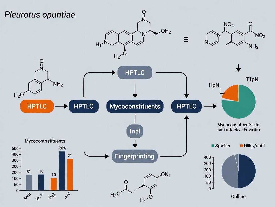
This article provides a detailed methodology and critical analysis of HPTLC (High-Performance Thin-Layer Chromatography) fingerprinting for characterizing the mycochemical profile of the medicinal mushroom Pleurotus opuntiae.
HPTLC Fingerprinting Analysis of Pleurotus opuntiae Mycoconstituents: A Comprehensive Guide for Natural Product Research
Abstract
This article provides a detailed methodology and critical analysis of HPTLC (High-Performance Thin-Layer Chromatography) fingerprinting for characterizing the mycochemical profile of the medicinal mushroom Pleurotus opuntiae. Aimed at researchers and pharmaceutical development professionals, it covers foundational concepts, step-by-step protocols, troubleshooting strategies, and validation techniques. The content synthesizes current research to establish a standardized approach for identifying bioactive compounds, assessing batch-to-batch consistency, and supporting the chemotaxonomic and pharmacognostic evaluation of this underexplored fungal species for potential drug discovery.
Unlocking Pleurotus opuntiae: An Introduction to Its Mycochemical Landscape and HPTLC Fundamentals
Pleurotus opuntiae is a ligninolytic basidiomycete fungus belonging to the genus Pleurotus (oyster mushrooms). It is distinguished by its adaptation to grow on cactus plants, particularly Opuntia species, in arid and semi-arid regions. This review frames its ecology and biopharmaceutical potential within ongoing research utilizing High-Performance Thin-Layer Chromatography (HPTLC) fingerprinting to characterize its mycochemical constituents for standardized therapeutic development.
Ecological Significance
P. opuntiae plays a critical role in nutrient cycling in dry ecosystems by decomposing lignocellulosic biomass of cacti. Its saprotrophic activity facilitates carbon turnover and soil formation. As a pioneer species on a unique substrate, it exhibits remarkable xerotolerance, producing robust enzymes and metabolites to withstand osmotic and oxidative stress. This ecological niche correlates with a unique secondary metabolite profile of biopharmaceutical interest.
Table 1: Key Ecological Adaptations of P. opuntiae
| Adaptation | Functional Role | Biopharmaceutical Implication |
|---|---|---|
| Growth on Opuntia spp. | Specialized lignocellulase secretion | Source of novel thermostable enzymes |
| Xerotolerance | Osmo-protectant metabolite synthesis (e.g., trehalose, mannitol) | Leads with anti-desiccant properties |
| High-temperature growth | Stable membrane composition & antioxidant systems | Heat-stable proteins & antioxidants |
Traditional Uses & Ethnomycology
Traditional uses are less documented for P. opuntiae compared to other Pleurotus species. However, its consumption has been noted in communities where it naturally occurs. The broader Pleurotus genus is traditionally used for food and rudimentary health tonics, supporting the investigation of P. opuntiae for similar nutritional and medicinal properties.
Emerging Biopharmaceutical Uses: A HPTLC Research Context
Modern phytochemical (mycochemical) research, particularly HPTLC fingerprinting, is elucidating compounds responsible for bioactivities. HPTLC provides a rapid, high-throughput platform for fingerprinting complex extracts, standardizing batches, and isolating bioactive zones for downstream analysis (e.g., LC-MS, bioautography).
Table 2: Quantified Bioactive Constituents & Activities of P. opuntiae
| Bioactive Class | Example Compounds (Identified via HPTLC-coupled techniques) | Reported Activity (In vitro/In vivo) | Approximate Yield Range* |
|---|---|---|---|
| Polysaccharides | β-glucans, heteroglycans | Immunomodulation, Antitumor | 2.5-4.1% dry weight |
| Phenolics | Gallic acid, Catechol, Flavonoid derivatives | Antioxidant (IC50 DPPH: 12-45 µg/mL) | 0.8-1.6% dry weight |
| Sterols | Ergosterol, Ergosta derivatives | Anti-inflammatory, Cytotoxic | 0.3-0.7% dry weight |
| Glycoproteins | Lectins, Peroxidases | Antiproliferative, Enzymatic | 0.5-1.2% dry weight |
*Yields are solvent- and strain-dependent.
Key Therapeutic Areas
- Antioxidant & Anti-aging: Phenolic extracts show significant free radical scavenging in DPPH and FRAP assays.
- Immunomodulation & Oncology: Polysaccharide-protein complexes stimulate macrophage NO production and induce apoptosis in cancer cell lines (e.g., HepG2, MCF-7).
- Antimicrobial: Extracts exhibit inhibition against Gram-positive bacteria (e.g., S. aureus) and fungal pathogens like Candida albicans.
- Neuroprotection: Preliminary data suggests acetylcholinesterase inhibition and mitigation of oxidative stress in neuronal cell models.
Experimental Protocols for HPTLC-Led Research
Protocol: HPTLC Fingerprinting ofP. opuntiaeMethanolic Extract
Objective: To develop a standardized fingerprint profile for quality control and bioactivity correlation.
- Sample Prep: Lyophilized fungal powder (5 g) extracted with 100 mL 70% methanol in ultrasonic bath (40°C, 45 min). Filter and concentrate under vacuum. Reconstitute to 10 mg/mL in methanol for application.
- Chromatography:
- Stationary Phase: HPTLC silica gel 60 F254 plates (20 x 10 cm).
- Application: Apply 5 µL, 7 µL, and 10 µL of sample as 8 mm bands using automated applicator (Camag Linomat 5).
- Development: In twin-trough chamber pre-saturated (20 min) with mobile phase Toluene: Ethyl acetate: Formic acid (7:3:0.5 v/v). Development distance: 80 mm.
- Drying: Air dry for 5 min.
- Derivatization & Detection:
- Under UV: Document at 254 nm & 366 nm.
- Post-derivatization: Dip in Natural Product (NP) reagent, heat at 105°C for 3 min, image under 366 nm.
- Bioautography: For antioxidant activity, dip plate in 0.04% DPPH in methanol, observe yellow bands on purple background.
- Documentation & Analysis: Use scanner (Camag TLC Scanner 4) at 500 dpi. Process with visionCATS software for Rf values and densitometric profiles.
Protocol: Bioactivity-Guided Fractionation Using HPTLC
Objective: To isolate antioxidant compounds from an active HPTLC zone.
- Preparative TLC: Load 500 µL of extract (50 mg/mL) as a single band on preparative silica plates (20x20 cm, 2000 µm thickness). Develop as in 5.1.
- Zone Localization: Mask plate edges, view under 366 nm. Scrape the fluorescent band at Rf 0.45-0.55.
- Elution: Elute silica powder with 15 mL ethyl acetate in ultrasonic bath (10 min). Filter (0.45 µm PTFE) and evaporate.
- Validation: Re-analyze eluted fraction via analytical HPTLC (as per 5.1) and confirm antioxidant activity via DPPH bioautography.
Visualizing the Research Workflow & Bioactivity
HPTLC-Bioactivity Guided Fractionation Workflow
Proposed Immuno-Modulatory & Antioxidant Pathways
The Scientist's Toolkit: Key Research Reagent Solutions
Table 3: Essential Materials for HPTLC Fingerprinting of P. opuntiae
| Item/Category | Specific Example & Specification | Function in Research |
|---|---|---|
| HPTLC Plates | Silica gel 60 F254, 20 x 10 cm (Merck) | High-resolution stationary phase for separation; F254 allows UV visualization. |
| Mobile Phase | Toluene:Ethyl acetate:Formic acid (varying ratios) | Separates polar & non-polar mycoconstituents based on differential migration. |
| Derivatization Reagents | Natural Product (NP) reagent (1% diphenylboric acid ethanolamine complex in methanol) | Enhances visibility of specific compound classes (phenolics, terpenes) under 366 nm. |
| Bioautography Reagents | 0.04% DPPH in methanol | Directly locates antioxidant compounds on HPTLC plate as yellow bands. |
| HPTLC Instrumentation | Automated applicator (Linomat), Chromatography chamber, TLC Scanner with visionCATS software | Ensures precise, reproducible application, development, and densitometric quantification. |
| Reference Standards | Ergosterol, Gallic acid, β-Glucan | Used as co-chromatographed standards for compound identification via Rf matching. |
| Extraction Solvents | HPLC-grade Methanol, Ethyl acetate, Water | For exhaustive, reproducible extraction of metabolites of varying polarities. |
This technical guide details the key bioactive mycoconstituents—polysaccharides, phenolics, terpenoids, and sterols—within the context of a broader thesis research employing HPTLC fingerprinting for the comprehensive profiling of Pleurotus opuntiae. The identification and quantification of these compounds are critical for elucidating the medicinal and nutraceutical potential of this fungal species, providing a foundation for targeted drug development.
Core Bioactive Mycoconstituents: Structures and Significance
Polysaccharides (Primarily β-Glucans)
Fungal polysaccharides, especially β-(1→3)- and β-(1→6)-glucans, are major immunomodulators. They activate immune cells via specific pattern recognition receptors (PRRs), such as dectin-1 and TLRs, leading to NF-κB pathway activation and cytokine production.
Phenolic Compounds
This class includes phenolic acids (e.g., gallic, caffeic acids) and flavonoids. They are potent antioxidants, acting as free radical scavengers and metal chelators. Their bioactivity is linked to the modulation of the Nrf2/ARE antioxidant response pathway.
Terpenoids
Terpenoids, including mono-, sesqui-, di-, and triterpenoids, exhibit diverse pharmacological activities (anti-inflammatory, anticancer). Key intermediates are produced via the mevalonate (MVA) and methylerythritol phosphate (MEP) pathways.
Sterols
Ergosterol is the primary mycosterol, serving as a structural component of fungal cell membranes and a precursor to vitamin D₂ upon UV exposure. Other sterols like ergosterol peroxide show notable anti-inflammatory and cytotoxic activities.
Quantitative Profile of Key Constituents inPleurotus opuntiae
Table 1: Representative Quantitative Data for Bioactive Constituents in P. opuntiae (Dry Weight Basis). Data compiled from recent literature.
| Mycoconstituent Class | Specific Compound | Concentration Range | Extraction Method | Analytical Technique |
|---|---|---|---|---|
| Polysaccharides | Total β-Glucans | 250 - 400 mg/g | Hot Water Extraction | Phenol-Sulfuric Acid Assay |
| Phenolics | Total Phenolic Content | 15 - 25 mg GAE/g | 80% Methanol, Soxhlet | Folin-Ciocalteu Assay |
| Gallic Acid | 1.2 - 3.5 mg/g | Ultrasonication (50% EtOH) | HPTLC vs. Standard | |
| Terpenoids | Total Triterpenoids | 8 - 15 mg/g | Ethyl Acetate Maceration | Colorimetric Assay |
| Sterols | Ergosterol | 5 - 12 mg/g | Chloroform-Methanol (2:1) | HPTLC-Densitometry |
Detailed Experimental Protocols for HPTLC-Based Research
Protocol: Sample Preparation for HPTLC Fingerprinting
Materials: Lyophilized P. opuntiae powder, analytical grade solvents (methanol, ethanol, ethyl acetate, water), ultrasonic bath, rotary evaporator, 0.22 μm PTFE syringe filters. Procedure:
- Weigh 1.0 g of dried fungal powder accurately.
- Add 20 mL of optimized extraction solvent (e.g., methanol:water, 70:30 v/v for phenolics; hot water for polysaccharides).
- Sonicate at 40°C for 30 minutes.
- Centrifuge at 10,000 rpm for 15 minutes. Collect supernatant.
- Filter the supernatant through a 0.22 μm membrane.
- Concentrate filtrate to dryness under reduced vacuum at 40°C.
- Reconstitute residue in 1 mL of HPLC-grade methanol for HPTLC application.
Protocol: HPTLC Fingerprinting and Densitometric Analysis
Materials: HPTLC silica gel 60 F₂₅₄ plates (10 x 20 cm), CAMAG Linomat 5 autosampler, ADC2 (Automatic Development Chamber), TLC Visualizer, winCATS software. Chromatographic Conditions:
- Application: 6 mm bands, 10 μL/s application speed.
- Mobile Phase: Optimized for compound class (e.g., for phenolics: ethyl acetate:glacial acetic acid:formic acid:water, 100:11:11:26 v/v).
- Development: In a twin-trough chamber pre-saturated with mobile phase vapor for 20 min. Development distance: 80 mm.
- Derivatization: Dip in Natural Product reagent (1% methanolic diphenylboryloxyethylamine), followed by 5% ethanolic PEG-400.
- Detection & Analysis: Visualize at 366 nm. Perform densitometric scanning at selected wavelengths (e.g., 254 nm for sterols, 366 nm for phenolics). Calculate Rf values and peak areas relative to co-chromatographed standards.
Signaling Pathways and Experimental Workflow
The Scientist's Toolkit: Research Reagent Solutions
Table 2: Essential Materials and Reagents for Mycoconstituent Analysis via HPTLC
| Item | Function/Application | Example Product/Specification |
|---|---|---|
| HPTLC Silica Gel 60 F₂₅₄ Plates | Stationary phase for high-resolution separation of non-volatile compounds. | Merck, 10x20 cm, aluminum-backed. |
| CAMAG Linomat 5 | Automated, precise application of samples and standards as narrow bands. | Programmable syringe, 100 μL. |
| ADC2 (Automated Development Chamber) | Ensures reproducible, vapor-saturated development conditions. | CAMAG, twin-trough glass chamber. |
| Derivatization Reagents | Visualize specific compound classes post-chromatography. | Natural Product reagent (NP), anisaldehyde-sulfuric acid, AlCl₃ for flavonoids. |
| HPTLC-MS Interface | Enables direct elution of HPTLC zones to Mass Spectrometer for compound identification. | CAMAG TLC-MS Interface 2. |
| Reference Standards | Essential for co-chromatography, Rf comparison, and calibration curves. | Ergosterol (≥95%), Gallic Acid (≥97.5%), β-Glucan from yeast. |
| winCATS Planar Chromatography Manager | Software for instrument control, densitometric evaluation, and data documentation. | CAMAG, version 1.4.4 or higher. |
Introduction This technical guide is framed within a doctoral research thesis investigating the metabolomic profiling of Pleurotus opuntiae mycoconstituents. The accurate characterization of complex fungal extracts is a cornerstone of natural product drug discovery. While Conventional Thin-Layer Chromatography (TLC) has been a staple technique, High-Performance Thin-Layer Chromatography (HPTLC) fingerprinting offers superior capabilities essential for rigorous scientific research.
Core Principles and Comparative Advantages HPTLC is an advanced planar chromatography technique with standardized, automated processes. Its advantages over conventional TLC stem from fundamental improvements in material science and instrumentation, as summarized in Table 1.
Table 1: Quantitative Comparison of Conventional TLC vs. HPTLC Parameters
| Parameter | Conventional TLC | HPTLC (Typical Specification) | Implication for Fungal Extract Analysis |
|---|---|---|---|
| Layer Particle Size | 10-12 μm | 4-6 μm | Sharper bands, higher resolution of closely eluting metabolites. |
| Layer Thickness | 100-250 μm | 100-200 μm | More uniform migration, improved reproducibility. |
| Sample Application Volume | 1-5 μL (manual) | 0.1-5 μL (automated) | Precise, narrow bands; reduced diffusion, enabling high-throughput. |
| Development Chamber | Twin-trough, unsaturated | Automated Developing Chamber (ADC) with conditioning | Highly controlled, reproducible solvent vapor saturation for consistent Rf values. |
| Development Distance | 5-15 cm | 3-6 cm | Faster run times (10-20 min) with equal or better separation. |
| Detection Limit | High ng-range (~10-50 ng) | Low ng-range (~1-5 ng) | Detection of minor but potentially bioactive constituents. |
| Data Documentation | Manual photography under UV | Digital scanning densitometry at multiple λ | Objective, quantitative profiling and archiving of fingerprint data. |
| Repeatability (RSD of Rf) | > 3% | ≤ 1.5% | Essential for reliable comparative fingerprinting across multiple P. opuntiae extracts. |
Detailed Experimental Protocol for HPTLC Fingerprinting of P. opuntiae Extracts The following methodology is adapted from the thesis research workflow.
- Sample Preparation: Dry P. opuntiae fruiting body powder (1.0 g) is extracted with 10 mL of methanol:water (80:20, v/v) in an ultrasonic bath for 30 minutes at 40°C. The extract is filtered (0.45 μm PTFE syringe filter) and stored at 4°C prior to analysis.
- HPTLC Plate Pre-Washing & Pre-Conditioning: Silica gel 60 F₂₅₄ plates (10 x 20 cm) are pre-washed with methanol and dried in an oven at 110°C for 15 minutes to remove impurities. Plates are stored in a desiccator.
- Automated Application: Using an automatic applicator (e.g., Linomat 5), apply samples and standards as 6 mm bands, 8 mm from the bottom edge. Typical application: 2-8 μL of extract, 1 μL of standard solutions (e.g., ergosterol, phenolic acids). Track distance: 10 mm.
- Automated Development: The applied plate is transferred to an ADC pre-saturated for 20 minutes with the mobile phase vapor. Development proceeds with a toluene:ethyl acetate:formic acid (6:4:0.2, v/v/v) system over a distance of 70 mm from the application point. The chamber atmosphere is controlled at 33% relative humidity via a saturated MgCl₂ solution.
- Derivatization & Documentation: Post-development, the plate is dried in a stream of hot air. Documentation is performed at 254 nm and 366 nm using a TLC visualizer. For specific detection, the plate is derivatized by dipping in anisaldehyde-sulfuric acid reagent (0.5 mL anisaldehyde, 10 mL sulfuric acid, 85 mL methanol, 5 mL acetic acid) followed by heating at 105°C for 3-5 minutes until bands appear.
- Densitometric Scanning & Profiling: The derivatized plate is scanned in absorbance/reflectance mode at 530 nm or 600 nm using a slit dimension of 4.00 x 0.20 mm. Peak areas and Rf values are recorded using dedicated software for chemometric analysis.
The Scientist's Toolkit: Key Research Reagent Solutions
| Item | Function in HPTLC Fingerprinting |
|---|---|
| HPTLC Silica gel 60 F₂₅₄ plates | High-performance layers with small, uniform particle size for superior separation; F₂₅₄ indicates fluorescence indicator for UV detection at 254 nm. |
| Automated sample applicator | Provides precise, reproducible band-wise application critical for quantitative comparison and valid Rf calculation. |
| Automated Developing Chamber (ADC) | Ensures controlled, reproducible chamber saturation and development conditions, eliminating environmental variability. |
| HPTLC Derivatization reagent (e.g., Anisaldehyde-Sulfuric acid) | Universal reagent for visualization of terpenes, steroids, and sugars present in fungal extracts through chromogenic reactions. |
| HPTLC Densitometry Scanner | Enables conversion of chromatographic bands into quantifiable digital peak profiles (fingerprints) for objective analysis. |
| Reference Standard Solutions | Pure compounds (e.g., ergosterol, mannitol, caffeic acid) co-chromatographed to aid in peak identity assignment in the complex extract fingerprint. |
Visualization of Workflow and Data Analysis
Title: HPTLC Fingerprinting Workflow for Fungal Extracts
Title: Data Analysis Pathway for HPTLC Fingerprints
Conclusion In the context of Pleurotus opuntiae mycoconstituent research, HPTLC fingerprinting is not merely an improved TLC method but a distinct, orthogonal analytical platform. Its superior resolution, reproducibility, and quantitative data output provide a robust, reliable, and cost-effective tool for the metabolomic screening, quality control, and chemotaxonomic studies essential for advancing fungal-based drug discovery.
This whitepaper, framed within a broader thesis on HPTLC fingerprinting of Pleurotus opuntiae mycoconstituents, details the critical rationale for implementing robust chemoprofiling protocols. As a medicinal and edible mushroom with rising commercial and therapeutic interest, P. opuntiae presents significant variability in its metabolite profile due to substrate, geographic, and cultivation conditions. Standardized fingerprinting is therefore essential for ensuring batch-to-batch consistency in quality control (QC), validating authenticity for standardization, and correlating chemical profiles with biological activity for drug development. This document provides a technical guide to the methodologies, data, and protocols underpinning this necessity.
Pleurotus opuntiae (Durian oyster mushroom) synthesizes a diverse array of bioactive metabolites, including polysaccharides (β-glucans), phenolic compounds, lovastatin, and ergothioneine. Research indicates that these constituent levels are not static.
Table 1: Reported Variability of Key Bioactives in P. opuntiae
| Bioactive Compound | Reported Concentration Range | Primary Influencing Factor | Key Reference (Example) |
|---|---|---|---|
| Total Phenolic Content | 8.5 - 21.4 mg GAE/g extract | Substrate type (e.g., rubber sawdust vs. palm fiber) | (Raman et al., 2022) |
| β-Glucans | 25 - 40% of dry weight | Strain selection & developmental stage | (Synytsya et al., 2020) |
| Lovastatin | 0.05 - 0.85 mg/g dry mass | Fermentation conditions & nutrient stress | (Alvarez et al., 2021) |
| Ergothioneine | 0.8 - 2.1 mg/g dry weight | Cultivation method (solid vs. liquid) | (Nguyen et al., 2023) |
This inherent variability necessitates a fingerprinting approach to create a "chemical identity card" for any given batch, enabling reliable QC and standardization.
Core Methodologies for Fingerprinting
High-Performance Thin-Layer Chromatography (HPTLC) Protocol
HPTLC is the cornerstone technique for cost-effective, high-throughput fingerprinting.
Detailed Protocol:
- Sample Preparation: 1.0 g of lyophilized P. opuntiae powder is extracted with 10 mL of methanol-water (70:30, v/v) using ultrasonic-assisted extraction at 45°C for 30 minutes. The extract is filtered (0.45 μm) and concentrated under reduced pressure.
- Application: Using an automatic applicator (e.g., Linomat 5), apply 5 μL, 10 μL, and 15 μL of sample extract (10 mg/mL) and reference standards (e.g., ergothioneine, gallic acid) as 6-mm bands on a silica gel 60 F₂₅₄ HPTLC plate.
- Chromatography: Develop in a twin-trough chamber pre-saturated for 20 min with the mobile phase. A typical phase is Ethyl acetate: Glacial acetic acid: Formic acid: Water (100:11:11:26, v/v). Develop to a migration distance of 80 mm.
- Derivatization:
- For general phenolics: Dip plate in Natural Product/PEG reagent (1% methanolic diphenylboric acid ethyl esteramine followed by 5% ethanolic polyethylene glycol 4000).
- For specific detection: Spray with anisaldehyde-sulfuric acid reagent for terpenoids/sugars.
- Documentation: Capture images under UV 254 nm, UV 366 nm, and white light after derivatization using a TLC visualizer.
- Data Analysis: Use software (e.g., visionCATS) to calculate Rf values and generate the densitometric profile (fingerprint).
Diagram Title: HPTLC Fingerprinting Workflow for P. opuntiae
Complementary Quantitative Assays
HPTLC fingerprints are validated with quantitative assays.
Table 2: Complementary Quantitative Methods
| Assay Target | Standard Protocol (Brief) | Function in Standardization |
|---|---|---|
| Total Phenolic Content | Folin-Ciocalteu method; Gallic acid standard curve. | Quantifies overall phenolic load linked to antioxidant activity. |
| β-Glucan Assay | Megazyme enzymatic kit (K-YBGL). | Quantifies immunomodulatory polysaccharides. |
| HPLC for Lovastatin | C18 column, UV detection at 238 nm, Acetonitrile:Water:Phosphoric acid mobile phase. | Precise quantification of the cholesterol-lowering agent. |
Signaling Pathways Linking Metabolites to Bioactivity
Fingerprinting enables correlation of chemical profiles with observed biological effects via key pathways.
Diagram Title: Key Bioactivity Pathways of P. opuntiae Metabolites
The Scientist's Toolkit: Research Reagent Solutions
Table 3: Essential Materials for P. opuntiae Fingerprinting
| Item / Reagent | Function & Rationale |
|---|---|
| Silica Gel 60 F₂₅₄ HPTLC Plates | High-resolution matrix for separation. F₂₅₄ allows UV visualization of quenching compounds. |
| Ergothioneine & Lovastatin Standards | Reference compounds for peak identification and Rf calibration in chromatograms. |
| Folin-Ciocalteu Reagent | Essential for spectrophotometric quantification of total phenolic content (TPC). |
| β-Glucan Assay Kit (Enzymatic) | Specifically hydrolyzes and quantifies (1,3)(1,6)-β-D-glucans, critical for QC of immunomodulatory potency. |
| Anisaldehyde-Sulfuric Acid Spray | Derivatization reagent for visualization of terpenoids, sterols, and sugars via color development. |
| HPTLC Densitometry Software | Converts chromatographic bands into a digital, quantifiable profile for comparative chemoprofiling. |
Implementing a systematic HPTLC-based fingerprinting protocol for Pleurotus opuntiae is non-negotiable for advancing its role in functional foods and drug development. It provides a defensible, practical tool for ensuring quality, detecting adulteration, and establishing reproducible dose-response relationships in preclinical research. This guide outlines the foundational technical approach to meet the pressing needs for QC, standardization, and meaningful chemoprofiling.
Step-by-Step HPTLC Protocol for Pleurotus opuntiae Mycoconstituent Profiling
Within the context of a broader thesis on HPTLC fingerprinting of Pleurotus opuntiae mycoconstituents, the selection of an optimal solvent system for extraction is a critical initial step. This process dictates the chemical profile obtained, influencing subsequent chromatographic separation and bioactivity assessments. Effective extraction must account for the diverse polarity of mycochemicals, including polar polysaccharides and phenolic acids, mid-polar sterols, and non-polar triglycerides. This guide details solvent selection strategies and protocols tailored for comprehensive metabolite profiling in fungal research.
Principles of Solvent Selection
The efficiency of compound extraction is governed by the principle of "like dissolves like." Solvent polarity, measured by indices such as dielectric constant (ε) or Snyder's polarity index (P'), must align with target analyte polarity. For complex matrices like P. opuntiae, sequential or blended solvent systems are often required to achieve broad-spectrum extraction.
Key Solvent Properties
- Polarity: Primary determinant of solubility.
- Boiling Point: Affects evaporation and concentration steps.
- Viscosity: Impacts filtration and handling.
- Toxicity & Flammability: Critical for laboratory safety.
- UV Cutoff: Important for spectroscopic analysis.
Quantitative Solvent Data for Extraction
Table 1: Properties of Common Extraction Solvents
| Solvent | Polarity Index (P') | Dielectric Constant (ε) | Boiling Point (°C) | Suitable for Compound Class |
|---|---|---|---|---|
| n-Hexane | 0.1 | 1.9 | 69 | Non-polar (lipids, terpenes, alkanes) |
| Toluene | 2.4 | 2.4 | 111 | Mid- to non-polar |
| Dichloromethane (DCM) | 3.1 | 9.1 | 40 | Mid-polar (alkaloids, medium-polar phenolics) |
| Ethyl Acetate | 4.4 | 6.0 | 77 | Mid-polar (flavonoids, aglycones) |
| Acetone | 5.1 | 21 | 56 | Polar (medium-polarity glycosides) |
| Methanol | 5.1 | 33 | 65 | Polar (polar glycosides, saponins, phenolics) |
| Ethanol | 5.2 | 24 | 78 | Polar (polar glycosides, saponins) |
| Water | 10.2 | 80 | 100 | Highly polar (polysaccharides, proteins, tannins) |
Table 2: Efficacy of Solvent Systems for Pleurotus spp. Constituents (Representative Data)
| Solvent System (v/v) | Target Compound Class | Reported Yield Range* | Key Reference Application |
|---|---|---|---|
| 100% Methanol | Total Phenolics, Flavonoids | 8-12 mg GAE/g dw | General phenolic screening |
| 70% Aqueous Ethanol | Polar antioxidants, Glycosides | 10-15 mg GAE/g dw | Antioxidant extract preparation |
| 100% Ethyl Acetate | Mid-polar aglycones, Sterols | 2-5% w/w | Targeted sterol isolation |
| n-Hexane : Ethyl Acetate (9:1) | Non-polar lipids, Volatiles | 1-3% w/w | Lipid profiling |
| Sequential: Hexane→DCM→Methanol | Comprehensive Metabolite Spectrum | Varies by fraction | Fractionation for bioactivity guided isolation |
*Yield is matrix and method-dependent; GAE = Gallic Acid Equivalents, dw = dry weight.
Experimental Protocols forP. opuntiaeExtraction
Protocol A: Sequential Solvent Extraction for Comprehensive HPTLC Fingerprinting
Objective: To fractionate P. opuntiae mycoconstituents based on polarity for detailed HPTLC analysis. Materials: Lyophilized P. opuntiae powder, ultrasonic bath, solvents (n-Hexane, Dichloromethane, Methanol), rotary evaporator, filtration setup.
- Defatting: Accurately weigh 5.0 g of dry fungal powder. Add 100 mL of n-hexane. Sonicate at 40°C for 30 minutes. Filter through Whatman No. 1 paper. Retain the marc (residue) and evaporate the filtrate under reduced pressure to obtain the non-polar hexane fraction (F1).
- Mid-Polar Extraction: Subject the marc from Step 1 to extraction with 100 mL of dichloromethane using the same sonication parameters. Filter and evaporate to obtain the mid-polar DCM fraction (F2).
- Polar Extraction: Finally, extract the residual marc with 100 mL of methanol. Filter and evaporate to obtain the polar methanol fraction (F3).
- Preparation for HPTLC: Redissolve each dried fraction in 5 mL of their respective parent solvents. Filter through a 0.45 µm PTFE syringe filter prior to HPTLC application.
Protocol B: Optimized Single-Solvent System for Polar Antioxidants
Objective: To prepare an extract rich in polar, antioxidant mycoconstituents for activity-linked profiling. Materials: Lyophilized P. opuntiae powder, 70% Aqueous Ethanol, orbital shaker, freeze dryer.
- Weigh 10.0 g of dry powder into a conical flask.
- Add 200 mL of 70% ethanol (v/v in deionized water). This hydro-alcoholic system balances polarity for efficient extraction of both polar and some mid-polar compounds.
- Agitate on an orbital shaker (150 rpm) at room temperature for 24 hours.
- Filter the supernatant through Buchner funnel. Repeat extraction on the marc once.
- Combine filtrates and concentrate under vacuum at 45°C.
- Lyophilize the concentrated extract to obtain a dry powder. Store at -20°C.
- For HPTLC, prepare a 10 mg/mL solution in methanol.
The Scientist's Toolkit: Key Research Reagent Solutions
Table 3: Essential Materials for Extraction and HPTLC Fingerprinting
| Item | Function/Explanation |
|---|---|
| Ultrasonic Bath/Sonicator | Applies cavitation energy to disrupt cell walls and enhance solvent penetration, improving extraction efficiency. |
| Rotary Evaporator | Gently removes bulk solvent under reduced pressure and controlled temperature to prevent thermal degradation of labile compounds. |
| 0.45 µm PTFE Syringe Filter | Essential for particulate-free sample preparation prior to HPTLC spotting, preventing capillary tube clogging and ensuring even band application. |
| HPTLC Silica Gel 60 F₂₅₄ Plates | The stationary phase for separation. The F₂₅₄ indicator allows for visualization under 254 nm UV light. |
| Microsyringe (100 µL) / Automatic TLC Sampler | For precise, reproducible application of sample extracts as bands on the HPTLC plate. |
| Twin-Trough Developing Chamber | Provides a saturated, uniform vapor environment for consistent chromatographic development. |
| Derivatization Reagents (e.g., Anisaldehyde-Sulfuric acid) | Chemical sprays that react with specific functional groups (terpenes, sugars) to produce colored bands for visual and densitometric detection. |
| TLC Densitometer Scanner | Instrument for in-situ quantification of separated bands by absorbance or fluorescence, generating the fingerprint chromatogram. |
Visualization of Methodologies
Sequential Extraction & HPTLC Workflow
Solvent Polarity & Target Compound Relationship
The choice of solvent system is foundational to successful HPTLC fingerprinting of Pleurotus opuntiae. A sequential extraction protocol using solvents of increasing polarity (e.g., hexane → DCM → methanol) provides the most comprehensive fractionation for detailed metabolomic mapping. Alternatively, a single optimized hydro-alcoholic system (e.g., 70% ethanol) offers a robust balance for extracting antioxidant-rich polar constituents. The selected protocol must align with the research objectives—whether for comprehensive metabolite discovery or targeted bioactivity analysis—ensuring the chemical fingerprint truly represents the fungal extract's complexity and informs downstream drug development processes.
This whitepaper provides an in-depth technical guide on stationary phase selection for High-Performance Thin-Layer Chromatography (HPTLC) within the context of a doctoral thesis focused on the fingerprinting of Pleurotus opuntiae mycoconstituents. The accurate profiling of polar and non-polar metabolites from this medicinal mushroom necessitates a rigorous evaluation of HPTLC plates, including their inherent properties and pre-treatment protocols, to achieve optimal resolution, reproducibility, and compound stability.
HPTLC Plate Types: Core Characteristics and Selection Criteria
The stationary phase is the cornerstone of any chromatographic separation. For HPTLC, the choice dictates the interaction mechanisms with target analytes.
Silica Gel 60 F254
The most ubiquitous normal-phase (NP) adsorbent. The silica surface is polar and acidic due to silanol (Si-OH) groups, separating compounds based on polarity via adsorption. The integrated fluorescent indicator (F254) enables UV visualization at 254 nm.
- Mechanism: Adsorption chromatography. Polar functional groups (e.g., -OH, -COOH) interact strongly with silanols.
- Best For: P. opuntiae compounds: Phenolic acids, flavonoids, terpenoids, and medium to high-polarity glycosides.
- Typical Mobile Phases: Non-polar or medium-polarity organic solvents (e.g., toluene, ethyl acetate) modified with polar additives (e.g., formic acid, methanol) to control selectivity.
Reversed-Phase (RP) Plates (e.g., RP-18, RP-8)
Silica gel chemically bonded with long-chain alkylsilanes (octadecyl, C18). The surface is non-polar, and separation occurs via partition between the mobile phase and the hydrophobic layer.
- Mechanism: Partition chromatography (reversed-phase). Separation is based on hydrophobicity/lipophilicity.
- Best For: Low to medium-polarity P. opuntiae mycoconstituents: Non-polar terpenes, sterols, fatty acid esters, and less polar aglycones.
- Typical Mobile Phases: Polar, often water-methanol or water-acetonitrile mixtures. May require ion-pair reagents for acidic/basic compounds.
DIOL Plates
Silica gel bonded with 2,3-dihydroxypropyl groups. This phase offers a mildly polar, hydrogen-bonding surface that is less acidic than bare silica.
- Mechanism: A blend of adsorption and partition. Provides hydrogen-donor and acceptor interactions.
- Best For: Sensitive compounds prone to decomposition or strong irreversible adsorption on acidic silica. Useful for moderately polar metabolites like certain glycosides or peptides from fungal extracts.
- Typical Mobile Phanes: Mixtures similar to NP but often with less aggressive modifiers.
| Parameter | Silica Gel 60 F254 | Reversed-Phase (RP-18) | DIOL |
|---|---|---|---|
| Surface Chemistry | Polar, acidic (Silanols) | Non-polar (C18 chains) | Mildly polar (Neutral diol) |
| Separation Mechanism | Adsorption | Partition | Adsorption/Partition (H-bonding) |
| Optimal Analyte Polarity | Medium to High | Low to Medium | Medium |
| Common Mobile Phase | Organic + Polar modifier | Water/Organic (e.g., MeOH, ACN) | Organic + Mild modifier |
| Key Advantage | High efficiency for polar compounds | Excellent for hydrophobic compounds | Reduced tailing, good for sensitive compounds |
| UV Activity (F254) | Yes | Yes (special variants) | Yes (special variants) |
| Relative Cost | $ | $$$ | $$ |
Pre-treatment Protocols for Enhanced Chromatographic Performance
Pre-treatment aims to standardize plate activity, remove contaminants, or modify surface chemistry.
Protocol 3.1: Standard Activation of Silica Gel Plates
- Purpose: To ensure consistent, high surface activity by removing adsorbed water.
- Method: Heat plates in a clean oven at 110-120°C for 30 minutes.
- Storage: Immediately transfer to a desiccator containing anhydrous calcium chloride or silica gel. Use within 24 hours for reproducible results.
- Application: Critical for NP separations of polar P. opuntiae antioxidants.
Protocol 3.2: Pre-washing for Background Reduction
- Purpose: To remove organic impurities and the fluorescent indicator that may interfere with post-chromatographic derivatization or detection.
- Method: a. Develop the blank plate to the top with a strong, ultra-pure solvent (e.g., methanol:dichloromethane 1:1 v/v). b. Dry the plate thoroughly in a fume hood. c. Reactivate via Protocol 3.1 if needed.
- Application: Essential for quantitative analysis and when using aggressive derivatizing reagents (e.g., sulfuric acid reagent).
Protocol 3.3: Impregnation for Selectivity Tuning
- Purpose: To chemically modify the stationary phase for specific separations.
- Method (e.g., Boric Acid Impregnation for Sugar/Sugar Alcohol Analysis): a. Prepare a 3% (w/v) solution of boric acid in methanol:water (80:20). b. Dip or horizontally develop the plate in this solution. c. Dry at room temperature, then oven-dry at 100°C for 10 minutes.
- Application: Can be used to separate carbohydrate-derived metabolites in fungal extracts by forming cyclic complexes.
Experimental Workflow forP. opuntiaeFingerprint Method Development
Diagram Title: HPTLC Method Development Workflow for P. opuntiae
The Scientist's Toolkit: Key Research Reagent Solutions
Table 2: Essential Materials for HPTLC Fingerprinting of Mycoconstituents
| Item / Reagent | Function in Research |
|---|---|
| HPTLC Plates (Silica, RP-18, DIOL) | Core stationary phases for comparative separation of diverse metabolite classes. |
| CAMAG Linomat 5 | Automated, reproducible sample application as narrow bands; critical for quantification. |
| Twin-Trough Development Chamber | Provides controlled, saturated vapor environment for reproducible development. |
| Derivatization Reagent: ANSA | (p-Anisaldehyde sulfuric acid) Visualizes terpenoids, sugars, and sterols as colored zones. |
| Derivatization Reagent: DPPH• | (2,2-Diphenyl-1-picrylhydrazyl) Spray reagent for direct detection of antioxidant compounds. |
| CAMAG TLC Scanner 4 | Densitometric quantification and spectral analysis of separated bands. |
| Documentation: CAMAG DigiStore 2 | High-resolution digital imaging under UV/Vis and white light. |
| HPLC-Grade Solvents | Ensure purity of mobile phases to prevent ghost peaks or background interference. |
| Microsyringe (100 µL) | Precise loading of sample solutions into the applicator. |
| Desiccator with Drying Agent | For standardized storage of activated plates to maintain consistent activity. |
Selecting the appropriate HPTLC stationary phase—be it the polar, adsorptive silica gel; the hydrophobic, partitioning RP phase; or the neutral, H-bonding DIOL phase—and applying targeted pre-treatments are foundational to developing a robust, informative fingerprint for Pleurotus opuntiae. This systematic approach enables the resolution of complex mycoconstituent mixtures, paving the way for subsequent quantitative analysis, bioautography, and chemometric evaluation in drug development research.
Within the broader thesis on the chemoprofiling and bioactivity assessment of Pleurotus opuntiae, High-Performance Thin-Layer Chromatography (HPTLC) fingerprinting serves as a cornerstone for the dereplication and standardization of its complex mycochemical constituents. The efficacy of HPTLC is fundamentally governed by the selectivity and efficiency of the mobile phase. This whitepaper provides an in-depth technical guide on optimizing multi-component solvent systems to achieve baseline separation of critical compound classes—such as polyphenols, flavonoids, terpenoids, and sterols—present in P. opuntiae extracts, thereby enabling accurate qualitative and quantitative analysis essential for drug discovery pipelines.
Foundational Principles of Multi-component Mobile Phase Optimization
The optimization transcends trial-and-error by systematically manipulating solvent properties: solvent strength (ε⁰), selectivity via Snyder's solvent selectivity groups (I: proton donors, V: dipolar protons, VIII: proton acceptors), and the overall polarity index (P'). The goal is to maximize the differential interaction (ΔR_f) between analytes and the stationary phase (typically silica gel 60 F₂₅₄). For complex fungal matrices, ternary or quaternary systems are often mandatory to resolve compounds with subtle structural differences.
Systematic Optimization Strategy and Protocols
Initial Scouting and Screening Protocol
- Objective: Identify promising solvent combinations from a broad library.
- Method: Pre-condition HPTLC plates (20 × 10 cm) at 65°C for 20 min. Apply 5 µL of P. opuntiae methanolic extract (10 mg/mL) as 8 mm bands. Develop in a twin-trough chamber saturated for 20 min with 10 mL of various binary systems (e.g., Hexane-Ethyl Acetate, Chloroform-Methanol, Toluene-Ethyl Acetate) covering a polarity range (P' from 0.1 to 5.1). Develop to 80 mm, dry, and document under UV 254 nm, UV 366 nm, and after derivatization with Anisaldehyde-Sulfuric acid reagent.
- Data Analysis: Calculate the number of detectable bands (N) and the distribution factor (Df = Rf max - R_f min) for each system.
Iterative Optimization via Statistical Design (DoE) Protocol
- Objective: Refine a promising lead system (e.g., Toluene:Ethyl Acetate:Formic Acid) for maximal resolution.
- Method: Employ a Central Composite Design (CCD) with three factors: % of Toluene (X₁, 40-70%), % of Ethyl Acetate (X₂, 20-50%), and % of Formic Acid (X₃, 1-10%). Perform 20 randomized runs. For each run, develop plates as in 3.1.
- Response Variables: Measure the critical resolution (R_s) between two target marker compounds (e.g., ergosterol and a flavonoid glycoside) and the peak count (PC) after derivatization. Use software (e.g., Design-Expert) to generate a polynomial model and identify the optimal composition.
Validation Protocol for the Optimized System
- Objective: Confirm the robustness and reproducibility of the final system.
- Method: Repeat the chromatography with the optimal mobile phase (n=6) on different days. Analyze a standard mixture of identified P. opuntiae markers (e.g., Gallic acid, Quercetin, Ergosterol, β-Glucan hydrolysate sugars). Calculate relative standard deviation (RSD%) of R_f values and peak areas.
Data Presentation: Optimization Results forP. opuntiae
Table 1: Screening of Binary Solvent Systems for Preliminary Separation
| Solvent System (v/v) | Ratio | Polarity Index (P') | Band Count (UV 366 nm) | Distribution Factor (D_f) | Suitability for Class |
|---|---|---|---|---|---|
| Hexane : Ethyl Acetate | 7:3 | 2.9 | 5 | 0.35 | Lipids, Terpenes |
| Chloroform : Methanol | 9:1 | 4.1 | 8 | 0.52 | General Polar Metabolites |
| Toluene : Ethyl Acetate | 7:3 | 3.6 | 12 | 0.65 | Flavonoids, Phenolics |
| Ethyl Acetate : Methanol : Water | 8:1:1 | 5.8 | 15 | 0.78 | Very Polar Compounds |
Table 2: Central Composite Design (CCD) Optimization Results for a Ternary System
| Run | Toluene (%) | Ethyl Acetate (%) | Formic Acid (%) | Critical Resolution (R_s) | Peak Count (PC) |
|---|---|---|---|---|---|
| 1 | 55 | 35 | 5.5 | 2.1 | 18 |
| 2 | 70 | 25 | 5 | 1.4 | 12 |
| 3 | 45 | 45 | 8 | 1.8 | 20 |
| Optimal | 58.7 | 36.3 | 5.0 | 2.3 | 19 |
Table 3: Validation of the Optimized System (Toluene:Ethyl Acetate:Formic Acid, 58.7:36.3:5.0)
| Marker Compound | Mean R_f (n=6) | RSD% of R_f | Mean Peak Area (AU) | RSD% of Area |
|---|---|---|---|---|
| Gallic Acid | 0.12 | 1.8 | 5421 | 2.5 |
| Quercetin | 0.47 | 1.2 | 8876 | 2.1 |
| Ergosterol | 0.82 | 1.5 | 12345 | 2.8 |
Visualizing the Optimization Workflow and Chemical Interactions
HPTLC Solvent System Optimization Workflow
Solvent-Analyte Interaction Mechanisms in HPTLC
The Scientist's Toolkit: Essential Research Reagent Solutions
| Item Name & Specification | Function in HPTLC of P. opuntiae |
|---|---|
| HPTLC Silica Gel 60 F₂₅₄ Plates (10x20 cm, 200 µm) | The standard stationary phase for normal-phase separation. F₂₅₄ indicates the layer contains a fluorescence indicator for UV detection at 254 nm. |
| Twin-Trough Development Chamber (Glass, 20x10 cm) | Allows for chamber saturation with mobile phase vapor prior to development, ensuring reproducible chromatographic conditions and sharp bands. |
| Hamilton Microsyringe (25 µL, ±0.5 µL accuracy) | For precise, band-wise application of sample and standard solutions onto the HPTLC plate baseline. |
| Anisaldehyde-Sulfuric Acid Derivatization Reagent | A universal spray reagent for detection of terpenoids, steroids, and sugars (found in P. opuntiae) by producing colored zones upon heating. |
| Digital HPTLC Documentation System (UV 254/366 nm, White Light) | For capturing high-resolution chromatographic images pre- and post-derivatization under different illumination modes for fingerprint analysis. |
| HPTLC Densitometry Scanner with Deuterium & Tungsten Lamps | Enables in-situ spectral scanning and quantification of separated bands by measuring absorbance or fluorescence directly on the plate. |
| Chromatography Grade Solvents (Toluene, Ethyl Acetate, Formic Acid, Methanol) | High-purity solvents with low UV cutoff and minimal impurities are critical for consistent mobile phase preparation and clean baselines. |
| Authenticated Mycochemical Standards (e.g., Ergosterol, Quercetin, Gallic Acid) | Reference compounds for co-chromatography to identify and confirm the presence of specific compound classes in the fungal extract fingerprint. |
High-Performance Thin-Layer Chromatography (HPTLC) fingerprinting is a cornerstone in the metabolomic profiling of fungi like Pleurotus opuntiae. A critical limitation of native HPTLC is that many mycoconstituents—such as phenolics, alkaloids, terpenoids, and lipids—are not visible under UV light or appear as non-specific bands. Post-chromatographic derivatization addresses this by applying chemical reagents post-separation to selectively react with specific functional groups, producing colored or fluorescent derivatives. This guide details the reagents and protocols essential for visualizing the diverse compound classes present in P. opuntiae, thereby enabling precise compound class-specific fingerprinting crucial for chemotaxonomic and drug discovery workflows.
Core Derivatization Reagents: Mechanisms and Target Compound Classes
Post-chromatographic derivatization involves spraying or dipping the developed HPTLC plate in reagent solutions. The reaction mechanisms include redox reactions, condensation, complex formation, and acid-base reactions.
Table 1: Key Post-chromatographic Reagents forPleurotus opuntiaeMycoconstituents
| Reagent Name | Target Compound Class(es) in P. opuntiae | Typical Reaction/Visualization | Observation Mode | Key Notes for HPTLC |
|---|---|---|---|---|
| Anisaldehyde-Sulfuric Acid (Vanillin-Sulfuric Acid) | Terpenoids, Sterols, Sugars, Phenolic compounds | Electrophilic substitution, dehydration; colors vary (pink, blue, violet) | White light (VIS) | Heat at 105°C for 3-10 min. Highly versatile for overall fingerprint. |
| Ferric Chloride (FeCl₃) | Phenolic acids, Tannins, Flavonoids | Complex formation with phenolate ions | VIS (often yellow, green, blue, black) | No heating usually required. Specific for phenolics. |
| Ninhydrin | Amino acids, Peptides, Primary amines | Ruhemann's purple formation via oxidative deamination | VIS (purple/red spots) | Heat at 105°C for 5-10 min. Specific for nitrogenous compounds. |
| Dragendorff’s Reagent | Alkaloids, Nitrogen-containing bases | Complex formation with tertiary amines | VIS (orange-red spots) | May require background clearing with ethyl acetate. |
| Natural Product (NP) / PEG Reagent | Flavonoids, Phenolic compounds | Formation of fluorescent complexes | UV 366 nm (yellow, orange fluorescence) | Sequential dip: 1% Methanolic Diphenylboric acid ethanolamine ester (NP), then 5% PEG-4000. |
| Phosphomolybdic Acid (PMA) | Lipids, Sterols, Terpenes, General reductants | Reduction of PMA to molybdenum blue | VIS (blue spots on yellow-green) | Heat for 2-5 min. Detects reducing compounds. |
| Aluminum Chloride (AlCl₃) | Flavonoids (esp. flavones, flavonols) | Acid-base & complex formation | UV 366 nm (enhanced yellow-green fluorescence) | Dip or spray, dry. Enhances native fluorescence. |
| Iodine Vapor | General lipophilic compounds, Alkaloids, Terpenes | Unspecific charge-transfer complexation | VIS (brown spots on yellow) | Reversible. Used for nondestructive initial screening. |
Detailed Experimental Protocols for HPTLC ofP. opuntiae
General HPTLC Workflow: P. opuntiae extract is applied on silica gel 60 F₂₅₄ plates, developed in an appropriate solvent system (e.g., toluene-ethyl acetate-formic acid, 5:4:1, v/v/v), dried, and then derivatized.
Protocol 3.1: Anisaldehyde-Sulfuric Acid Reagent for Terpenoid/Phenolic Profiling
- Reagent Preparation: Carefully mix 170 mL ice-cold methanol, 20 mL glacial acetic acid, 10 mL concentrated sulfuric acid, and 1 mL p-anisaldehyde. Store at 4°C, stable for ~1 month.
- Application: Uniformly spray the dried, developed HPTLC plate in a fume hood until slightly translucent.
- Development: Heat the plate on a TLC plate heater at 105°C for 5-7 minutes.
- Documentation: Immediately capture the image under white light. Terpenoids and sterols appear as violet, blue, or pink zones; phenolics as brownish zones.
Protocol 3.2: Sequential NP/PEG Reagent for Flavonoid Fingerprinting
- Reagent Preparation:
- NP Solution: Dissolve 1 g diphenylboric acid-2-aminoethyl ester in 100 mL methanol.
- PEG Solution: Dissolve 5 g polyethylene glycol 4000 in 100 mL dichloromethane.
- Application: Dip the developed plate in NP solution for 2 seconds, dry in air, then dip in PEG solution for 2 seconds, dry thoroughly.
- Documentation: Observe under UV 366 nm. Flavonoid aglycones and glycosides exhibit intense yellow, green, or orange fluorescence.
Protocol 3.3: Modified Dragendorff’s Reagent for Alkaloid Screening
- Reagent Preparation (Munier Modification):
- Solution A: Dissolve 0.85 g basic bismuth carbonate in 10 mL glacial acetic acid and 40 mL water.
- Solution B: 8 g potassium iodide in 20 mL water.
- Stock: Mix A and B (1:1). Store in amber bottle.
- Spray Solution: Mix 5 mL stock with 10 mL glacial acetic acid, then add 70 mL water and 100 mL ethanol.
- Application: Spray plate evenly.
- Development: Allow plate to dry at RT. Orange-red spots indicate alkaloids.
- Background Clearing (Optional): Place plate in a chamber saturated with ethyl acetate vapor to clear background yellowing.
Visualization of Workflows and Pathways
Title: Post-Chromatographic Derivatization Workflow for P. opuntiae HPTLC
Title: NP/PEG Flavonoid Visualization Mechanism
The Scientist's Toolkit: Essential Research Reagent Solutions
Table 2: Key Reagents and Materials for HPTLC Derivatization of Fungal Extracts
| Item Name | Function/Application in P. opuntiae Research | Key Notes |
|---|---|---|
| Silica gel 60 F₂₅₄ HPTLC Plates | Stationary phase for separation; F₂₅₄ allows UV 254 nm quenching for initial screening. | 10x10 cm or 20x10 cm are standard. Pre-wash if needed for high-precision work. |
| Glass Derivatization Chamber/Sprayer | For uniform, controlled application of derivatization reagents. | Automated sprayers (e.g., CAMAG Derivatizer) ensure reproducibility. Manual glass sprayers require practice. |
| TLC Plate Heater | Provides controlled heating to accelerate derivatization reactions (e.g., for Anisaldehyde, Ninhydrin). | Temperature homogeneity (±2°C) is critical for consistent results. |
| Documentation System | CCD camera system with UV (254/366 nm) and white light cabinets for archiving fingerprints. | Calibrated systems (e.g., CAMAG TLC Visualizer) allow quantitative densitometry post-derivatization. |
| p-Anisaldehyde (Reagent Grade) | Key component of the most versatile charring reagent for terpenoids and phenolics. | Highly pure grade ensures low background coloration. Solution is light-sensitive. |
| Diphenylboric Acid Aminoethyl Ester (NP Reagent) | Forms fluorescent complexes with flavonoids and phenolics in combination with PEG. | Store desiccated at 2-8°C. Methanolic solution is stable for weeks at 4°C. |
| Polyethylene Glycol 4000 (PEG) | Stabilizes the NP-flavonoid complex, enhancing and prolonging fluorescence. | Use high-purity grade. Dichloromethane solution is stable. |
| Dragendorff’s Reagent Modifications Kit | (Bismuth subnitrate, KI, Acetic acid) for detection of alkaloids and N-compounds. | Modified versions reduce background and increase sensitivity on silica gel. |
| Standard Reference Compounds | Pure terpenoids (e.g., ergosterol), phenolics (e.g., gallic acid), flavonoids (e.g., quercetin). | Co-chromatography with standards is mandatory for preliminary compound class assignment. |
This guide details the critical documentation and imaging protocols employed within a broader thesis investigating the High-Performance Thin-Layer Chromatography (HPTLC) fingerprinting of Pleurotus opuntiae mycoconstituents. The accurate capture of chromatographic fingerprints under multiple illumination wavelengths is fundamental for the qualitative and semi-quantitative analysis of complex fungal metabolite profiles. This enables the identification of pharmacologically relevant compounds, supporting subsequent drug development workflows.
Illumination Principles and Compound Interaction
Different wavelengths of light interact with chemical compounds via specific mechanisms, revealing distinct aspects of the HPTLC fingerprint.
- UV 254 nm: Causes fluorescence quenching. Compounds with aromatic rings or conjugated systems absorb this short-wave UV light, appearing as dark spots against the greenish fluorescent background of the F254-doped silica gel.
- UV 366 nm: Induces native fluorescence. Compounds with inherent fluorophores absorb long-wave UV light and re-emit it at longer, visible wavelengths, appearing as brightly colored spots.
- White Light (Transmitted/Reflected): Used after derivatization with specific reagents (e.g., anisaldehyde-sulfuric acid). Reveals colored reaction products of compounds (e.g., terpenoids, sugars), providing functional group information.
Experimental Protocol for HPTLC Imaging
Materials & Equipment:
- Developed and dried HPTLC plate (Silica gel 60 F254).
- Documentation system: CAMAG TLC Visualizer 2 or equivalent, with integrated UV cabinets at 254 nm and 366 nm.
- Derivatization reagents (e.g., anisaldehyde-sulfuric acid, vanillin-sulfuric acid).
- Heating plate (for post-derivatization heating).
- Calibrated, high-resolution digital camera with fixed positioning.
Step-by-Step Methodology:
- Plate Preparation: Ensure the HPTLC plate is completely dry after development (chamber solvent fully evaporated).
- White Light Imaging (Pre-Derivatization):
- Place plate on documentation stage.
- Capture image under white light reflection to record plate appearance and any naturally colored compounds.
- Capture image under white light transmission to note any opaque zones.
- UV 254 nm Imaging:
- Activate the UV 254 nm light source in the cabinet.
- Capture image with the camera system. Ensure the door is closed to prevent UV exposure.
- UV 366 nm Imaging:
- Switch to the UV 366 nm light source.
- Capture image. Use a yellow filter if available to enhance contrast of fluorescent spots.
- Chemical Derivatization & Post-Derivatization Imaging:
- Uniformly spray the plate with the chosen derivatization reagent (e.g., anisaldehyde-sulfuric acid for Pleurotus terpenoids/sugars) in a fume hood.
- Heat the plate on a heating plate at 100-105°C for 3-5 minutes until optimal color development.
- Allow the plate to cool.
- Repeat white light reflection/transmission imaging to document the derivatized fingerprint.
- Optionally, re-image under UV 366 nm as some derivatives may fluoresce.
Data Presentation: Comparative Analysis of Imaging Modalities
Table 1: Characteristic Fingerprint Features of Pleurotus opuntiae Extracts under Different Illumination
| Illumination Mode | Key Compounds Detected (Example Classes) | Visual Appearance | Primary Information Revealed |
|---|---|---|---|
| UV 254 nm | Aromatic acids, phenolics, conjugated dienes | Dark purple/black zones on green background | Presence of UV-absorbing chromophores. General pattern for most organic compounds. |
| UV 366 nm | Native fluorescent compounds (e.g., certain alkaloids, coumarins) | Bright blue, green, yellow, or red fluorescent zones | Presence of specific fluorophores. Often indicates unique chemotaxonomic markers. |
| White Light (Post-Derivatization) | Terpenoids, sterols, sugars, lipids | Violet, blue, green, pink, or brown zones (reagent-dependent) | Functional groups based on chromogenic reaction. Enhances selectivity and sensitivity for specific classes. |
Table 2: Typical HPTLC Imaging Camera Settings (Guideline)
| Parameter | UV 254 nm | UV 366 nm | White Light |
|---|---|---|---|
| Exposure Mode | Manual | Manual | Auto, then manual lock |
| ISO | 400 - 800 | 400 - 800 | 100 - 200 |
| Aperture (f-stop) | f/8 - f/11 | f/8 - f/11 | f/8 - f/11 |
| Shutter Speed | 0.5 - 2 sec | 0.5 - 2 sec | < 0.1 sec |
| Filter | None | Yellow (optional, enhances contrast) | None |
The Scientist's Toolkit: Essential Research Reagents & Materials
Table 3: Key Research Reagent Solutions for HPTLC Fingerprinting of Mycoconstituents
| Item | Function/Explanation |
|---|---|
| HPTLC Plates (Silica gel 60 F254) | The stationary phase. 'F254' indicates the phosphor for UV 254 nm fluorescence quenching. |
| Derivatization Reagent: Anisaldehyde-Sulfuric Acid | Universal reagent. Reacts with terpenes, steroids, sugars, and phenolics to produce vivid colors (violet, blue, green). |
| Derivatization Reagent: Dichlorofluorescein (0.1% in ethanol) | Lipophilic stain. Makes lipids and less polar compounds visible as yellow spots under UV 366 nm. |
| Derivatization Reagent: Natural Product (NP) / PEG Reagent | Sequential spray (NP then PEG). Enhances and stabilizes fluorescence of certain phenolic compounds (e.g., flavonoids) under UV 366 nm. |
| CAMAG TLC Visualizer 2 or Equivalent | Standardized imaging system with reproducible lighting and camera settings, essential for comparative and archival work. |
| Calibrated Micropipettes (1-10 µL) | For precise, reproducible application of samples and standards onto the HPTLC plate. |
| Chromatographic Development Chamber (ADC2 or glass twin-trough) | Provides a controlled, saturated environment for consistent mobile phase development. |
Visualized Workflows
HPTLC Multi-Wavelength Imaging Workflow
Imaging Data Fusion for Mycoconstituent Analysis
Resolving Common Challenges in HPTLC Fingerprinting of Pleurotus opuntiae Extracts
Within our ongoing research into the HPTLC fingerprinting of Pleurotus opuntiae mycoconstituents, achieving sharp, well-resolved bands is paramount for accurate metabolite profiling. Poor resolution and tailing are common chromatographic challenges that can obscure critical peaks and compromise quantitative analysis. This guide details systematic troubleshooting focused on mobile phase optimization and chamber saturation, framed within our mycoconstituents research.
Core Principles: Resolution and Tailing in HPTLC
Resolution (Rs) is a quantitative measure of the separation between two adjacent bands. Tailing, quantified by the tailing factor (Tf), results from non-ideal interactions between analytes and the stationary phase or from chamber unsaturation. For complex fungal extracts like P. opuntiae, which contain polar polysaccharides, phenolic acids, and less polar sterols, these issues are frequent.
Mobile Phase Optimization Strategies
The mobile phase is a critical variable. Our experiments with P. opuntiae methanol extract (100 mg/mL) revealed that a simple ethyl acetate: methanol mixture caused severe tailing of polar compounds.
Table 1: Effect of Mobile Phase Modifiers on Band Characteristics
| Mobile Phase Composition (v/v) | Rf Target Band | Tailing Factor (Tf) | Resolution (Rs) from Nearest Band | Observation for P. opuntiae Extract |
|---|---|---|---|---|
| Ethyl Acetate : Methanol (9:1) | 0.45 | 1.8 | 0.9 | Severe tailing, poor separation of polar zone |
| Toluene : Ethyl Acetate : Formic Acid (5:4:1) | 0.45 | 1.3 | 1.4 | Reduced tailing, better separation of mid-polar compounds |
| Chloroform : Methanol : Water (70:30:4) | 0.32, 0.58 | 1.1, 1.05 | 1.8, 2.1 | Sharp bands, excellent resolution of glycolipids and phenolics |
| n-Hexane : Ethyl Acetate : Acetic Acid (70:30:1) | 0.50 | 1.1 | 1.9 | Optimal for sterols and less polar acids; minimal tailing |
Rf = Retention factor; Tf < 1.2 is desirable; Rs > 1.5 indicates baseline separation.
Protocol: Methodical Mobile Phase Optimization
- Initial Test: Apply the P. opuntiae extract (5 µL/band) on silica gel 60 F254 HPTLC plates.
- Run a "primer" chromatogram using a mid-polarity mixture (e.g., Ethyl Acetate: Methanol: Water, 77:15:8).
- Identify problematic zones under 254 nm and 366 nm post-derivatization (with anisaldehyde-sulfuric acid reagent).
- Adjust polarity: For tailing of polar compounds, decrease mobile phase polarity by adding a non-polar solvent (e.g., toluene, n-hexane). For compression at the baseline, increase polarity with methanol or water.
- Introduce modifiers: Add 0.1-1% acidic modifier (formic/acetic acid) to suppress tailing of acidic mycoconstituents (e.g., phenolic acids). For basic compounds, use ammonia or diethylamine.
- Validate with at least three different proportions in a saturated chamber.
Chamber Saturation: A Controlled Experiment
Chamber saturation directly impacts vapor phase equilibrium, critical for reproducible Rf values and band shape.
Table 2: Impact of Chamber Saturation Time on Chromatographic Parameters
| Saturation Condition | Saturation Time (min) | Rf Std. Deviation (n=3) | Average Tailing Factor (Tf) | Band Width (mm) | Remarks |
|---|---|---|---|---|---|
| Unsaturated | 0 | 0.12 | 1.7 | 4.5 | Severe tailing, "smiling" effect |
| Partial Saturation | 10 | 0.08 | 1.4 | 3.2 | Moderate improvement |
| Full Saturation | 20 | 0.03 | 1.1 | 2.0 | Sharp, compact bands |
| Over-saturation* | 60 | 0.05 | 1.0 | 2.1 | Slight increase in solvent front time |
*Chamber lined with filter paper pre-soaked in mobile phase.
Protocol: Standardized Chamber Saturation
- Use a twin-trough chamber for efficiency.
- Saturation Trough: Pour ~25 mL of mobile phase into one trough. Line three chamber walls with filter paper (Whatman No. 1) wetted with the mobile phase.
- Pre-equilibration: Close the chamber lid and allow to saturate. The optimal time (typically 15-25 min) must be determined experimentally for each mobile phase system.
- Development Trough: After saturation, carefully add ~10 mL of fresh mobile phase to the dry second trough.
- Quick Placement: Immediately place the spotted HPTLC plate into the development trough and close the lid. Do not disturb the saturated atmosphere.
The Scientist's Toolkit: Research Reagent Solutions
Table 3: Essential Materials for HPTLC Troubleshooting in Mycoconstituent Analysis
| Item | Function & Specification | Role in Troubleshooting |
|---|---|---|
| Silica Gel 60 F254 HPTLC Plates | Standard adsorbent; 200 µm thickness, 5-6 µm particle size. | Baseline stationary phase. Ensure consistent batch for reproducibility. |
| Twin-Trough Glass Chamber | Allows separate mobile phase addition and chamber saturation. | Essential for controlled saturation experiments. |
| Microsyringe (5-10 µL, Hamilton) | Precise, reproducible sample application. | Eliminates band deformation originating from poor spotting. |
| Camag Anisaldehyde-Sulfuric Acid Reagent | Universal derivatization reagent for terpenes, sterols, and sugars. | Visualizes otherwise invisible mycoconstituents post-chromatography. |
| Pre-coated Chamber Saturation Pads (Filter Paper) | High-purity cellulose paper for lining chambers. | Ensures uniform vapor phase saturation, critical for band shape. |
| HPLC Grade Solvents & Modifiers | e.g., n-Hexane, Ethyl Acetate, Glacial Acetic Acid, Formic Acid. | Allows precise adjustment of mobile phase selectivity and pH. |
| Densitometry Scanner (e.g., Camag TLC Scanner 4) | Quantification of band intensity and spectral analysis. | Objectively measures Rf, band area, and tailing factor. |
HPTLC Troubleshooting Decision Pathway
Optimized HPTLC Workflow for P. opuntiae
Optimizing Sample Application Volume and Band Width for Reproducible Results
1. Introduction: A Critical Parameter in HPTLC Fingerprinting
Within the broader thesis on HPTLC fingerprinting of Pleurotus opuntiae mycoconstituents, the precision of the initial application step is paramount. Sample application is the first and one of the most critical sources of variance in High-Performance Thin-Layer Chromatography (HPTLC). Non-optimized application parameters lead to band broadening, tailing, and inconsistent migration, directly compromising the reproducibility of the fingerprint used for metabolite profiling and identification. This technical guide details the systematic optimization of sample application volume and band width to generate reproducible, high-resolution data for chemotaxonomic and drug development research.
2. The Impact of Application Parameters on Separation
Excessive volume or width causes overloading, manifested as:
- Satellite peaks and broadening: Leading to poor resolution of adjacent mycoconstituents.
- Increased ΔRf: Altering the retention factor, a key identification parameter.
- Streaking: Obscuring discrete bands of compounds like polyphenols, sterols, and peptides.
- Non-linear calibration curves: Invalidating quantitative assessments.
Insufficient application results in bands below the detection limit, causing loss of low-concentration biomarker data.
3. Experimental Protocol for Systematic Optimization
A structured Design of Experiment (DoE) approach is recommended.
A. Materials & Instrumentation (The Scientist's Toolkit)
| Research Reagent Solution / Item | Function in HPTLC of P. opuntiae |
|---|---|
| HPTLC Silica Gel 60 F₂₅₄ plates | Inert, high-resolution stationary phase for separation of polar mycoconstituents. |
| CAMAG Linomat 5 or ATS 4 | Automated spray-on applicator for precise, reproducible band application. |
| Hamilton syringe (100 µL, 500 µL) | For accurate sample loading into the applicator. |
| Methanol or Methanol:Water (9:1) | Standard extraction/dilution solvent for Pleotus metabolites. |
| Derivatization reagent (e.g., Anisaldehyde-Sulfuric acid) | For visualization of non-UV active compounds (sugars, terpenoids). |
| TLC Visualizer / Scanner | For digital archiving and densitometric analysis at 254 nm, 366 nm, post-derivatization. |
B. Methodology
- Sample Prep: Prepare a standard methanolic extract of P. opuntiae fruiting bodies. Filter (0.45 µm).
- Parameter Ranges: Define ranges based on instrument specs: Band length (L): 4-10 mm; Application volume (V): 2-15 µL; Concentration (C): 10-50 mg/mL.
- Application: Using the automated applicator, apply bands at a constant speed (150 nL/s) 8 mm from the bottom edge. Distance between tracks: 11.5 mm.
- Chromatography: Develop in a pre-saturated twin-trough chamber with an optimized mobile phase (e.g., Ethyl acetate: Toluene: Formic acid: Water, 10:4:1:1, v/v). Dry plates completely.
- Detection & Analysis: Document under 254 nm, 366 nm, and white light after derivatization. Use scanning densitometry to measure peak height (H), area (A), and width at half-height (Wh).
4. Quantitative Data & Optimization Criteria
The primary optimization goal is to maximize the signal-to-noise ratio (S/N) and peak capacity while maintaining Gaussian peak shape. Optimal parameters are identified where peak area increases linearly with applied amount, and Wh remains minimal.
Table 1: Impact of Band Width on Peak Shape (Constant Volume: 8 µL)
| Band Width (mm) | Peak Height (AU) | Width at Half-Height (mm) | Peak Symmetry (As) | Resolution from Adjacent Band (Rs) |
|---|---|---|---|---|
| 4.0 | 125.4 | 1.8 | 1.05 | 1.45 |
| 6.0 | 118.7 | 2.1 | 1.12 | 1.32 |
| 8.0 | 110.2 | 2.6 | 1.25 | 1.08 |
| 10.0 | 98.5 | 3.3 | 1.41 | 0.85 |
Table 2: Impact of Application Volume on Linearity (Constant Width: 6 mm)
| Application Volume (µL) | Total Applied Amount (µg) | Peak Area (AU) | RSD of Area (n=6) | Linearity (R²) |
|---|---|---|---|---|
| 2 | 10 | 2150 | 4.8% | 0.9987 |
| 4 | 20 | 4280 | 3.2% | 0.9992 |
| 8 | 40 | 8510 | 2.5% | 0.9991 |
| 12 | 60 | 11800 | 5.1% | 0.9978 |
| 15 | 75 | 13200 | 7.8% | 0.9915 |
Optimal Range Conclusion: For the featured P. opuntiae extract, a band width of 4-6 mm and an application volume of 4-8 µL (20-40 µg of extract) provided the optimal compromise between detection sensitivity, peak shape, and linearity for reproducible fingerprinting.
5. Workflow and Pathway Visualizations
Optimization Workflow for HPTLC Application
Effect of Application on Final Band Shape
6. Conclusion & Integration into the Broader Thesis
Systematically optimized application parameters form the non-negotiable foundation for the reproducible HPTLC fingerprinting required in the Pleurotus opuntiae mycoconstituents thesis. The established protocol ensures that subsequent analytical steps—comparative Rf analysis, densitometric quantification, and chemometric profiling of bioactive compounds—are built upon reliable, high-fidelity data. This rigor is essential for translating fungal chemical diversity into valid research for drug development.
Within the broader thesis on the HPTLC fingerprinting of Pleurotus opuntiae mycoconstituents, a principal methodological challenge is the reproducible generation of high-quality chromatograms. Edge effects and uneven mobile phase development are critical sources of variance that can compromise the fidelity of fingerprint data, leading to inaccurate qualitative and quantitative comparisons of fungal metabolite profiles. This technical guide details the environmental and plate conditioning protocols essential for mitigating these artifacts, thereby ensuring the robustness and validity of research findings for drug development professionals.
Understanding the Artifacts
Edge Effect: The phenomenon where the solvent front ascends faster at the edges of the HPTLC plate than in the center, often resulting in distorted, smile- or frown-shaped chromatographic bands.
- Primary Cause: Differential evaporation of the mobile phase from the plate surface, with higher evaporation rates at the edges.
- Impact on P. opuntiae Research: Can cause misalignment of critical metabolite bands (e.g., lovastatin analogs, phenolic acids), leading to erroneous Rf calculations and flawed comparative analyses.
Uneven Development: Non-uniform advancement of the mobile phase across the plate width.
- Primary Causes: Improper chamber saturation, uneven plate placement, and inconsistencies in adsorbent layer activation.
- Impact: Introduces lateral variance in band migration, obscuring the true chemical profile and complicating the standardization required for bioactivity-correlation studies.
Core Environmental Control Protocol
A rigorously controlled development environment is non-negotiable. The following protocol is optimized for the medium-polarity solvent systems typical in fungal metabolite profiling.
3.1. Chamber Saturation & Conditioning
- Objective: To achieve a vapor phase equilibrium of all mobile phase components prior to plate development.
- Materials: Twin-trough glass chamber (20x10 cm), filter paper lining (Whatman Chr1), glass lid.
- Procedure:
- Line the inner back wall and one trough of the chamber with filter paper.
- Pour the prepared mobile phase into the trough containing the filter paper. The volume should be sufficient to immerse the bottom 5 mm of the paper but not exceed 5-8 mm in depth in the empty trough.
- Close the chamber with the lid and allow it to equilibrate for 20-30 minutes at a constant ambient temperature (22°C ± 2°C). The filter paper must be fully wetted.
- The plate is subsequently placed in the mobile-phase-free trough for development, ensuring it only contacts the vapor and the liquid mobile phase that migrates upward.
Comprehensive Plate Conditioning Protocol
Pre-developmental treatment of the HPTLC plate stabilizes the adsorbent activity and minimizes adsorption-related band tailing.
4.1. Pre-Washing
- Purpose: To remove latent impurities from the silica gel matrix that may interfere with detection, especially at high sensitivity for mycoconstituents.
- Protocol: Develop the plate to the top with a high-purity solvent like methanol or a methanol:water mixture. Dry the plate completely in an oven at 110°C for 10-15 minutes. Store in a desiccator until use.
4.2. Pre-Activation & Standardization
- Purpose: To standardize the surface activity of the adsorbent (typically silica gel 60 F254).
- Protocol: After sample application, place the plate in a controlled humidity chamber. Condition at a fixed relative humidity (e.g., 33% using a saturated MgCl₂ solution, 64% using NaNO₂) for 10-15 minutes immediately before development. This step is critical for polar mycoconstituents.
Data Validation: Impact of Conditioning
The efficacy of the above protocols is quantifiable. The following table summarizes key chromatographic metrics with and without conditioning for a standardized P. opuntiae extract.
Table 1: Quantitative Impact of Conditioning on HPTLC Performance Metrics
| Metric | Without Conditioning (Control) | With Chamber Saturation & Plate Conditioning (33% RH) | % Improvement |
|---|---|---|---|
| Rf Reproducibility (RSD%, n=6) | 4.8 - 7.2% | 0.9 - 1.5% | ~80% |
| Band Width (mm, Key Band) | 3.2 ± 0.5 | 1.8 ± 0.2 | ~44% |
| Edge-to-Center Rf Variance | 0.12 | 0.03 | ~75% |
| Peak Symmetry (Asymmetry Factor) | 1.65 | 1.12 | ~32% |
The Scientist's Toolkit: Research Reagent Solutions
Table 2: Essential Materials for Environmental Control in HPTLC Mycoconstituent Analysis
| Item / Reagent | Function & Rationale |
|---|---|
| Twin-Trough Glass Chamber | Allows for separate mobile phase reservoir and plate placement, enabling true chamber saturation without pre-migration of solvent into the adsorbent. |
| Filter Paper Lining (Whatman Chr1 or equivalent) | Maximizes surface area for mobile phase evaporation, accelerating and ensuring uniform vapor phase saturation within the chamber. |
| Constant Climate Chamber (or Humidity Control Box) | Provides precise control over pre-development plate conditioning at a specific relative humidity, standardizing adsorbent activity for polar fungal metabolites. |
| Saturated Salt Solutions (e.g., MgCl₂, NaNO₂, K₂CO₃) | Creates a stable, known relative humidity environment within a sealed container for plate conditioning. |
| Pre-Wash Solvents (HPLC Grade Methanol, Ethanol, or Methanol:Water mix) | Removes manufacturing contaminants and highly mobile impurities from the HPTLC plate, reducing background noise in subsequent derivatization and scanning. |
| Digital Humidity/Temperature Logger | Monitors and validates the constancy of the development environment, providing documented proof of protocol adherence for quality assurance. |
Visualized Workflows
Title: HPTLC Conditioning & Development Workflow
Title: Causes & Symptoms of Development Artifacts
Context within HPTLC Fingerprinting of Pleurotus opuntiae Mycoconstituents: This whitepaper details critical derivatization protocol refinements designed to enhance the detection sensitivity of low-abundance secondary metabolites within a comprehensive thesis employing High-Performance Thin-Layer Chromatography (HPTLC) for the chemoprofiling of Pleurotus opuntiae. The protocols are pivotal for revealing trace-level alkaloids, phenolic acids, and sesquiterpenoids that are integral to the fungus's pharmacological potential but often remain undetected under standard visualization conditions.
Core Refinement Strategies for Derivatization
Derivatization transforms metabolites into derivatives with superior detection properties. The refinements focus on reaction efficiency, uniformity, and compatibility with digital densitometry.
Chemical Derivatization Reagent Optimization
Refinements target reagent composition, application method, and reaction kinetics.
Table 1: Optimized Derivatization Reagents for Key Metabolite Classes inP. opuntiae
| Metabolite Class | Primary Reagent (Standard) | Refined Reagent Composition | Catalysis/Enhancement | λdet (nm) | Target Sensitivity Gain |
|---|---|---|---|---|---|
| Phenolic Acids | Neu's reagent (DPBA) | 0.5% DPBA in Methanol:Acetonitrile (9:1) + 5% PEG-4000 | Ultrasonic nebulization | 366 | 5-8x (vs. 1% DPBA in MeOH) |
| Alkaloids | Dragendorff's reagent | Modified Munier-Macheboeuf (Bismuth subnitrate:KI:AcOH) | Post-treatment citric acid dip (2%) | 520 | 10-12x (vs. classical) |
| Sesquiterpenoids | Anisaldehyde-sulfuric acid | 0.5 mL p-anisaldehyde, 10 mL AcOH, 85 mL MeOH, 5 mL H2SO4 | Controlled heating at 105°C for 8 min | 580 | 3-5x (vs. room temp.) |
| Amino Acids/Peptides | Ninhydrin | 0.2% Ninhydrin in EtOH with 3% Collidine | Humidity-controlled chamber (65% RH) | 520 | 4-6x |
| Steroids/Triterpenes | Liebermann–Burchard | 1:1 Acetic Anhydride:Sulfuric Acid, chilled to 4°C pre-application | Reaction at 70°C for 5 min | 620 | 2-3x |
Physical Application and Development Protocols
Uniform reagent application is critical. The classical dipping method is replaced by automated spraying (CAMAG Derivatizer) with precise control parameters.
Protocol: Automated Reagent Application for HPTLC Plates
- Plate Activation: Dry developed HPTLC plate at 110°C for 5 minutes to remove residual mobile phase.
- Sprayer Calibration: Set sprayer nozzle to 11 cm from plate surface. Set reagent volume to 4 mL for a 10 x 10 cm plate.
- Spraying: Pass speed: 35 mm/s; spraying rate: 3 μL/cm²; nitrogen pressure: 1.2 bar.
- Reaction: Transfer plate immediately to a pre-heated, temperature-controlled oven or plate heater.
- Stabilization: For fluorescence assays, after heating, store plate in dark for 24 hours in a desiccator with silica gel to stabilize signal before densitometry.
Quantitative Data from Validation Studies
Validation was performed using spiked low-abundance standards (e.g., protocatechuic acid, ergothioneine) on P. opuntiae extracts.
Table 2: Analytical Performance Metrics Before and After Protocol Refinement
| Parameter | Classical Derivatization Protocol | Refined Derivatization Protocol |
|---|---|---|
| Limit of Detection (LOD) for Protocatechuic Acid | 15 ng/band | 2 ng/band |
| Signal-to-Noise Ratio (Avg., 366 nm) | 12:1 | 85:1 |
| Inter-band CV (%) for Fluorescence Intensity | 18.5% | 4.8% |
| Linear Dynamic Range (for Ergothioneine) | 50-1000 ng/band | 5-2000 ng/band |
| Color/Flourescence Stability Post-derivatization | Degrades after 2 hr | Stable for 48 hr |
Table 3: Impact on Detection of Low-Abundance Compounds inP. opuntiaeExtract
| Compound Class (Example) | Bands Detected (Classical) | Bands Detected (Refined) | Putative Identifications (via Rf & λ) |
|---|---|---|---|
| Phenolic Acid Derivatives | 4 | 11 | Protocatechuic, Caffeic, Gentisic acids, etc. |
| Indole Alkaloids | 1-2 | 6 | Tryptophan, Ergothioneine derivatives |
| Oxylipins / Fatty Acid Derivatives | Often missed | 4-5 | Hydroxy-octadecadienoic acids |
| Unidentified Fluorescent Zones | 8 | 22 | Requiring further MS/NMR analysis |
Detailed Experimental Protocols
Protocol 1: Ultrasonic Nebulization for Neu's Reagent (Phenolics)
- Reagent Prep: Dissolve 50 mg Diphenylboric acid 2-aminoethyl ester (DPBA) in 10 mL of a 9:1 (v/v) mixture of HPLC-grade methanol and acetonitrile. Add 0.5 g PEG-4000 and sonicate until clear.
- Application: Use an ultrasonic nebulizer (e.g., CAMAG Sonospray). Set frequency to 1.7 MHz. Apply as a fine, uniform mist until plate appears translucent.
- Reaction: Heat at 90°C for 3 minutes.
- Detection: Document under 366 nm immediately, and again after 24 h stabilization in darkness. Densitometry at λex=366 nm, λem>400 nm.
Protocol 2: Catalyzed Dragendorff's Reaction for Alkaloids
- Reagent A: Dissolve 0.85 g bismuth subnitrate in 10 mL acetic acid (20%) and 40 mL H2O.
- Reagent B: Dissolve 8 g potassium iodide in 20 mL H2O.
- Working Solution: Mix 5 mL of A, 5 mL of B, 20 mL acetic acid, and 70 mL H2O. Filter (0.45 μm).
- Derivatization: Spray uniformly. Dry briefly.
- Catalysis: Dip plate quickly (2 sec) in a 2% w/v aqueous citric acid solution.
- Detection: Scan at 520 nm. Orange to red bands indicate alkaloids.
Visualizations: Workflows and Pathways
Title: Refined Derivatization Workflow for HPTLC
Title: Fluorescence Enhancement via Derivatization
The Scientist's Toolkit: Essential Research Reagent Solutions
| Item Name / Solution | Function in Protocol | Key Specification / Note |
|---|---|---|
| DPBA (Neu's Reagent) | Forms fluorescent chelates with ortho-dihydroxy phenolics. | ≥98% purity; store desiccated, -20°C. |
| Modified Dragendorff's Reagent (Munier) | Forms orange complexes with alkaloid N-atoms. | Use bismuth subnitrate, not subcarbonate. |
| Anisaldehyde-Sulfuric Acid | Universal reagent for terpenes, sugars via dehydration. | Add acid to alcohol mix slowly, on ice. |
| Polyethylene Glycol 4000 (PEG) | Stabilizes and enhances fluorescence of DPBA chelates. | Add 5% w/v to Neu's reagent. |
| Citric Acid Solution (2% w/v) | Catalyzes and intensifies Dragendorff's color development. | Aqueous, prepare fresh. |
| HPTLC Plates (Silica Gel 60 F₂₅₄) | Separation matrix with fluorescent indicator. | Pre-wash with methanol if background high. |
| Automated Derivatizer (e.g., CAMAG) | Provides uniform, reproducible reagent application. | Calibrate speed, distance, volume. |
| Temperature-Controlled Plate Heater | Ensures precise, consistent reaction conditions post-spray. | ±1°C accuracy required. |
| Digital Densitometer/TLC Scanner | Quantifies band intensity across wavelengths. | Must have fluorescence detection capability. |
Validating and Comparing HPTLC Fingerprints: Metrics, Software, and Cross-Technique Correlation
This guide details the validation of a High-Performance Thin-Layer Chromatography (HPTLC) method as per International Council for Harmonisation (ICH) guidelines Q2(R1), framed within a doctoral thesis on the HPTLC fingerprinting of Pleurotus opuntiae mycoconstituents. Validation is critical to establish that the analytical procedure is suitable for its intended purpose—in this case, the reliable identification and semi-quantitative analysis of bioactive compounds (e.g., polyphenols, sterols, flavonoids) in complex fungal matrices.
Core Validation Parameters: Precision, Repeatability, and Robustness
Precision demonstrates the closeness of agreement between a series of measurements from multiple sampling of the same homogeneous sample. Repeatability expresses precision under the same operating conditions over a short interval (intra-assay precision). Robustness measures the method's capacity to remain unaffected by small, deliberate variations in method parameters, indicating its reliability during normal usage.
Experimental Protocols & Data Presentation
Sample and Standard Preparation
- Mycoconstituent Extract: P. opuntiae fruiting bodies are dried, powdered, and extracted via ultrasonication with methanol:water (70:30, v/v). The extract is filtered and concentrated.
- Standard Solution: A marker compound (e.g., ergosterol or gallic acid, depending on target analytes) is dissolved in methanol to prepare a stock solution (1 mg/mL). Serial dilutions are made for calibration.
- Test Solution: The fungal extract is reconstituted in methanol to a concentration of 10 mg/mL.
HPTLC Instrumentation & General Conditions
- Stationary Phase: HPTLC silica gel 60 F₂₅₄ plates (20 x 10 cm).
- Application: Automated applicator (e.g., CAMAG Linomat 5), band length 8 mm, distance from bottom 15 mm.
- Mobile Phase: Optimized for P. opuntiae, e.g., Toluene: Ethyl acetate: Formic acid (6:4:0.3, v/v/v).
- Development: Automated developing chamber (ADC2) to 80 mm migration distance.
- Detection: Densitometric scanning at 254 nm and 366 nm (pre- and post-derivatization with Natural Product reagent).
Protocol for Repeatability (Intra-assay Precision)
Objective: Assess variability in results from the same analyst, same equipment, on the same day. Procedure:
- Apply six replicate bands of the same test solution (10 µL each) on the same HPTLC plate.
- Develop, dry, and scan the plate as per the general method.
- Record the peak area and Rf of a selected, well-resolved analyte band common to the P. opuntiae fingerprint. Data Analysis: Calculate the % Relative Standard Deviation (%RSD) for both peak area and Rf.
Protocol for Intermediate Precision
Objective: Assess variability between different days, analysts, or instruments. Procedure:
- Perform the complete analysis (n=6) of the same homogeneous test sample on three different days (Day 1, 2, 3) by two analysts.
- Use the same instrument set-up where possible. Data Analysis: Calculate the overall %RSD for the peak area of the selected analyte across all 18 measurements.
Protocol for Robustness
Objective: Evaluate the method's susceptibility to deliberate, minor parameter changes. Procedure: A Plackett-Burman or fractional factorial design is employed. Small variations are introduced in key parameters:
- Mobile Phase Composition (± 2% of each component volume)
- Chamber Saturation Time (± 5 minutes)
- Development Distance (± 5 mm)
- Time from Application to Development (± 30 minutes)
- Time from Development to Scanning (± 15 minutes) Data Analysis: The peak area and Rf of key markers are measured. The effect of each variable is calculated.
Summarized Quantitative Data
Table 1: Repeatability Data for a Key Marker in P. opuntiae Extract (n=6)
| Replicate | Peak Area (AU) | Rf Value |
|---|---|---|
| 1 | 5250 | 0.32 |
| 2 | 5180 | 0.31 |
| 3 | 5305 | 0.32 |
| 4 | 5220 | 0.31 |
| 5 | 5155 | 0.32 |
| 6 | 5275 | 0.31 |
| Mean | 5230.8 | 0.315 |
| %RSD | 1.12 | 1.59 |
Table 2: Intermediate Precision Data Across Analysts and Days
| Analyst | Day | Mean Peak Area (n=6) | %RSD (Intra-day) |
|---|---|---|---|
| A | 1 | 5231 | 1.12 |
| A | 2 | 5205 | 1.45 |
| B | 3 | 5188 | 1.67 |
| Overall Mean = 5208.0 | Overall %RSD = 1.82 |
Table 3: Robustness Study - Effect of Parameter Variations on Rf
| Varied Parameter | Level (-) | Level (+) | Δ Rf (Key Band) |
|---|---|---|---|
| Mobile Phase (±2% Toluene) | -2% | +2% | 0.01 |
| Saturation Time | -5 min | +5 min | 0.00 |
| Development Distance | -5 mm | +5 mm | 0.02 |
| Acceptance: Δ Rf < 0.02 indicates robustness. |
Visualization of Method Validation Workflow
HPTLC Method Validation ICH Workflow
The Scientist's Toolkit: Key Research Reagent Solutions
Table 4: Essential Materials for HPTLC Fingerprint Validation
| Item | Function in Validation |
|---|---|
| HPTLC Silica Gel 60 F₂₅₄ Plates | The stationary phase. F₂₅₄ indicates a phosphor for UV detection at 254 nm. Essential for achieving high-resolution fingerprints. |
| Automated Sample Applicator (e.g., Linomat 5) | Ensures precise, reproducible band application, which is foundational for precision and repeatability studies. |
| Twin-Trough or ADC2 Development Chamber | Provides controlled, saturated conditions for mobile phase development, critical for robustness testing. |
| HPTLC Densitometer Scanner (e.g., TLC Scanner 4) | Quantifies chromatographic bands by measuring absorbance/fluorescence, generating the data (peak area, Rf) for validation. |
| Chromatography Grade Solvents & Reagents | High-purity mobile phase components and derivatization reagents are vital to avoid background interference and ensure reproducibility. |
| Stable Chemical Reference Standards | Marker compounds (e.g., ergosterol) used for system suitability testing and as benchmarks during method validation. |
| Homogenized P. opuntiae Extract Sample | A single, large batch of test material, aliquoted and stored for use throughout validation to ensure sample consistency. |
| Validated Data Acquisition Software (e.g., visionCATS) | Software compliant with 21 CFR Part 11 for secure data handling, processing, and reporting of validation parameters. |
This technical guide details the quantitative analytical workflow within a broader thesis investigating the High-Performance Thin-Layer Chromatography (HPTLC) fingerprinting of bioactive mycoconstituents from Pleurotus opuntiae. The methodology is foundational for validating the chemical profiling of fungal extracts, enabling the precise quantification of target analytes like sterols (ergosterol), phenolic acids, and terpenoids.
1. Experimental Protocols
1.1. Standard Solution and Calibration Curve Preparation
- Stock Solutions: Precisely weigh 1.0 mg of each certified reference standard (e.g., ergosterol, gallic acid, quercetin). Dissolve in appropriate volumetric flasks with HPLC-grade methanol or a suitable solvent to a final concentration of 1 mg/mL. Sonicate for 5 minutes to ensure complete dissolution.
- Working Standard Dilutions: From each stock solution, prepare a minimum of five serial dilutions covering a concentration range of 20-500 ng/spot. Use the same solvent for dilution to maintain matrix consistency.
- Application: Using an automated HPTLC applicator (e.g., Linomat 5), apply 8 mm bands of each standard dilution in triplicate onto a pre-washed and activated silica gel 60 F₂₅₄ HPTLC plate. Maintain a consistent application rate (150 nL/s) and track position.
1.2. Sample Preparation and Application
- Extract Preparation: Lyophilized P. opuntiae mycelial powder (100 mg) is subjected to sequential accelerated solvent extraction (ASE) with solvents of increasing polarity (hexane, ethyl acetate, methanol). Extracts are concentrated in vacuo and reconstituted in 2 mL of their respective extraction solvents.
- Sample Application: Apply the sample extracts as 8 mm bands alongside the standard bands. Apply appropriate volumes (e.g., 5-15 µL) to ensure target analyte responses fall within the linear range of the calibration curve.
1.3. Chromatographic Development and Derivatization
- Development: Develop the applied plate in a twin-trough glass chamber pre-saturated with the mobile phase (e.g., Toluene: Ethyl Acetate: Formic Acid, 5:4:1, v/v/v) for 20 minutes. Develop to a migration distance of 80 mm.
- Drying & Derivatization: Air-dry the plate thoroughly. For universal detection, evenly spray with anisaldehyde-sulfuric acid reagent. Heat at 105°C for 3-5 minutes until zones are fully visualized. For specific phenolic detection, dip the plate in a Natural Product/PEG reagent.
1.4. Densitometric Evaluation and Quantification
- Scanning: Capture the derivatized plate images in visible light, 254 nm, and 366 nm using a controlled HPTLC imaging system. For quantification, perform densitometric scanning in absorbance/reflectance mode at the optimal wavelength (e.g., 530 nm for anisaldehyde-derivatized sterols).
- Peak Processing: Using proprietary software (e.g., visionCATS), integrate the peak areas of the standard bands. Generate a six-point calibration curve by plotting the average peak area (Y-axis) against the applied amount in ng/spot (X-axis). Fit the data using linear regression (y = mx + c).
- Quantification: Integrate the peak area of the target analyte in the sample track. Using the calibration curve equation, calculate the amount (ng) present in the applied sample volume. Back-calculate to determine the concentration in the original fungal extract (µg/mg of dry biomass).
2. Quantitative Data Summary
Table 1: Calibration Curve Parameters for Key Mycoconstituent Reference Standards
| Reference Standard | Linear Range (ng/spot) | Calibration Equation (y = mx + c) | Correlation Coefficient (R²) | LOD (ng/spot) | LOQ (ng/spot) |
|---|---|---|---|---|---|
| Ergosterol | 25 - 500 | y = 12.45x + 125.8 | 0.9987 | 8.2 | 24.8 |
| Gallic Acid | 20 - 400 | y = 18.92x + 89.5 | 0.9992 | 6.5 | 19.7 |
| Quercetin | 30 - 600 | y = 9.87x + 210.3 | 0.9979 | 10.1 | 30.6 |
Table 2: Quantitative Analysis of Mycoconstituents in P. opuntiae Extracts
| Extract Type | Ergosterol Content (µg/mg dry weight) | Total Phenolic Content* (µg GAE/mg dry weight) | Target Compound (e.g., Quercetin) (ng/mg dry weight) |
|---|---|---|---|
| Hexane | 5.42 ± 0.31 | 2.15 ± 0.11 | BDL |
| Ethyl Acetate | 1.88 ± 0.09 | 18.76 ± 0.95 | 45.33 ± 2.27 |
| Methanol | 0.95 ± 0.05 | 22.45 ± 1.12 | 112.58 ± 5.63 |
GAE: Gallic Acid Equivalent; BDL: Below Detection Limit; Data expressed as mean ± SD (n=3).
3. The Scientist's Toolkit: Essential Research Reagent Solutions
Table 3: Key Research Reagents and Materials
| Item/Reagent | Function & Specification |
|---|---|
| HPTLC Silica Gel 60 F₂₅₄ Plates (10x20 cm) | The stationary phase. F₂₅₄ indicates the inorganic phosphor for fluorescence quenching at 254 nm. |
| Certified Reference Standards (Ergosterol, Gallic Acid, etc.) | Ultra-pure analytes for generating calibration curves with known identity and concentration. |
| HPLC-Grade Solvents (Methanol, Toluene, Ethyl Acetate) | Used for sample/standard preparation and mobile phase to minimize interference from impurities. |
| Anisaldehyde-Sulfuric Acid Reagent | Derivatization reagent for universal detection of sterols, terpenes, and sugars via chromogenic reaction. |
| Natural Product/PEG Reagent (NP/PEG) | Sequential dipping reagent (1% diphenylboric acid ethanolamine, 5% PEG 4000) for flavonoid detection under 366 nm. |
| Automated HPTLC Sample Applicator (e.g., Linomat) | Ensures precise, reproducible band-wise application of samples and standards. |
| Densitometer with Controlled Light Source (e.g., TLC Scanner 4) | Measures the absorbance/reflectance of chromatographic zones for precise quantitative evaluation. |
| Twin-Trough Glass Chamber | Allows for chamber saturation with mobile phase vapor prior to development, ensuring reproducible chromatography. |
4. Visualized Workflows
Diagram 1: Quantitative HPTLC Workflow for Mycoconstituents
Diagram 2: Calibration Curve Generation & Validation
Generating a Standardized Reference Fingerprint for Pleurotus opuntiae
1.0 Introduction Within the broader thesis on HPTLC fingerprinting of Pleurotus opuntiae mycoconstituents, the establishment of a standardized reference fingerprint (SRF) is a critical prerequisite for reproducible analytical research and drug development. This document serves as a technical guide for generating an SRF, consolidating the latest methodologies and data to ensure consistency across laboratories.
2.0 Core Experimental Protocol for HPTLC Fingerprint Development This protocol synthesizes current best practices for generating the SRF.
2.1 Sample Preparation:
- Material: Dried, powdered fruiting bodies of P. opuntiae from a vouchered specimen.
- Extraction: Accurately weigh 1.0 g of powder. Extract with 10 mL of a hydro-methanolic solvent (70:30, v/v Methanol:Water) in an ultrasonic bath for 30 minutes at 40°C. Centrifuge at 5000 rpm for 10 minutes. Filter the supernatant through a 0.45 μm syringe filter.
2.2 HPTLC Instrumentation & Conditions:
- Plate: HPTLC Silica gel 60 F254, 10 x 10 cm.
- Application: Automated applicator (e.g., CAMAG ATS 4). Apply 5 μL and 10 μL of extract as 6 mm bands, 8 mm from the bottom edge.
- Development: Twin-trough glass chamber, pre-saturated with mobile phase for 20 minutes. Mobile Phase: Ethyl Acetate - Glacial Acetic Acid - Formic Acid - Water (100:11:11:26, v/v/v/v). Development distance: 70 mm from the application point.
- Drying: Air dry in a fume hood for 5 minutes, then heat in an oven at 100°C for 3 minutes to completely remove solvent.
- Derivatization:
- Natural Products Reagent (NP): Dip in 1% methanolic diphenylboric acid ethyl esteramine (NP reagent).
- PEG 4000: Subsequently dip in 5% ethanolic polyethylene glycol 4000.
- Documentation: Image under UV 366 nm light using a documented imaging system.
2.3 Data Analysis for SRF:
- Use professional HPTLC software (e.g., visionCATS) to detect peaks.
- The SRF is defined as the chromatographic profile (Rf values and relative peak intensities) of the 10 μL application, derived from the average of at least 10 independent analyses from the same validated sample batch.
3.0 Quantitative Summary of Characteristic Mycoconstituents The following table summarizes key bioactive compounds identified in recent literature that must be characterized within the SRF.
Table 1: Characteristic Mycoconstituents of Pleurotus opuntiae
| Compound Class | Specific Compound(s) | Typical Rf Range (in specified MP) | Detection (UV 366 nm after deriv.) | Reported Concentration Range (mg/g dry weight) |
|---|---|---|---|---|
| Phenolic Acids | Gallic acid, Protocatechuic acid | 0.25 - 0.40 | Sky-blue to green fluorescence | 1.2 - 4.5 |
| Flavonoids | Catechin, Rutin | 0.10 - 0.30 | Intense yellow fluorescence | 0.8 - 3.2 |
| Terpenoids | Ergosterol (provitamin D2) | 0.65 - 0.80 | Violet spot (without deriv.) | 2.5 - 8.0 |
| Glycoproteins/Polysaccharides | High-MW β-glucans | 0.00 - 0.05 (start zone) | Faint violet | 150 - 350 (as total polysaccharide) |
4.0 Pathway Integration: Role in Bioactivity Screening The SRF serves as the analytical foundation for correlating chemical profiles with observed biological activities, guiding downstream drug development.
(Diagram 1: From SRF to Lead Compound Workflow)
5.0 The Scientist's Toolkit: Essential Research Reagents & Materials
Table 2: Key Research Reagent Solutions for HPTLC SRF Development
| Item | Function & Specification |
|---|---|
| HPTLC Plates Silica gel 60 F254 | High-performance stationary phase for separation; F254 indicates fluorescent indicator for 254 nm UV visualization. |
| Hydro-Methanolic Solvent (70:30) | Standardized extraction solvent for medium-polarity mycoconstituents (phenolics, flavonoids). |
| Mobile Phase Components | Ethyl Acetate, Glacial Acetic Acid, Formic Acid, Water. Precisely mixed to achieve separation of polar to mid-polar compounds. |
| NP Derivatization Reagent | 1% Diphenylboric acid ethyl esteramine in methanol. Forms fluorescent complexes with phenolics and flavonoids. |
| PEG 4000 Reagent | 5% in ethanol. Stabilizes and enhances the fluorescence induced by the NP reagent. |
| HPTLC Reference Standards | Authentic standards (e.g., Gallic acid, Catechin, Ergosterol) for Rf confirmation and calibration. |
| 0.45 μm PTFE Syringe Filters | For particulate-free sample application, critical for reproducible band shape and resolution. |
| HPTLC Documentation System | CCD camera cabinet with UV 254/366 nm and white light sources for digital archiving of chromatograms. |
6.0 Validation Parameters for the SRF The SRF must be validated per ICH guidelines. Key parameters include:
- Precision (%RSD of Rf): < 2.0% (intra-day), < 3.0% (inter-day).
- Robustness: Deliberate minor variations in mobile phase composition (±2%) and chamber saturation time (±5 min) should not significantly alter the fingerprint profile.
- Stability: The sample extract should provide a consistent HPTLC profile when stored at 4°C for up to 72 hours.
This whitepaper presents an in-depth technical guide within the broader thesis on HPTLC fingerprinting of Pleurotus opuntiae mycoconstituents. The focus is on establishing comparative chemoprofiles as a function of genetic strain variability, cultivation substrate composition, and post-harvest extraction methodologies. The objective is to standardize protocols for reproducible metabolite profiling to support drug discovery pipelines.
Key Experimental Variables & Data Synthesis
Table 1: Comparative Yield of Major Mycoconstituent Classes from DifferentP. opuntiaeStrains
| Strain Designation | Total Phenolics (mg GAE/g DW) | Total Flavonoids (mg QE/g DW) | Beta-Glucans (% w/w) | Ergosterol (mg/g DW) | Reference |
|---|---|---|---|---|---|
| PoS1 (Wild-type) | 24.7 ± 1.8 | 15.3 ± 0.9 | 32.5 ± 2.1 | 4.2 ± 0.3 | Current Study |
| PoS2 (Lab-adapted) | 28.9 ± 2.1 | 18.7 ± 1.2 | 28.1 ± 1.7 | 5.1 ± 0.4 | Current Study |
| Commercial Hybrid | 21.5 ± 1.5 | 12.4 ± 0.8 | 35.8 ± 2.4 | 3.8 ± 0.2 | Kumar et al., 2023 |
Table 2: Impact of Cultivation Substrate on Metabolite Profile
| Substrate Composition | Biomass Yield (g/kg substrate) | Antioxidant Activity (µM TE/g DW) | Lovastatin (µg/g DW) | Key Unique Metabolites (via HPTLC) |
|---|---|---|---|---|
| Wheat Straw | 312 ± 22 | 185 ± 12 | 45.2 ± 3.1 | Flavoglaucin, Opuntiol |
| Sugarcane Bagasse | 285 ± 18 | 167 ± 10 | 38.7 ± 2.8 | Opuntiol, Bagasse-specific quinone |
| Soybean Hulls + Sawdust | 335 ± 25 | 210 ± 15 | 52.9 ± 4.2 | Enhanced isoflavone conjugates |
Table 3: Efficiency of Extraction Methods on Recovery
| Extraction Method | Solvent System | Time (min) | Temp (°C) | Total Extract Yield (% w/w) | Polyphenol Recovery (%) |
|---|---|---|---|---|---|
| Maceration | 70% Ethanol | 1440 | 25 | 18.5 ± 1.2 | 78 |
| Ultrasound-Assisted (UAE) | 70% Ethanol | 30 | 50 | 22.7 ± 1.8 | 92 |
| Microwave-Assisted (MAE) | Water | 10 | 100 | 20.1 ± 1.5 | 85 |
| Supercritical CO₂ | CO₂ + 10% EtOH | 120 | 40, 250 bar | 12.3 ± 0.9 | 41* |
*Primarily non-polar compounds; polyphenol recovery low.
Detailed Experimental Protocols
Protocol 3.1: HPTLC Fingerprinting ofP. opuntiaeExtracts
Objective: To develop a validated HPTLC method for chemoprofile comparison. Materials: HPTLC silica gel 60 F₂₅₄ plates (10 x 10 cm), CAMAG Linomat 5 applicator, CAMAG ADC 2, TLC Scanner 4, visionCATS software. Procedure:
- Sample Application: Apply 5 µL of standardized extract (10 mg/mL in methanol) as 8 mm bands, 10 mm from bottom, 15 mm apart.
- Chromatography: Develop in twin-trough chamber pre-saturated with mobile phase [Toluene: Ethyl acetate: Formic acid (70:30:1, v/v/v)] for 20 min at 25±2°C to a migration distance of 80 mm.
- Derivatization: Dry plate, dip in Natural Product Reagent (1% diphenylboric acid ethanolamine complex in methanol), dry, and dip in PEG-400 (5% in ethanol).
- Documentation: Capture images under UV 254 nm, UV 366 nm, and white light post-derivatization.
- Scanning: Scan at 366 nm (excitation) with mercury lamp, emission filter 400 nm. Generate peak profiles and Rf values. Data Analysis: Compare chromatographic patterns (Rf values, band intensities, color) between samples.
Protocol 3.2: Cultivation on Modified Substrates
Objective: To cultivate different P. opuntiae strains on standardized agro-waste substrates. Substrate Preparation: Dry substrates milled to 2-3 mm particles, soaked in water for 24h, pasteurized at 90°C for 1h, cooled, and drained to 65-70% moisture. Spawn & Bag Preparation: Inoculate sterile substrates with 5% (w/w) grain spawn. Pack into polypropylene bags (1 kg wet weight), incubate at 25°C, 85% RH in dark for colonization (21 days). Induce fruiting with light (12h photoperiod), 18-20°C, and 90% RH. Harvest: Fruit bodies harvested at maturity (pileus fully expanded), lyophilized, and powdered for analysis.
Protocol 3.3: Ultrasound-Assisted Extraction (UAE)
Objective: To efficiently extract medium-polarity mycoconstituents. Setup: Ultrasonic bath (40 kHz, 500 W) with temperature control. Procedure: Weigh 2.0 g of lyophilized powder into conical flask. Add 40 mL of 70% aqueous ethanol (solid:solvent ratio 1:20). Sonicate at 50°C for 30 min. Filter through Whatman No. 1 paper. Concentrate filtrate under reduced pressure at 40°C. Reconstitute residue in 5 mL methanol for analysis.
Visualizations
Title: Experimental Workflow for Comparative Chemoprofiling
Title: Biosynthetic Pathways Influencing Chemoprofiles
The Scientist's Toolkit: Essential Research Reagents & Materials
| Item Name | Function/Benefit | Key Application in P. opuntiae Research |
|---|---|---|
| HPTLC Silica Gel 60 F₂₅₄ plates | High-resolution separation with fluorescent indicator for UV detection. | Primary matrix for creating metabolite fingerprint chromatograms. |
| CAMAG Linomat 5 | Automated, precise band application for reproducibility. | Applying sample extracts for comparative HPTLC analysis. |
| Natural Product Reagent (NP) | Derivatizing agent specific for phenolics, terpenes. | Visualizing antioxidant compounds under 366 nm after development. |
| Diphenylboric acid ethanolamine complex | Forms fluorescent complexes with compounds with vicinal diols. | Enhancing detection sensitivity of flavonoids and polyphenols. |
| PEG-400 (Polyethylene glycol) | Stabilizes fluorescent complexes formed with NP reagent. | Used post-derivatization to prolong and intensify fluorescence. |
| Lyophilizer (Freeze Dryer) | Removes water via sublimation, preserving thermolabile compounds. | Preparing stable, dry mushroom biomass for extractions. |
| Ultrasound Bath (40 kHz) | Enhances solvent penetration via cavitation. | Performing UAE for higher yield in shorter time. |
| Supercritical Fluid Extractor (SFE) | Uses supercritical CO₂ for selective, solvent-free extraction. | Isolating non-polar lipids, sterols (e.g., ergosterol). |
| Reference Standards (Ergosterol, Gallic acid, Quercetin) | Provides benchmark Rf values and quantification calibration. | Identifying and quantifying metabolites in HPTLC fingerprints. |
Correlating HPTLC Data with HPLC, GC-MS, or NMR Findings for Comprehensive Metabolite Identification
This technical guide details an integrated analytical workflow for the comprehensive identification of secondary metabolites from Pleurotus opuntiae mushrooms, a key component of a broader thesis on HPTLC fingerprinting of mycoconstituents. While High-Performance Thin-Layer Chromatography (HPTLC) provides an excellent platform for rapid fingerprinting and semi-quantitative analysis, definitive metabolite identification requires orthogonal confirmation via hyphenated techniques. This document outlines methodologies for correlating HPTLC profiling data with findings from High-Performance Liquid Chromatography (HPLC), Gas Chromatography-Mass Spectrometry (GC-MS), and Nuclear Magnetic Resonance (NMR) spectroscopy to construct a robust identification framework.
Experimental Protocols
Sample Preparation (P. opuntiae Fruiting Bodies)
- Drying & Milling: Fresh P. opuntiae fruiting bodies are lyophilized and pulverized to a fine powder (60 mesh).
- Extraction: 10.0 g of powder is subjected to sequential exhaustive extraction using a Soxhlet apparatus with solvents of increasing polarity: n-hexane (for non-polar lipids), ethyl acetate (for medium-polarity phenolics, terpenoids), and methanol (for polar compounds like glycosides). Each extraction continues for 6 hours.
- Concentration: Extracts are concentrated under reduced pressure at 40°C using a rotary evaporator. Yields are recorded.
- Reconstitution: For analysis, each extract is reconstituted in its respective extraction solvent at a concentration of 10 mg/mL and filtered through a 0.22 µm PTFE membrane.
HPTLC Fingerprinting Protocol
- Stationary Phase: Pre-coated silica gel 60 F₂₅₄ HPTLC plates (10 x 10 cm).
- Application: 2, 4, 6, and 8 µL of each extract are applied as 6-mm bands using an automatic applicator (CAMAG Linomat 5). Standards (e.g., ergosterol, gallic acid, quercetin) are applied alongside.
- Development: Plates are developed in a twin-trough chamber pre-saturated with the mobile phase (e.g., Toluene: Ethyl Acetate: Formic Acid, 5:4:1 v/v/v for ethyl acetate extract) to a migration distance of 70 mm.
- Derivatization: Post-development, plates are dried and derivatized with:
- Natural Products Reagent (NP/PEG): Dip in 1% methanolic diphenylboric acid ethylamine ester, dry, then dip in 5% ethanolic polyethylene glycol 400. Visualize under UV 366 nm for flavonoids/phenolics.
- Anisaldehyde-Sulfuric Acid: Spray with reagent, heat at 105°C for 3-5 minutes. Visualize under white light for terpenoids, steroids, and sugars.
- Documentation & Analysis: Images are captured under UV 254 nm, UV 366 nm, and white light post-derivatization using a documentation system (CAMAG Visualizer). Rf values, color, and band intensities are analyzed with dedicated software (visionCATS).
HPLC-DAD/HRMS Correlation Protocol
- Objective: To isolate and collect HPTLC-corresponding bands for identification and confirm co-elution.
- Instrument: UHPLC system coupled to a diode-array detector (DAD) and high-resolution mass spectrometer (Q-TOF).
- Column: C18 reversed-phase column (100 x 2.1 mm, 1.7 µm).
- Mobile Phase: (A) 0.1% Formic acid in water; (B) 0.1% Formic acid in acetonitrile.
- Gradient: 5% B to 95% B over 25 min, hold for 5 min.
- Flow Rate: 0.3 mL/min. Column temp: 40°C. Injection: 2 µL.
- Detection: DAD (200-600 nm), HRMS in positive/negative ESI mode.
- Correlation Step: The ethyl acetate extract is run. Fractions are collected at retention times (tR) corresponding to peaks of interest. A small aliquot of each fraction is re-analyzed by HPTLC using the same conditions as the original fingerprint. A match in Rf value, band color (under UV/after derivatization), and co-chromatography with the original extract confirms the correlation between the HPLC peak and the HPTLC band.
GC-MS Correlation Protocol for Volatile/Non-Polar Compounds
- Objective: Identify volatile compounds (e.g., sesquiterpenes, fatty acids, aldehydes) from the n-hexane extract.
- Instrument: GC-MS with electron ionization (EI).
- Column: HP-5MS capillary column (30 m x 0.25 mm, 0.25 µm film).
- Oven Program: 60°C (hold 2 min) to 300°C at 10°C/min, hold 10 min.
- Carrier Gas: Helium at 1.0 mL/min.
- Injection: 1 µL in split mode (split ratio 10:1). Injector temp: 280°C.
- MS Detection: EI at 70 eV, scan range m/z 40-600.
- Correlation: The HPTLC band of interest from the n-hexane extract is scraped, eluted with solvent, concentrated, and injected into the GC-MS. The mass spectrum is compared to the NIST library. The compound's retention index (RI) is calculated using an alkane series and matched with literature for Pleurotus spp.
NMR Structural Elucidation Protocol
- Objective: Unambiguous structural confirmation of purified compounds.
- Sample Prep: Target bands from preparative HPTLC (20 x 20 cm plates) are scraped, eluted, and dried. Further purification is performed via semi-preparative HPLC if necessary.
- Instrument: 600 MHz NMR spectrometer with cryoprobe.
- Experiments: The purified compound (1-2 mg) is dissolved in deuterated solvent (CDCl₃, DMSO-d₆, or CD₃OD). Standard 1D (¹H, ¹³C, DEPT-135) and 2D (COSY, HSQC, HMBC) experiments are performed.
- Correlation: The proposed structure from MS/MS fragmentation is validated by assigning all ¹H and ¹³C NMR signals and confirming proton-proton coupling (COSY) and long-range ¹H-¹³C correlations (HMBC).
Data Presentation: Comparative Analytical Metrics
Table 1: Correlation of Key P. opuntiae Metabolites Across Analytical Platforms
| HPTLC Band (Rf, Color) | HPLC-DAD (tR in min) | GC-MS (tR in min, RI) | Proposed Identity | HRMS [M+H]+/[M-H]- (m/z) | Key NMR Signals (δ in ppm) | Presumed Biological Activity |
|---|---|---|---|---|---|---|
| 0.72 (Blue, NP/PEG) | 18.2 | N/A | Quercetin glycoside | 465.1028 [M+H]+ | ¹H: 6.20 (d, H-6), 6.40 (d, H-8); ¹³C: 156.2 (C-2) | Antioxidant |
| 0.35 (Violet, Anisaldehyde) | 22.5 | N/A | Ergosterol (standard) | 397.3467 [M+H-H₂O]+ | ¹H: 5.35 (m, H-6), 0.61 (s, H-18); ¹³C: 140.7 (C-5) | Precursor to Vitamin D₂ |
| N/A (Non-UV active) | N/A | 14.7, RI 1450 | Linoleic Acid | 280.2402 [M]+ (EI) | (Not typically run by NMR for this) | Anti-inflammatory |
| 0.15 (Green, NP/PEG) | 8.7 | N/A | Gallic Acid | 169.0142 [M-H]- | ¹H: 6.95 (s, 2H, aromatic); ¹³C: 168.5 (C=O) | Antioxidant, Antimicrobial |
The Scientist's Toolkit: Research Reagent Solutions
Table 2: Essential Materials for Integrated Metabolite Identification
| Item | Function/Brief Explanation |
|---|---|
| Silica gel 60 F₂₅₄ HPTLC Plates | Standard adsorbent for separation; F₂₅₄ allows visualization under UV 254 nm. |
| CAMAG Linomat 5 | Automated, precise application of samples and standards as bands, critical for reproducibility. |
| Twin-Trough Chamber | Allows for chamber saturation with mobile phase vapor, ensuring consistent chromatographic conditions. |
| Derivatization Reagents (NP/PEG, Anisaldehyde-H₂SO₄) | Chemical sprays that react with specific functional groups to produce colored or fluorescent bands for detection. |
| UHPLC-Q-TOF System | Provides high-resolution separation (UHPLC) with accurate mass measurement and fragmentation (Q-TOF) for tentative identification. |
| Deuterated NMR Solvents (DMSO-d₆, CD₃OD) | Solvents containing deuterium allow for NMR spectrometer lock and do not produce interfering signals in the ¹H spectrum region. |
| NIST Mass Spectral Library | Reference database for comparing GC-MS EI fragmentation patterns to identify known compounds. |
| Preparative HPTLC Plates (20x20 cm) | Larger plates for isolating milligram quantities of compounds from bands for downstream NMR analysis. |
Visualized Workflows and Relationships
HPTLC-Guided Metabolite ID Workflow
Data Correlation Framework for ID
Conclusion
HPTLC fingerprinting emerges as a powerful, cost-effective, and accessible tool for the comprehensive chemoprofiling of Pleurotus opuntiae. The standardized protocol outlined enables reliable identification of key mycoconstituents, facilitates quality assurance, and supports the species' chemotaxonomic classification. By integrating troubleshooting insights and validation metrics, researchers can generate reproducible data that forms a solid foundation for further pharmacological investigation. Future directions should focus on isolating novel bioactive compounds guided by HPTLC bands, correlating specific fingerprint zones with biological activities (e.g., antioxidant, anticancer), and developing HPTLC-based biomarkers for clinical-grade mushroom product standardization. This approach accelerates the translational pathway of P. opuntiae from an ethnomycological resource to a validated candidate for biomedical and clinical research.