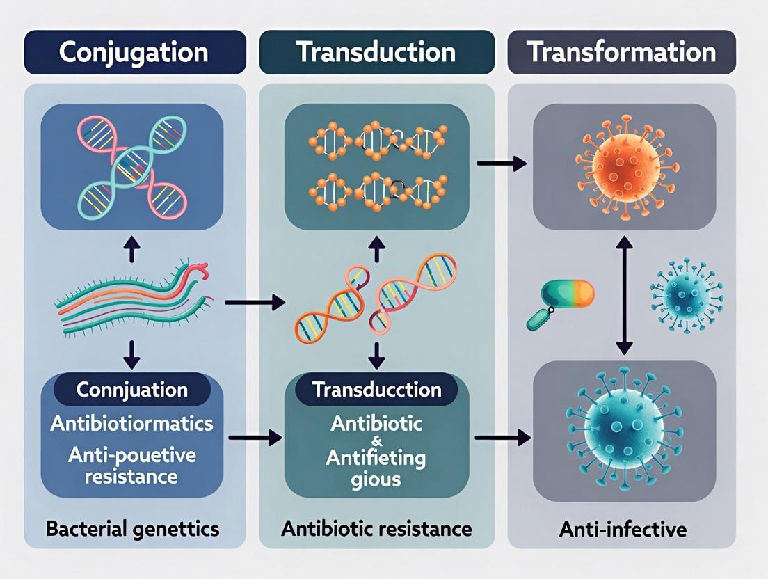
Horizontal Gene Transfer Mechanisms: How Conjugation, Transduction, and Transformation Drive Antibiotic Resistance in Bacteria
This article provides a comprehensive analysis of the three primary horizontal gene transfer (HGT) mechanisms—conjugation, transduction, and transformation—and their critical role in the dissemination of antibiotic resistance genes (ARGs).
Horizontal Gene Transfer Mechanisms: How Conjugation, Transduction, and Transformation Drive Antibiotic Resistance in Bacteria
Abstract
This article provides a comprehensive analysis of the three primary horizontal gene transfer (HGT) mechanisms—conjugation, transduction, and transformation—and their critical role in the dissemination of antibiotic resistance genes (ARGs). Tailored for researchers, scientists, and drug development professionals, it explores the molecular foundations of each process, details advanced methodologies for their study, addresses common experimental challenges and optimization strategies, and offers comparative validation of techniques. The synthesis underscores how understanding these pathways is essential for developing novel strategies to combat the global antimicrobial resistance (AMR) crisis.
The Molecular Triad: Deconstructing Conjugation, Transduction, and Transformation in AMR Spread
Horizontal Gene Transfer (HGT) is the non-hereditary movement of genetic information between organisms, often across species boundaries. Within the critical field of antibiotic resistance research, HGT—specifically via conjugation, transduction, and transformation—is the principal mechanism accelerating the global spread of multi-drug resistance (MDR) in bacterial pathogens. This whitepaper provides an in-depth technical analysis of HGT mechanisms, their quantitative contribution to MDR, standardized experimental protocols for their study, and essential research tools.
Mechanisms of HGT in Antibiotic Resistance Dissemination
Three primary mechanisms facilitate HGT, each with distinct pathways for mobilizing antibiotic resistance genes (ARGs).
Conjugation
Conjugation involves the direct, cell-to-cell transfer of mobile genetic elements (MGEs) like plasmids and integrative conjugative elements (ICEs) via a pilus. It is considered the most prevalent and efficient route for ARG spread.
Transduction
Transduction is bacteriophage-mediated gene transfer. During phage replication and assembly, bacterial DNA (including ARGs) can be mistakenly packaged into a phage capsid and injected into a new host.
Transformation
Transformation is the uptake and incorporation of free environmental DNA (released from lysed cells) by naturally competent bacteria.
Table 1: Quantitative Impact of HGT Mechanisms on MDR Spread
| Mechanism | Primary MGEs Transferred | Estimated Contribution to Clinical ARG Spread* | Key Bacteria Affected |
|---|---|---|---|
| Conjugation | Plasmids, ICEs | ~70-80% | Enterobacteriaceae, Enterococcus, Pseudomonas |
| Transduction | Phage genomes, genomic islands | ~10-20% | Staphylococcus aureus, Salmonella |
| Transformation | Free DNA fragments | ~5-10% | Streptococcus pneumoniae, Neisseria, Acinetobacter |
Note: Estimates based on current literature review; contributions vary by ecological niche and bacterial species.
Experimental Protocols for Studying HGT
Protocol: Filter Mating Assay for Conjugation
Objective: Quantify plasmid-mediated conjugation frequency between donor and recipient strains. Materials: Donor (with plasmid-borne resistance marker), Recipient (with chromosomal counterselection marker), sterile nitrocellulose filters, appropriate agar plates. Method:
- Grow donor and recipient cultures separately to mid-exponential phase.
- Mix donor and recipient cells at a standardized ratio (e.g., 1:10 donor:recipient) and concentrate by centrifugation.
- Resuspend cell mixture in small volume and apply to a sterile nitrocellulose filter placed on a non-selective agar plate.
- Incubate for a defined conjugation period (e.g., 2-18 hours).
- Resuspend cells from the filter in buffer and perform serial dilutions.
- Plate dilutions onto selective agar plates containing antibiotics that select for transconjugants (recipients that have acquired the plasmid) while counterselecting against donors and recipients.
- Calculate conjugation frequency = (Number of transconjugants) / (Number of recipient cells).
Protocol: Phage-Mediated Transduction Assay
Objective: Demonstrate transfer of an antibiotic resistance marker via a bacteriophage. Materials: Donor bacterial strain (carrying ARG), recipient strain, specific bacteriophage, calcium/magnesium solution (for phage adsorption), soft agar. Method:
- Propagate phage on the donor strain to create a lysate potentially carrying packaged ARGs.
- Treat lysate with DNase to destroy any free extracellular DNA, ensuring any gene transfer is phage-mediated.
- Mix phage lysate with recipient cells in the presence of Ca²⁺/Mg²⁺ to facilitate adsorption.
- Add mixture to soft agar and pour onto a base agar plate for incubation.
- Plate aliquots on selective media containing the relevant antibiotic. Colonies represent transductants.
- Include controls: recipient alone, phage lysate alone, and DNase-treated free DNA from donor.
Protocol: Natural Transformation Assay
Objective: Assess uptake of free DNA carrying an ARG by a naturally competent bacterium. Materials: Competent bacterial strain (e.g., A. baylyi), purified donor DNA containing ARG, DNase I. Method:
- Induce competence in the recipient strain using specific growth conditions (e.g., nutrient limitation).
- Divide culture into two tubes. To the experimental tube, add purified donor DNA. To the control tube, add donor DNA followed immediately by DNase I.
- Incubate to allow for DNA uptake and integration.
- Plate cultures on selective media. Transformants should appear only in the experimental tube, not in the DNase-treated control.
- Calculate transformation frequency = (Number of transformants) / (Total viable cell count).
Visualization of HGT Pathways and Workflows
Title: Bacterial Conjugation Process for Plasmid Transfer
Title: Generalized Transduction Cycle for Gene Transfer
Title: Natural Transformation via DNA Uptake
Title: General Workflow for HGT Experiments
The Scientist's Toolkit: Key Research Reagent Solutions
Table 2: Essential Materials for HGT & MDR Research
| Item | Function & Application | Example/Notes |
|---|---|---|
| Selective Agar Plates | Counterselection of donor/recipient and selection for transconjugants/transformants. Critical for quantifying HGT events. | LB agar + Antibiotic A (donor selection) + Antibiotic B (recipient/transconjugant selection). |
| Broad-Host-Range Plasmid Vectors | Model conjugative plasmids to study transfer kinetics and host range. | RP4, pKM101, IncF, IncN group plasmids. |
| Phage Lysates (Generalized) | Essential reagents for transduction studies to confirm phage-mediated ARG transfer. | P22 phage for Salmonella, 80α phage for S. aureus. |
| DNase I (RNase-free) | Control enzyme to degrade free extracellular DNA in transformation/transduction assays, confirming mechanism. | Used in transformation negative controls and to treat phage lysates. |
| Competence-Inducing Media | Specific growth media to induce the natural competent state in bacteria like Bacillus or Acinetobacter. | M-IV media for A. baylyi, Competence media for S. pneumoniae. |
| Chromosomal DNA Extraction Kits | To purify high-quality donor DNA for transformation assays or for PCR confirmation of transferred genes. | Phenol-chloroform or commercial column-based kits. |
| PCR Master Mix & ARG-Specific Primers | To confirm the presence and identity of transferred antibiotic resistance genes in transconjugants/transductants. | Primers for common ARGs (e.g., blaCTX-M, mecA, vanA). |
| Fluorescent Antibiotic Probes (e.g., Bocillin FL) | Visualize antibiotic accumulation and efflux in strains pre- and post-HGT to confirm functional resistance. | Used in microscopy and flow cytometry. |
| Bioinformatics Pipelines (e.g., ARIBA, ABRicate) | In silico identification of ARGs and MGEs in whole genome sequence data to trace HGT events. | For analyzing sequencing data from donor, recipient, and output strains. |
This whitepaper provides an in-depth technical examination of bacterial conjugation, the primary mechanism for horizontal gene transfer (HGT) via a pilus. Framed within the critical context of antibiotic resistance research, this process is a principal driver for the dissemination of resistance genes, virulence factors, and other adaptive traits encoded on mobile genetic elements (MGEs). Understanding the molecular machinery of conjugation is paramount for developing strategies to curb the spread of multidrug-resistant pathogens.
Molecular Machinery of Conjugation
Conjugation systems are classified by secretion system type (Type IV secretion system - T4SS) and mobility (self-transmissible vs. mobilizable plasmids). The core apparatus consists of:
- The Relaxosome: A nucleoprotein complex assembled at the origin of transfer (oriT). It includes the plasmid DNA, a site-specific DNA-binding protein, and the relaxase, which nicks the DNA at nic site to initiate transfer.
- The Mating Pair Formation (Mpf) Complex: A multiprotein T4SS spanning the inner and outer membranes, forming the conjugative pilus. The pilus, often composed of VirB2 pilin proteins, initiates contact with recipient cells.
- The Coupling Protein (T4CP): Links the relaxosome to the Mpf complex, facilitating the translocation of single-stranded DNA (ssDNA) into the recipient.
Key Mobile Genetic Elements in Conjugation
| MGE Type | Key Features | Common Resistance Genes Carried | Transfer Frequency (Approx. Range) |
|---|---|---|---|
| Broad-Host-Range IncP Plasmids | Self-transmissible, promiscuous, robust T4SS. | blaTEM, aac, tet(A), sul1 | 10-2 – 10-5 per donor |
| Narrow-Host-Range IncF Plasmids | Common in Enterobacteriaceae, often carry multiple AMR genes. | blaCTX-M, blaNDM, qnr, erm(B) | 10-3 – 10-6 per donor |
| Integrative Conjugative Elements (ICEs) | Chromosomally integrated, excise to form circular transfer intermediate. | mef(A), tet(M), vanA | 10-4 – 10-7 per donor |
| Conjugative Transposons | Similar to ICEs; classic example: Tn916. | tet(M), erm(B) | 10-5 – 10-8 per donor |
Quantitative Data on Plasmid Transfer Dynamics
Factors influencing conjugation efficiency are critical for modeling resistance spread.
| Factor | Experimental Condition | Impact on Transfer Frequency (Log10 Change) | Notes |
|---|---|---|---|
| Growth Phase | Early Exponential vs. Stationary | +2.0 to +3.0 | Highest frequency in early exponential phase. |
| Antibiotic Presence | Sub-MIC of Tetracycline | +1.0 to +2.0 | SOS response induction can upregulate T4SS genes. |
| Temperature | 37°C vs. 25°C | +1.5 to +2.5 | Optimal at host physiological temperature. |
| Surface vs. Liquid | Solid Agar vs. Liquid Broth | +1.0 to +3.0 | Surface mating drastically more efficient. |
| Donor:Recipient Ratio | 1:1 vs. 1:10 | -0.5 to -1.0 | Slight decrease with excess recipients. |
Detailed Experimental Protocols
Standard Solid-Surface Conjugation Assay
Purpose: To quantify the transfer frequency of a conjugative plasmid between donor and recipient strains.
Materials: See Scientist's Toolkit. Protocol:
- Culture Overnight: Grow donor (with plasmid, resistant to Antibiotic A) and recipient (chromosomally resistant to Antibiotic B) in separate broth cultures.
- Normalize & Mix: Harvest cells, wash, and resuspend in fresh medium. Mix donor and recipient cells at a 1:1 ratio (e.g., 100 µL each). For controls, plate donor and recipient alone.
- Mate on Filter: Pipette 200 µL of the mix onto a sterile membrane filter (0.22 µm pore) placed on non-selective agar. Incubate for a defined mating period (1-18 hours) at optimal temperature.
- Resuspend Cells: Transfer the filter to a tube with fresh broth. Vortex vigorously to resuspend the cell mass.
- Plate for Transconjugants: Serially dilute the suspension and plate on selective agar containing both Antibiotic A and Antibiotic B. Only transconjugants (recipients that received the plasmid) will grow.
- Plate for Viable Counts: Plate dilutions on selective agar for donor (Antibiotic A) and recipient (Antibiotic B) counts.
- Calculate Frequency: Transfer Frequency = (Number of Transconjugants) / (Number of Donors). Typically reported as transconjugants per donor.
Liquid Mating Assay
Purpose: To assess conjugation in broth, relevant for plasmid transfer in liquid environments like bloodstream or industrial fermenters. Protocol: Steps 1-2 as above. In step 3, mix cells directly in liquid broth (no filter) with mild agitation. Proceed with steps 4-7. Frequencies are typically lower than solid surface.
Visualization of Pilus Expression (Fluorescent Labeling)
Purpose: To detect and localize conjugative pili. Protocol: Induce expression of pilus genes. Label cells with a primary antibody against a major pilin protein (e.g., VirB2), followed by a fluorophore-conjugated secondary antibody. Wash and visualize using fluorescence or super-resolution microscopy.
Diagrams
Title: Molecular Steps in Pilus-Mediated Conjugation
Title: Solid-Surface Conjugation Assay Workflow
The Scientist's Toolkit: Research Reagent Solutions
| Item | Function/Application in Conjugation Research |
|---|---|
| Selective Agar Plates | Contain specific antibiotics to selectively grow donors, recipients, or transconjugants. Critical for quantifying transfer frequency. |
| Membrane Filters (0.22/0.45 µm) | Provide a solid surface for cell-to-cell contact during mating assays. Pores allow nutrient diffusion while trapping bacteria. |
| Antibiotics (Clinical & Lab Grade) | Used for selection pressure, to maintain plasmids, and to study the effect of sub-inhibitory concentrations on transfer. |
| Chromosomal & Plasmid-Borne Fluorescent Reporters (e.g., GFP, mCherry) | Enable visualization of donor, recipient, and transconjugant populations via fluorescence microscopy or flow cytometry. |
| Anti-Pilin Primary Antibodies | For immunofluorescence detection and quantification of conjugative pilus expression on bacterial surfaces. |
| MOPS or Other Defined Minimal Media | Used to control for metabolic states and to eliminate unknown variables present in rich media like LB. |
| DNaase I / RNase A | Controls in liquid mating assays to confirm transfer requires cell contact (DNAse degrades free DNA, ruling out transformation). |
| Conjugation Inhibitors (e.g., unsaturated fatty acids, synthetic peptides) | Experimental compounds that disrupt pilus assembly or function; used to probe mechanism and potential therapeutics. |
Within the triad of horizontal gene transfer (HGT) mechanisms—conjugation, transduction, and transformation—transduction represents a critical and efficient pathway for the dissemination of antibiotic resistance genes (ARGs). This whitepaper positions transduction, specifically via bacteriophage vectors, within the broader research thesis on HGT-driven antibiotic resistance. While conjugation involves direct cell-to-cell contact and transformation entails uptake of free DNA, transduction leverages bacterial viruses (phages) as natural shuttles, packaging bacterial DNA, including ARGs, and injecting it into new host cells. This process facilitates ARG spread across diverse bacterial genera, even in the absence of selective pressure, posing a significant challenge to public health and drug development.
Mechanisms of Transductive ARG Transfer
Generalized vs. Specialized Transduction
Generalized transduction occurs during the lytic cycle when phage machinery erroneously packages random fragments of bacterial chromosomal or plasmid DNA into a phage capsid, creating a transducing particle. Specialized transduction occurs during the lysogenic cycle when a prophage excises incorrectly, carrying adjacent bacterial genes (which can include ARGs if located near the phage integration site).
Diagram 1: Mechanisms of Phage-Mediated ARG Transduction
Key Pathways for ARG Capture
Mobile Genetic Elements (MGEs) like plasmids and transposons often carry ARGs. Phages can transduce entire plasmids (plasmid transduction) or can pick up ARGs integrated into the chromosome near phage integration sites or within moron regions (phage-encoded genes that can carry ARGs like qnr).
Quantitative Data on Phage-Mediated ARG Transfer
Table 1: Documented ARGs Transferred via Bacteriophage Vectors
| ARG Class | Specific Gene(s) | Phage Family/Type | Bacterial Host(s) | Transfer Frequency (Range) | Key Reference (Example) |
|---|---|---|---|---|---|
| β-lactamases | blaTEM-1, blaCTX-M | Myoviridae, Siphoviridae | E. coli, Salmonella | 10⁻⁸ – 10⁻⁶ per plaque-forming unit (PFU) | Colomer-Lluch et al., 2011 |
| Quinolone Resistance | qnrA, qnrS | Podoviridae | E. coli, Klebsiella | 10⁻⁷ – 10⁻⁵ per PFU | Wang et al., 2018 |
| Tetracycline Resistance | tet(A), tet(M) | Myoviridae | Enterococcus, Staphylococcus | 10⁻⁹ – 10⁻⁷ per PFU | Zhang et al., 2019 |
| Macrolide Resistance | erm(B), mef(A) | Siphoviridae | Streptococcus, Enterococcus | 10⁻⁸ – 10⁻⁶ per PFU | Haaber et al., 2016 |
| Vancomycin Resistance | vanA | Myoviridae | Enterococcus faecium | ~10⁻⁹ per PFU | Fillol-Salvà et al., 2022 |
| Colistin Resistance | mcr-1 | Inovirus (filamentous) | E. coli | Not fully quantified; demonstrated in situ | Wang et al., 2020 |
Table 2: Environmental Metagenomic Studies of Phage-Encoded ARGs
| Environment | Sample Type | Dominant ARG Classes in Virome | Relative Abundance (ARGs per Gb metagenome) | Common MGE Association |
|---|---|---|---|---|
| Wastewater Treatment | Influent, Effluent | β-lactam, multidrug efflux | 0.05 – 0.5 | Integrons, plasmid fragments |
| Animal Husbandry | Manure, Soil | Tetracycline, sulfonamide | 0.1 – 1.2 | Transposase genes |
| Human Gut | Feces | Macrolide, tetracycline | 0.01 – 0.1 | CRISPR spacer matches |
| River Water | Surface water | Multidrug, quinolone | 0.001 – 0.05 | Integrase genes |
Experimental Protocols
Protocol: Induction and Concentration of Transducing Phages from Environmental Samples
Objective: Isulate phage particles capable of transducing ARGs from complex samples like wastewater or feces.
Materials:
- Sample (e.g., 100mL wastewater)
- Chloroform
- DNase I (1 µg/mL) and RNase A (1 µg/mL)
- PEG 8000 (10% w/v)
- NaCl (0.5 M)
- SM Buffer
- 0.22 µm pore-size filters (PES)
- Ultracentrifuge and CsCl gradient materials (optional)
Procedure:
- Pre-treatment: Clarify sample by low-speed centrifugation (6,000 x g, 10 min, 4°C). Filter supernatant through 0.22 µm filter to remove bacteria.
- Nuclease Treatment: Add DNase I and RNase A to filtered supernatant to final concentration 1 µg/mL. Incubate 1h at 37°C to degrade free nucleic acids not protected within capsids.
- Phage Precipitation: Add NaCl to 0.5 M, mix. Add PEG 8000 to 10% w/v. Dissolve and incubate overnight at 4°C. Pellet phages by centrifugation (12,000 x g, 30 min, 4°C). Resuspend pellet in 1-2 mL SM Buffer.
- Optional Purification: Purify further via CsCl density gradient ultracentrifugation (e.g., 1.45-1.5 g/mL CsCl, 150,000 x g, 24h). Collect opalescent band.
- Storage: Store phage concentrate at 4°C with a drop of chloroform or at -80°C in 15% glycerol.
Protocol:In VitroTransduction Assay for ARG Transfer
Objective: Quantify the frequency of ARG transfer from a donor bacterial strain to a recipient strain via phage lysate.
Materials:
- Donor strain: Antibiotic-resistant bacterium (e.g., carries blaCTX-M on chromosome).
- Recipient strain: Antibiotic-sensitive, selectively marked (e.g., Rifampicin-resistant).
- Phage lysate propagated on donor strain (from Protocol 4.1, but using donor culture).
- Appropriate antibiotic plates: Agar containing antibiotic selecting for transduced ARG (e.g., Ceftazidime) and antibiotic selecting for recipient (e.g., Rifampicin).
- Calcium chloride (10mM)
- Soft agar (0.7%)
Procedure:
- Prepare Recipient Cells: Grow recipient strain to mid-exponential phase (OD600 ~0.4). Wash and resuspend in broth with 10mM CaCl₂ (enhances phage adsorption).
- Transduction Mix: Combine 100 µL recipient cells, 100 µL phage lysate (titer known), and 200 µL broth. Include a no-phage control (broth instead of lysate).
- Adsorption: Incubate mixture at host-specific temperature (e.g., 37°C for E. coli) for 30 min to allow phage adsorption/injection.
- Eliminate Free Phage/Donor Cells: Treat mixture with phage antiserum or vortex with chloroform to kill any remaining phage and donor cells. Centrifuge, plate supernatant.
- Selection: Plate entire mixture or dilutions onto double-selection plates (Antibiotic A [recipient marker] + Antibiotic B [transduced ARG]). Also plate on recipient-marker-only plates to determine viable recipient count.
- Calculation: Incubate plates 24-48h. Transduction frequency = (CFU on double-selection plates) / (total viable recipients on single-selection plates) OR / (PFU added). Report as transductants per PFU.
Diagram 2: In Vitro Transduction Assay Workflow
Protocol: Metagenomic Analysis of Virome-Encoded ARGs
Objective: Identify and quantify ARGs within viral fractions of environmental or clinical samples.
Materials:
- Phage concentrate (from Protocol 4.1)
- Meta-Vic Nucleic Acid Extraction Kit (optimized for viral particles)
- Multiple Displacement Amplification (MDA) kit (e.g., REPLI-g)
- Illumina DNA sequencing library prep kit
- Bioinformatics pipelines: VirSorter, MetaPhinder, ARG databases (CARD, ResFinder)
Procedure:
- Viral DNA Extraction: Extract total nucleic acids from phage concentrate using a kit designed for viral particles, incorporating an internal DNA standard for quantification.
- MDA: Perform MDA on extracted DNA to generate sufficient material for sequencing. Include no-template controls.
- Library Prep & Sequencing: Prepare Illumina shotgun sequencing library from MDA product. Sequence on HiSeq/NovaSeq platform to achieve >10 Gb data.
- Bioinformatics: a. Quality Control & Host Depletion: Trim reads (Trimmomatic), remove reads mapping to bacterial genomes (Bowtie2). b. Viral Sequence Identification: Assemble reads (MEGAHIT). Identify viral contigs using VirSorter and MetaPhinder. c. ARG Annotation: Predict open reading frames (Prodigal). BlastP against CARD database using stringent thresholds (e-value <1e-10, coverage >80%, identity >70%). d. Quantification: Normalize ARG hit counts by sequencing depth and internal standard.
The Scientist's Toolkit: Key Research Reagent Solutions
Table 3: Essential Materials for Transduction & ARG Research
| Item/Category | Example Product/Strain | Function/Explanation |
|---|---|---|
| Model Phage-Bacteria Systems | Phage P1 (generalized), Phage λ (specialized), Phage Φ80 | Well-characterized transduction models for E. coli; control experiments. |
| Antibiotic-Marked Recipient Strains | E. coli MG1655 Rif⁸, S. aureus RN4220 Rif⁸ | Recipient with chromosomal resistance to antibiotic (e.g., Rifampicin) for positive selection in transduction assays. |
| Broad-Host-Range Phage Propagator | Pseudomonas phage ΦKZ, Salmonella phage P22 | Used to generate high-titer lysates from diverse Gram-negative donors. |
| Viral Metagenome Extraction Kit | Norgen's Meta-Vic Nucleic Acid Kit | Optimized for low-biomass, high-inhibitor environmental viral concentrates. |
| Multiple Displacement Amplification Kit | Qiagen REPLI-g Single Cell Kit | Whole-genome amplification of minute viral DNA amounts pre-sequencing. |
| DNase I (RNase-free) | Thermo Scientific EN0521 | Critical for degrading free DNA in phage concentrates to ensure viral-origin signal. |
| Phage Buffer with Ca²⁺/Mg²⁺ | SM Buffer (NaCl, MgSO₄, Tris, Gelatin) with 10mM CaCl₂ | Stabilizes phage particles; Ca²⁺ promotes adsorption to cell walls. |
| Density Gradient Medium | OptiPrep (Iodixanol) or Cesium Chloride | For ultracentrifuge-based purification of intact phage particles from lysates. |
| Selective Agar with Antibiotics | Mueller-Hinton Agar + defined antibiotics | For plating transductants under selection; use CLSI-recommended concentrations. |
| Bioinformatics Database | Comprehensive Antibiotic Resistance Database (CARD), ACLAME (MGEs) | Curated reference for annotating ARGs and associated mobile elements. |
Implications and Future Research Directions
The role of bacteriophages as shuttles for ARGs underscores a complex ecological dimension to the antibiotic resistance crisis. Future research must focus on:
- Quantifying in situ transduction rates in human, animal, and environmental microbiomes.
- Elucidating the molecular signals that promote packaging of ARG-bearing MGEs into phage capsids.
- Exploring phage therapy implications: understanding if therapeutic phages could inadvertently mobilize ARGs.
- Developing novel inhibitors that specifically target phage-mediated gene transfer without driving resistance.
Integrating transduction dynamics into the broader HGT framework (conjugation, transformation) is essential for a holistic understanding of ARG dissemination and for developing effective strategies to mitigate it.
Abstract Within the critical research framework of horizontal gene transfer (conjugation, transduction, transformation) and antibiotic resistance dissemination, natural competence stands as a fundamental mechanism. This in-depth guide examines the molecular machinery, regulatory networks, and experimental methodologies underpinning the transformation process, wherein bacteria actively uptake environmental DNA fragments. Emphasis is placed on the integration of this process into the resistome, providing researchers and drug development professionals with a technical foundation for understanding and investigating this pathway of genetic exchange.
1. Introduction and Molecular Framework Natural competence is a genetically programmed physiological state enabling bacteria to bind, uptake, and recombine extracellular DNA. This process is a direct contributor to the spread of antibiotic resistance genes (ARGs) among bacterial populations, complementing conjugation and transduction. Competence is typically tightly regulated by quorum-sensing and nutritional stress signals, ensuring expression only under favorable conditions.
Diagram: Core Regulatory Pathway for Natural Competence Induction
2. The DNA Uptake Machinery: A Multi-Step Process The process can be broken down into distinct, quantifiable stages: DNA binding, processing, translocation across membranes, and recombination.
Table 1: Key Stages and Quantitative Parameters of Natural Transformation
| Stage | Key Components | Function | Representative Kinetic Data (Model: Streptococcus pneumoniae) |
|---|---|---|---|
| DNA Binding & Processing | ComEA, EndA (nuclease) | Binds extracellular dsDNA; degrades one strand. | Uptake rate: ~100 bp/sec; Processivity: >10 kbp fragments preferred. |
| Pilus Assembly & Retraction | ComGC, ComGD, etc. (Type IV pilus-like) | Forms pseudopilus; retracts to pull DNA. | Pilus length: ~0.5-1 µm; Retraction force: ~20 pN. |
| Translocon | ComEC | Forms transmembrane pore for ssDNA import. | Pore size: ~2.2 nm; Voltage-gated. |
| Cytoplasmic Protection | SsbB (SSB protein) | Coats incoming ssDNA. | Binds ssDNA with high affinity (Kd ~10⁻⁹ M). |
| Recombination | RecA, DprA | Mediates homologous recombination. | Requires ~20-50 bp homology; Efficiency: ~1-10% of uptake events. |
Diagram: Workflow of DNA Uptake and Integration
3. Essential Experimental Protocols
3.1. Induction and Quantification of Natural Competence
- Principle: Trigger competence via synthetic competence-stimulating peptide (CSP) or growth phase monitoring, followed by exposure to selectable marker DNA.
- Protocol (CSP Induction in Streptococci):
- Grow target strain to mid-exponential phase (OD₆₀₀ ~0.05-0.1) in appropriate broth.
- Add synthetic CSP at empirically determined concentration (typically 50-200 ng/mL). Include a no-CSP control.
- Incubate for 10-15 minutes at 37°C.
- Add purified donor DNA (e.g., genomic DNA containing an antibiotic resistance marker, 100-500 ng/mL).
- Incubate for 30-60 minutes to allow uptake and expression.
- Plate on selective agar containing the relevant antibiotic. Include controls for DNA-only and recipient-only viability.
- Calculate transformation frequency: CFU on selective plate / total viable CFU on non-selective plate.
3.2. Measuring DNA Uptake Directly via qPCR
- Principle: Quantify internalized DNA fragments that are protected from external DNase.
- Protocol:
- Induce competence as in 3.1.
- Add exogenous DNA (e.g., a specific amplicon) to culture.
- After uptake period, treat aliquots with DNase I (1 U/µL, 15 min, 37°C) to degrade all external DNA.
- Stop DNase with EDTA (5 mM final).
- Lyse cells (e.g., with lysozyme & heat).
- Purify total nucleic acid. Treat with RNase if needed.
- Perform qPCR using primers specific to the added DNA fragment.
- Quantify using a standard curve of known DNA copies. Express as copies per CFU or per mL.
4. The Scientist's Toolkit: Key Research Reagent Solutions
Table 2: Essential Materials for Competence & Transformation Research
| Reagent/Material | Function/Description | Key Considerations |
|---|---|---|
| Synthetic Competence Peptides (CSP) | Chemically defined inducer of competence in Streptococcus, Bacillus spp. | Species/strain-specific; requires purity >95%. |
| DNase I (RNase-free) | Degrades extracellular DNA post-uptake to distinguish internalized DNA. | Critical for qPCR uptake assays; control activity with Mg²⁺/Ca²⁺. |
| Quantitative PCR (qPCR) Master Mix | Quantifies low-copy number internalized DNA fragments. | Use high-efficiency, SYBR Green or probe-based kits. |
| Homologous Donor DNA | Genomic DNA or PCR amplicons containing a selectable marker (e.g., erm, cat). | Requires sufficient flanking homology (>500 bp) for recombination. |
| Competence-Specific Reporter Plasmids | Plasmid with GFP/luciferase under control of a competence-specific promoter (e.g., comX). | Enables real-time monitoring of competence development in populations. |
| RecA Inhibitors (e.g., curcumin analogs) | Chemical inhibitors to block the final recombination step. | Tool to dissect uptake from integration; can affect cell viability. |
| Fluorescently-labeled DNA (Cy3/dUTP) | Visualize DNA binding and uptake kinetics via microscopy or flow cytometry. | Allows single-cell analysis of competence heterogeneity. |
1. Introduction: Framing within Antibiotic Resistance Research
Horizontal Gene Transfer (HGT)—via conjugation, transduction, and transformation—is the principal engine driving the rapid dissemination of antibiotic resistance genes (ARGs) among bacterial pathogens. Isolating and characterizing the genomic "scar tissue" left by these events is a cornerstone of modern comparative genomics. This whitepaper provides a technical guide for identifying the hallmark sequences of HGT, underpinning a broader thesis on understanding and interrupting the mobilization pathways of ARGs.
2. Hallmark Genomic Signatures of HGT
HGT events leave distinct imprints on the recipient genome. Comparative analysis seeks these signatures against a genomic background.
Table 1: Hallmark Sequence Signatures of HGT Mechanisms
| HGT Mechanism | Primary Hallmark | Supporting Signatures | Associated ARG Vectors |
|---|---|---|---|
| Conjugation | Presence of mobile genetic element (MGE) machinery (e.g., tra, trb, virB operons) and an origin of transfer (oriT). | Flanking insertion sequences (IS); tRNA/phage integration sites; plasmid partitioning (par) genes. | Conjugative plasmids, genomic islands (ICEs). |
| Transduction | Phage-related genes (capsid, integrase, terminase) flanking the candidate region. | Direct terminal repeats (attL/attR); elevated GC content vs. host; integration at tRNA loci. | Phages (temperate), phage-plasmids (phagemids). |
| Transformation | Mosaic patches of high homology to distant species, lacking MGE signatures. | Uptake signal sequences (USS) in Neisseria, Haemophilus; competence (com) genes nearby; blunt edges. | Free DNA from lysed cells. |
3. Core Computational Identification Pipeline
Experimental Protocol 1: In Silico HGT Region Prediction
Objective: To identify putative horizontally acquired regions in a bacterial genome assembly. Input: Genome sequence (FASTA), annotated GFF file (optional). Software: Command-line tools (BLAST+, HMMER), scripting (Python/R).
- Baseline Establishment:
- Calculate whole-genome metrics: GC content, k-mer frequency, codon usage bias.
- Comparative Analysis:
- Perform BLASTn/BLASTp against a curated non-redundant database (e.g., NCBI nr).
- Use DIAMOND for accelerated protein searches.
- MGE Detection:
- Screen against MGE databases (ACLAME, ICEberg, PHASTER) using HMMER (hidden Markov models).
- Synteny Disruption:
- Compare to closely related non-pathogenic reference genomes using Mauve or progressiveMauve to identify genomic rearrangements.
- Integration & Scoring:
- Use dedicated integrators like IslandViewer 4 or Pathogenomics to combine signals (GC deviation, MGE hits, integration sites) and predict genomic islands.
HGT Prediction Computational Workflow
4. Experimental Validation of Predicted HGT Regions
Experimental Protocol 2: PCR-Based Amplicon Sequencing for Junction Verification
Objective: To experimentally confirm the insertion points and structure of a predicted genomic island.
- Primer Design:
- Design outward-facing primer pairs: One binding within the predicted HGT element, the other binding in the adjacent core genome. This amplifies across the junction.
- PCR Amplification:
- Reaction Mix: 1X PCR buffer, 2.5 mM MgCl₂, 0.2 mM dNTPs, 0.5 µM each primer, 1.25 U high-fidelity DNA polymerase, 50 ng genomic DNA template.
- Cycling: Initial denaturation: 98°C, 30s; 35 cycles: 98°C (10s), 60°C (15s), 72°C (1 min/kb); Final extension: 72°C, 5 min.
- Amplicon Analysis:
- Run PCR product on 1% agarose gel. Purify band of expected size.
- Sanger sequence the purified amplicon using the same primers.
- Validation:
- Align sequence data to the in silico assembly. Exact match confirms prediction. Discrepancy may indicate assembly error or strain variation.
The Scientist's Toolkit: Key Research Reagent Solutions
Table 2: Essential Materials for HGT Identification & Validation
| Reagent / Solution | Function | Example / Specification |
|---|---|---|
| High-Fidelity DNA Polymerase | Accurate amplification of junction regions for sequencing. | Platinum SuperFi II, Q5 Hot Start. |
| Gel Extraction Kit | Purification of PCR amplicons from agarose gels. | Qiagen QIAquick, Zymoclean Gel DNA Recovery Kit. |
| Sanger Sequencing Service | Verification of PCR amplicon sequence. | In-house ABI sequencer or commercial service (Eurofins). |
| Next-Gen Sequencing Kit | For whole-genome sequencing of novel isolates. | Illumina DNA Prep, Oxford Nanopore Ligation Kit. |
| MGE-Specific Databases | In silico identification of mobile element components. | ACLAME (MGE proteins), ICEberg (Integrative Conjugative Elements). |
| Comparative Genomics Suite | Synteny analysis and visualization. | Mauve aligner, BRIG for circular genome comparisons. |
5. Case Study: Identifying a Resistance Island in Klebsiella pneumoniae
Scenario: A clinical K. pneumoniae isolate shows resistance to carbapenems. Sequencing reveals a putative 30-kb genomic island.
Analysis Steps:
- Detection: IslandViewer 4 predicts an island with GC content of 52% (vs. host 57%).
- Annotation: Prokka/Roary annotation identifies a blaKPC carbapenemase gene and an intact int integrase gene.
- MGE Context: HMMER scan against ACLAME reveals a match to a Tn4401-like transposon structure.
- Synteny: Mauve alignment with susceptible strain shows the 30-kb insertion disrupts a ygeG homolog locus.
- Validation: Junction PCR with primers in blaKPC and the core ygeG region yields a 1.2-kb product, sequenced to confirm a perfect Tn4401 insertion.
Resistance Island Structure in K. pneumoniae
6. Conclusion
Methodical identification of HGT hallmarks—through integrated computational prediction and experimental validation—is critical for mapping the resistance mobilome. This workflow directly informs the broader thesis on conjugation, transduction, and transformation by providing the foundational evidence needed to track ARG origin, vector, and dissemination routes, ultimately guiding targeted drug and therapeutic development.
The Integrative Role of Genomic Islands and Resistance Cassettes in ARG Assembly
This whitepaper explores the molecular machinery driving the assembly and dissemination of antibiotic resistance genes (ARGs). Framed within the broader thesis on horizontal gene transfer (HGT) mechanisms—conjugation, transduction, and transformation—this guide details how genomic islands (GIs), particularly integrative and conjugative elements (ICEs), and integrons with their resistance gene cassettes, serve as foundational platforms for ARG acquisition and recombination. Their integrative function is central to the evolution of multidrug-resistant (MDR) pathogens, presenting a critical challenge for drug development.
Core Mechanisms of ARG Assembly and Integration
Genomic Islands (GIs) as ARG Hubs
GIs are discrete, horizontally acquired DNA segments integrated into bacterial chromosomes. Their role in ARG assembly is characterized by:
- Integration: Site-specific recombination via integrases into tRNA or other loci.
- Mobility: Many are ICEs, containing genes for conjugation, excision, and integration.
- ARG Carriage: They often harbor clusters of ARGs alongside other adaptive genes.
Table 1: Key Features of Major Genomic Island Types in ARG Dissemination
| Island Type | Key Integrase/Recombinase | Primary Attachment Site | Common ARG Examples | Mobility Mechanism |
|---|---|---|---|---|
| ICEs (e.g., Tn916, SXT/R391) | Tyrosine or Serine Integrase | tRNA, rlmH, etc. | tet(M), erm(B) | Conjugation |
| PAIs (Pathogenicity Islands) | Phage-like Integrase | tRNA, leuX | Often linked to virulence | Variable, often phage-mediated |
| GI-Sym (Symbiosis Islands) | P4-type Integrase | phe-tRNA | Rarely carry ARGs | Conjugation |
Integrons and Resistance Gene Cassettes
Integrons are genetic capture systems that assemble arrays of promoterless gene cassettes. Their structure is fundamental to ARG assembly:
- Platform: A stable attI xss site and an integrase gene (intI).
- Cassettes: Circular, mobile gene units containing an ARG and an attC xss site.
- Assembly: The integrase catalyzes recombination between attI and attC, inserting the cassette in a specific orientation for expression from a common promoter, Pc.
Table 2: Quantitative Data on Major Integron Classes and Cassette Prevalence
| Integron Class | Integrase Type | Estimated Known Cassettes | Most Common ARG Cassettes (Examples) | Primary Host Context |
|---|---|---|---|---|
| Class 1 | IntI1 | ~130+ | aadA (aminoglycoside), dfrA (trimethoprim), blaVIM/NDM (carbapenem) | Plasmids, Transposons, Chromosomes |
| Class 2 | IntI2 | ~40 | dfrA1, sat2, aadA1 | Tn7 transposons |
| Class 3 | IntI3 | ~10 | blaGES (carbapenemase) | Plasmids |
| Chromosomal | Diverse (e.g., IntI9) | Hundreds | Variable, often of unknown function | Bacterial chromosome (e.g., Vibrio spp.) |
Integrative Synergy: How Islands and Cassettes Cooperate
The synergy between these systems creates powerful ARG assembly lines:
- Cassette Capture by Islands: ICEs and other GIs frequently carry complete integrons or single cassettes. For example, the Vibrio cholerae SXT ICE contains a Class 1 integron.
- Island Mobilization of Cassettes: Once integrated into a mobile GI, an entire cassette array becomes mobilizable via conjugation or transduction.
- Nested Recombination: Cassettes within integrons, located on ICEs, can be further rearranged, creating combinatorial ARG diversity on a single, transmissible element.
Diagram 1: ARG Assembly and HGT via Integron-Island Synergy.
Key Experimental Protocols for Investigation
Protocol: Mapping Cassette Arrays in Genomic Islands
Objective: Identify and characterize integron cassette arrays within a sequenced GI (e.g., an ICE). Method:
- In Silico Prediction: Using a bacterial genome assembly, screen for intI genes (BLAST). Extract the downstream region.
- attC Site Detection: Use the ATTACC program or manual search for conserved inverse core sites (RYYYAAC) and stem-loop structures.
- Cassette Delineation: Define cassette boundaries from the attC site of one cassette to the attC of the next.
- ARG Annotation: Annotate each open reading frame using ResFinder or CARD databases.
- PCR & Sanger Sequencing Validation: Design primers flanking the predicted array and internal cassette primers. Amplify, sequence, and compare to in-silico results.
Protocol: Tracking Island Excision and Transfer
Objective: Quantify excision frequency of an ICE and confirm its conjugative transfer of ARGs. Method:
- Excision PCR: Design primers outward-facing from the predicted ICE boundaries (targeting the empty attachment site). Amplify from genomic DNA.
- Quantitative PCR (qPCR): Use one primer within the ICE and one in the flanking chromosome to quantify the excised circular form relative to the chromosomal form.
- Conjugation Assay (Filter Mating): a. Mix donor (ICE+, recipient) strains at a set ratio. b. Filter onto a membrane, incubate on non-selective agar. c. Resuspend cells, plate on selective media containing antibiotics that select for the ICE-borne ARG in the recipient and counterselect the donor. d. Calculate transfer frequency (transconjugants per donor).
The Scientist's Toolkit: Essential Research Reagents & Materials
Table 3: Key Reagent Solutions for ARG Assembly Research
| Reagent / Material | Function / Application | Key Characteristics / Example |
|---|---|---|
| High-Fidelity DNA Polymerase (e.g., Q5, Phusion) | Accurate amplification of integron arrays and island boundaries for sequencing. | Low error rate, GC-rich buffer capability. |
| Long-Range PCR Kits | Amplification of large GI or cassette array regions (>10 kb). | Enzyme blends optimized for processivity. |
| Transposon Mutagenesis Kits (e.g., EZ-Tn5) | Functional genomics to identify genes essential for island excision/conjugation. | In-vitro transposome complexes. |
| Chromosomal DNA Extraction Kit (Gram-Negative/Gram-Positive specific) | Pure, high-molecular-weight DNA for sequencing and PCR. | Includes lysozyme/mutanolysin for Gram-positives. |
| Gateway or Gibson Assembly Cloning Kits | Cloning large integron or island fragments for functional studies. | Enables seamless assembly of multiple fragments. |
| Conjugation Counterselection Antibiotics | Essential for filter mating assays to select transconjugants. | e.g., Nalidixic Acid for counterselecting E. coli, Rifampicin. |
| attC-Specific PCR Primers | Detection of free circular gene cassettes. | Designed against conserved attC stem-loop sequences. |
| Integrase Expression Vectors | In-vitro assay of integrase activity on attI x attC recombination. | IPTG-inducible expression (e.g., pET system). |
Visualization of Experimental Workflows
Diagram 2: Workflow for Analyzing ARG Assembly Systems.
Advanced Techniques: Tracking and Quantifying HGT in Laboratory and Clinical Settings
Horizontal Gene Transfer (HGT) is a primary driver of antibiotic resistance (AMR) dissemination among bacterial populations. Within the broader thesis investigating the mechanisms of conjugation, transduction, and transformation, in vitro conjugation assays serve as the foundational experimental system for quantifying and characterizing the direct cell-to-cell transfer of plasmids, particularly those harboring antimicrobial resistance genes (ARGs). This technical guide details two core methodologies—filter mating and liquid mating—which are indispensable for studying plasmid mobility, host range, and the efficacy of potential conjugation inhibitors in AMR research and drug development.
Core Principles of Bacterial Conjugation
Conjugation is a type of HGT mediated by conjugative plasmids or integrative conjugative elements (ICEs). It requires direct contact between a donor cell (harboring the conjugative element) and a recipient cell. The process involves:
- Mating pair formation (via pili or adhesins).
- DNA mobilization and transfer (initiation at oriT, strand transfer).
- DNA replication and establishment in the recipient.
Experimental Protocols
General Preparation
- Bacterial Strains: Donor: Contains a conjugative plasmid with selectable marker(s) (e.g., Amp^R). Recipient: Contains a chromosomally-encoded, distinct selectable marker (e.g., Rif^R, Str^R). Use appropriate, well-characterized lab strains (e.g., E. coli).
- Media: Use non-selective, rich media (e.g., LB broth) for pre-culture growth. Use appropriate selective agar plates for selection of transconjugants and controls.
- Controls: Essential for data validation (see Table 1).
Protocol A: Filter Mating Assay
Filter mating provides a solid support for efficient cell-cell contact, often yielding higher conjugation frequencies.
Detailed Methodology:
- Overnight Cultures: Grow donor and recipient strains separately in LB broth with appropriate antibiotics (for donor maintenance) at optimal temperature (e.g., 37°C, 200 rpm).
- Cell Harvesting: Subculture 1:100 into fresh, antibiotic-free LB and grow to mid-exponential phase (OD600 ~0.4-0.6).
- Washing: Pellet 1 mL of each culture by centrifugation (e.g., 5,000 x g, 2 min). Wash cell pellets twice with 1 mL of fresh, pre-warmed LB or phosphate-buffered saline (PBS) to remove residual antibiotics.
- Mixing: Resuspend pellets in LB. Mix donor and recipient cells at a defined ratio (typically 1:10 donor:recipient) in a final volume of 100-200 µL. Common ratios range from 1:1 to 1:100.
- Filtration: Pipette the mixture onto a sterile membrane filter (0.22 µm or 0.45 µm pore size, cellulose nitrate or acetate) placed on a vacuum filtration manifold.
- Incubation: Aseptically transfer the filter, bacteria-side-up, onto the surface of a pre-warmed, non-selective LB agar plate. Incubate for a defined mating period (e.g., 1-18 hours) at the desired temperature.
- Resuspension: After incubation, transfer the filter to a tube containing 1-2 mL of sterile saline or LB. Vortex vigorously to resuspend the bacterial cells from the filter.
- Plating and Selection: Perform serial 10-fold dilutions of the mating mixture. Plate aliquots onto:
- Selective Agar A: Antibiotics selecting for the recipient marker only. Count colonies to determine recipient (R) CFU/mL.
- Selective Agar B: Antibiotics selecting for the plasmid marker only. Count colonies to determine donor (D) CFU/mL.
- Selective Agar C: Antibiotics selecting for BOTH donor plasmid and recipient markers. Count colonies to determine transconjugant (T) CFU/mL.
- Calculation:
- Conjugation Frequency = (Transconjugant CFU/mL) / (Recipient CFU/mL)
Protocol B: Liquid Mating Assay
Liquid mating occurs in broth, simulating a more planktonic environment and is often faster but can yield lower frequencies.
Detailed Methodology:
- Steps 1-3: Follow the same culture growth, subculture, and washing steps as in the Filter Mating protocol.
- Mixing and Incubation: Mix washed donor and recipient cells at the desired ratio directly in a tube containing 1 mL of fresh, pre-warmed, antibiotic-free LB broth.
- Incubation: Incubate the static or shaking tube for the mating period (typically 1-2 hours).
- Plating and Selection: Vortex the mating mixture briefly. Perform serial dilutions and plate on the three selective agar types as described in Step 8 of the Filter Mating protocol.
- Calculation: Use the same formula for conjugation frequency.
Notes: For both protocols, plate appropriate dilutions of the initial donor and recipient cultures alone on selective agars to confirm antibiotic sensitivity profiles.
Data Presentation: Comparison of Assay Parameters
Table 1: Key Parameters and Controls for In Vitro Conjugation Assays
| Parameter | Filter Mating Assay | Liquid Mating Assay | Purpose / Rationale |
|---|---|---|---|
| Typical Mating Time | 2 - 18 hours | 0.5 - 2 hours | Optimize for plasmid type; longer times may increase frequency but also growth. |
| Donor:Recipient Ratio | 1:1 to 1:10 | 1:1 to 1:100 | Affects contact probability. 1:10 often standard. |
| Conjugation Frequency Range | 10^-1 to 10^-6 | 10^-3 to 10^-7 | Plasmid-dependent. Filter mating is generally more efficient. |
| Negative Control | Donor alone plated on transconjugant-selective agar. | As for filter mating. | Confirms donor cannot grow without recipient's chromosomal marker. |
| Viability Control | Recipient alone plated on recipient-selective agar. | As for filter mating. | Confirms recipient viability and antibiotic resistance. |
| Selective Agar Types | Agar A (Recipient marker), Agar B (Donor marker), Agar C (Both markers). | Identical to filter mating. | Distinguishes donor, recipient, and transconjugant populations. |
| Key Advantage | Maximizes cell contact; higher efficiency; standardized contact time. | Simpler/faster; mimics liquid environments; amenable to high-throughput. | |
| Key Limitation | Requires extra materials (filters, manifold); less suited for very high throughput. | Lower efficiency; mating time conflated with growth. |
Diagrams
Conjugation Assay Workflow: Filter vs. Liquid Mating
Molecular Pathway of Plasmid Conjugation
The Scientist's Toolkit: Research Reagent Solutions
Table 2: Essential Materials and Reagents for Conjugation Assays
| Item / Reagent | Function / Purpose in Conjugation Assays | Key Considerations |
|---|---|---|
| Selective Antibiotics | To selectively grow donor, recipient, and transconjugant populations. Critical for quantification. | Validate minimal inhibitory concentration (MIC) for all strains. Use fresh stocks. Avoid cross-resistance. |
| Membrane Filters (0.22/0.45 µm) | In filter mating, provides a solid surface for bacterial aggregation and mating pair stabilization. | Sterile, mixed cellulose esters are common. Ensure pore size retains bacteria but allows nutrient diffusion. |
| Rich Agar/Broth (e.g., LB) | Standard medium for bacterial growth during pre-culture and mating. | Avoid sugars if studying certain plasmid systems (e.g., F-plasmid fertility inhibition). |
| Sterile Saline or PBS | For washing cells to remove antibiotics and for serial dilution of mating mixtures. | Maintains osmotic balance while stopping further conjugation during processing. |
| Conjugative Plasmid Vectors | Self-transmissible plasmids (e.g., RP4, pKM101) or mobilizable plasmids with helper. | Defined origin of transfer (oriT), resistance markers, and host range. |
| Antibiotic-Resistant Recipient Strains | Provide the counter-selection marker necessary to identify transconjugants. | Chromosomal resistance to rifampicin, nalidixic acid, or streptomycin is common. |
| Potential Conjugation Inhibitors | Test compounds (e.g., unsaturated fatty acids, biocides) that may disrupt pilus formation, mating pair stability, or DNA transfer. | Include solvent controls (e.g., DMSO). Add at sub-inhibitory concentrations to avoid killing. |
This technical guide details the methodologies for measuring bacteriophage propagation and transduction efficiency, specifically via plaque assays. Within the broader research on horizontal gene transfer (conjugation, transduction, transformation) and the dissemination of antibiotic resistance genes, transduction—mediated by bacteriophages—represents a critical vector. Accurate quantification of phage infectivity and transducing particle frequency is fundamental to understanding the dynamics of resistance gene transfer in clinical, environmental, and research settings.
Core Concepts: Plaque Assays and Transduction
A plaque assay is the standard method for quantifying viable, lytic bacteriophages. A single infectious phage particle infects a bacterial cell, undergoes lytic replication, and lyses the host, releasing progeny that infect neighboring cells. After several cycles, this results in a clear zone, or plaque, in a bacterial lawn. For specialized transduction (where phage integrates into the host genome and excises with adjacent host DNA) or generalized transduction (where host DNA is packaged into phage capsids during the lytic cycle), the plaque assay is adapted to measure the frequency of transducing particles among the total viral population.
Key Quantitative Data: Typical Efficiencies and Titers
The following tables summarize standard quantitative benchmarks in phage research, crucial for contextualizing experimental results in antibiotic resistance transduction studies.
Table 1: Typical Phage Titers and Transduction Efficiencies
| Phage-Bacterial System | Typical Plaque-Forming Unit (PFU) Titer (per mL lysate) | Typical Transducing Particle Frequency (per PFU) | Key Transduced Markers (e.g., Antibiotic Resistance) |
|---|---|---|---|
| Lambda phage (λ) - E. coli | 10^9 - 10^11 | 10^-5 - 10^-7 (Specialized) | gal, bio, bla (if engineered) |
| P1 phage - E. coli | 10^8 - 10^10 | 10^-5 - 10^-6 (Generalized) | Antibiotic resistance cassettes, genomic DNA |
| T4 phage - E. coli | 10^10 - 10^12 | <10^-8 (Rare Generalized) | Limited, due to degradation of host DNA |
| Φ80 - E. coli | 10^9 - 10^10 | ~10^-6 (Specialized) | tonB, trp |
| PBS1/PBS2 - B. subtilis | 10^8 - 10^9 | ~10^-5 (Generalized) | met, thy, antibiotic resistance |
Table 2: Critical Parameters for Plaque Assay Optimization
| Parameter | Optimal Range / Typical Value | Impact on Assay Outcome |
|---|---|---|
| Host Cell Growth Phase | Mid-log phase (OD600 ~0.4-0.6) | Maximizes infection efficiency; stationary phase cells reduce plating efficiency. |
| Top Agar Concentration | 0.3% - 0.7% (commonly 0.5%) | Too soft: plaques run; too hard: phage diffusion inhibited, plaques small. |
| Incubation Temperature | Host-dependent (e.g., 37°C for E. coli) | Affects phage replication cycle speed and host metabolism. |
| Plaque Development Time | 6-24 hours | Under-incubation: plaques too small; over-incubation: lawn lyses completely. |
| Multiplicity of Infection (MOI) in Transduction | <1 (typically 0.01-0.1) | Prevents multiple infections of a single cell, which can artifactually lower transduction frequency counts. |
Experimental Protocols
Protocol 4.1: Standard Double-Layer Agar Plaque Assay for Phage Titration
Objective: To determine the concentration of infectious phage particles (PFU/mL) in a lysate.
Materials: See "The Scientist's Toolkit" below. Procedure:
- Prepare Host Culture: Grow the susceptible bacterial host to mid-log phase (OD600 ≈ 0.4-0.6) in appropriate broth.
- Prepare Dilutions: Serially dilute the phage lysate (e.g., 10-fold dilutions in SM buffer or broth) across 6-8 tubes.
- Mix Phage and Bacteria: For each dilution, combine 100 µL of bacterial culture with 100 µL of phage dilution in a sterile tube. Include a bacteria-only control.
- Adsorb: Incubate the mixture for 5-15 minutes at the host's growth temperature to allow phage adsorption.
- Add Top Agar: Add 3-5 mL of molten, tempered (45-50°C) soft agar to each phage-bacteria mixture. Vortex briefly and immediately pour onto a pre-warmed, dry base agar plate. Swirl gently to distribute evenly.
- Solidify and Incubate: Allow the top agar to solidify completely (10-15 minutes at room temperature). Invert plates and incubate at the appropriate temperature for 6-24 hours.
- Enumerate: Count plaques on plates yielding 20-200 discrete plaques. Calculate PFU/mL using the formula: PFU/mL = (Plaque count) / (Dilution factor × Volume plated in mL).
Protocol 4.2: Transduction Efficiency Assay (Generalized Transduction using P1)
Objective: To measure the frequency of transducing particles carrying a specific antibiotic resistance marker.
Materials: As above, plus selective agar plates containing the relevant antibiotic. Procedure:
- Generate Donor Lysate: Grow a donor bacterial strain (carrying the antibiotic resistance marker, e.g., Kan^R) to mid-log phase. Infect with P1 phage at a low MOI (~0.01) and allow full lysis. Clarify the lysate by centrifugation and filtration (0.45 µm) to remove bacterial debris. This lysate contains both phage (PFU) and transducing particles.
- Titer the Lysate: Perform a standard plaque assay (Protocol 4.1) on the susceptible recipient strain (Kan^S) to determine the total PFU/mL.
- Perform Transduction: Mix 100 µL of the recipient culture (Kan^S, mid-log phase) with 100 µL of the P1 donor lysate at an MOI of ~0.1. Include controls: recipient only, recipient + phage only (no donor marker).
- Adsorb and Recover: Adsorb for 20-30 minutes at 37°C. Add 1 mL of broth, incubate for 1 hour to allow expression of the antibiotic resistance gene.
- Plate for Transductants: Pellet cells, resuspend in a small volume, and plate all of the resuspension onto selective agar plates containing kanamycin. Also, plate dilutions on non-selective agar to determine the total viable recipient count (CFU/mL).
- Incubate and Count: Incubate selective plates for 24-48 hours. Count the resulting colonies (Kan^R transductants).
- Calculate Efficiency: Transduction Frequency = (Number of Kan^R transductant colonies) / (Total number of PFU plated in the transduction mix). Alternatively, report as transductants per PFU or transductants per recipient cell.
Visualizations
Plaque Formation Cycle
Transduction Efficiency Assay Workflow
The Scientist's Toolkit: Research Reagent Solutions
| Item | Function & Rationale |
|---|---|
| Soft Agar (Top Agar) | Low-concentration (0.3-0.7%) agar allows for even pouring of bacterial lawns and facilitates phage diffusion for plaque formation. Typically contains nutrients to support transient bacterial growth. |
| Base Agar Plates | Standard concentration (1.2-1.5%) nutrient agar plates provide a solid support for the soft agar overlay and sustained nutrient supply. |
| SM Buffer or Lambda Dil | A stable, saline-magnesium buffer used for phage storage and serial dilution, preserving phage infectivity and preventing adsorption to tube walls. |
| Calcium & Magnesium Salts (e.g., CaCl₂, MgSO₄) | Divalent cations (often 2-10 mM) are critical for the adsorption of many phages (e.g., λ, P1) to their host receptors and are added to media/buffers. |
| Chloroform | Used to lyse bacterial cells and release intracellular phage during lysate preparation. Also sterilizes lysates of living bacteria without harming many phage capsids. |
| DNase I & RNase A | Added during lysate preparation to degrade unpackaged host nucleic acid, reducing viscosity and preventing DNA-induced clumping that can lower transduction efficiency. |
| Selective Agar Plates | Contain specific antibiotics (e.g., kanamycin, ampicillin) to select for transductants that have acquired resistance genes from the donor via phage transduction. |
| Sodium Pyrophosphate/Citrate | Used to treat phage lysates (e.g., P1) to disaggregate phage clumps, ensuring an accurate PFU count by promoting a uniform particle distribution. |
Inducing and Measuring Natural/Artificial Transformation in Model and Pathogenic Strains
This guide provides a technical framework for studying bacterial transformation, a critical horizontal gene transfer (HGT) mechanism. Within the broader thesis on conjugation, transduction, and transformation driving antibiotic resistance dissemination, this document focuses specifically on transformation—both natural competence and artificially induced methods. Mastery of these techniques is essential for researchers and drug development professionals to model resistance acquisition, study genetic regulation, and develop strategies to counteract HGT.
Fundamentals of Natural vs. Artificial Transformation
- Natural Transformation: An encoded, regulated physiological process in naturally competent bacteria (e.g., Streptococcus pneumoniae, Neisseria gonorrhoeae, Bacillus subtilis). It involves competence pilus assembly, DNA binding, uptake, processing, and recombination.
- Artificial Transformation: A laboratory-induced process in non-competent cells (e.g., Escherichia coli, Staphylococcus aureus) using physical (electroporation) or chemical (CaCl₂) methods to permeabilize the cell membrane for DNA entry.
Inducing Natural Competence: Mechanisms & Protocols
Competence is tightly regulated by quorum-sensing and nutritional cues.
Diagram 1: Natural Competence Signaling in S. pneumoniae
Protocol 1: Inducing Competence in Streptococcus pneumoniae (Strain D39)
- Culture Conditions: Inoculate bacteria in C+Y medium (pH 8.0) pre-warmed to 37°C. Grow without aeration to an OD₆₀₀ of ~0.1.
- Competence Peptide Addition: Add synthetic competence-stimulating peptide (CSP-1, final concentration 100-200 ng/mL).
- Incubation: Incubate at 37°C for 10-15 minutes to allow competence development.
- DNA Addition: Add donor DNA (200 ng/mL - 1 µg/mL). Incubate for 30-45 minutes.
- Selection: Plate on selective blood agar plates containing appropriate antibiotic. Include controls lacking CSP or DNA.
Artificial Transformation: Key Methodologies
Protocol 2: Chemical Transformation of E. coli (CaCl₂ Method)
- Cell Preparation: Grow a culture to mid-log phase (OD₆₀₀ ~0.5). Chill on ice.
- Competent Cell Prep: Pellet cells, resuspend gently in ice-cold 0.1M CaCl₂. Incubate on ice for 30 min. Pellet and resuspend in a smaller volume of ice-cold 0.1M CaCl₂.
- Transformation: Mix 100 µL cells with 1-100 ng plasmid DNA. Incubate on ice 30 min.
- Heat Shock: Heat at 42°C for exactly 30-45 seconds. Immediately return to ice for 2 min.
- Recovery & Plating: Add 1 mL LB broth. Shake at 37°C for 1 hour. Plate on selective media.
Protocol 3: Electroporation for Pseudomonas aeruginosa and Other Gram-negatives
- Cell Wash: Grow culture to OD₆₀₀ ~0.5-0.8. Pellet, wash thoroughly 2-3x with ice-cold, sterile 10% (v/v) glycerol to remove ions.
- Electroporation: Mix 50 µL cells with DNA (<100 ng, in low-salt buffer). Transfer to ice-cold 2 mm electroporation cuvette. Apply pulse (e.g., 2.5 kV, 25 µF, 200 Ω for P. aeruginosa).
- Recovery: Immediately add 1 mL SOC broth. Transfer to tube, shake at 37°C for 1-2 hours. Plate.
Measurement, Quantification, and Key Assays
Table 1: Quantitative Metrics for Transformation Efficiency
| Strain & Method | Typical Donor DNA | Common Selection | Expected Efficiency Range | Key Influencing Factor |
|---|---|---|---|---|
| S. pneumoniae (Natural) | Genomic DNA (rifampicin-R allele) | Rifampicin (10 µg/mL) | 10⁻⁴ - 10⁻² transformants/viable cell | CSP concentration, growth phase |
| B. subtilis (Natural) | Plasmid or genomic DNA | Chloramphenicol (5 µg/mL) | 10⁻⁵ - 10⁻³ transformants/viable cell | Acetate starvation, ComK expression |
| E. coli (Chemical) | Plasmid (pUC19, 2.7 kb) | Ampicillin (100 µg/mL) | 10⁶ - 10⁸ CFU/µg DNA | CaCl₂ purity, heat-shock duration |
| P. aeruginosa (Electro) | Plasmid (pUCP18, 4.7 kb) | Carbenicillin (300 µg/mL) | 10⁷ - 10¹⁰ CFU/µg DNA | Wash buffer ionic strength, field strength |
Core Measurement Protocol: Calculating Transformation Frequency (TF)
- Perform transformation as described.
- Plate appropriate dilutions on selective media (to count transformants) and non-selective media (to determine total viable count).
- Incubate plates until colonies form.
- Calculate: TF = (Number of transformants) / (Total number of viable cells plated).
- For plasmid transformation: Efficiency (CFU/µg) = (Number of transformants) / (Amount of DNA in µg).
The Scientist's Toolkit: Essential Research Reagents
Table 2: Key Reagent Solutions for Transformation Studies
| Reagent/Solution | Primary Function | Critical Application/Note |
|---|---|---|
| Competence-Stimulating Peptide (CSP) | Induces natural competence regulon via quorum sensing. | Specific to species/complex. Requires aliquoting to prevent degradation. |
| C+Y Medium (pH 8.0) | Defined medium for pneumococcal competence induction. | Precisely adjusted pH is crucial for reliable competence development. |
| Calcium Chloride (0.1M, ice-cold) | Neutralizes charge repulsion between DNA & cell membrane; permeabilizes. | Must be ice-cold, high-purity, sterile-filtered for chemical transformation. |
| 10% Glycerol (ice-cold) | Low-ionic strength wash/preservation buffer for electrocompetent cells. | Essential for removing conductive ions prior to electroporation. |
| SOC Recovery Broth | Rich medium for outgrowth post-transformation. | Contains nutrients and Mg²⁺ to boost cell wall repair and expression of resistance markers. |
| DNase I (Control) | Degrades free extracellular DNA. | Critical negative control in natural transformation to confirm uptake is required. |
Diagram 2: Experimental Workflow for Transformation Studies
1. Introduction This technical guide details the application of high-throughput sequencing (HTS) in metagenomics to monitor the flux of antibiotic resistance genes (ARGs) within complex microbial communities. This work is situated within the critical research framework of horizontal gene transfer (HGT) mechanisms—conjugation, transduction, and transformation—which are the primary drivers for the dissemination of ARGs across microbiomes, undermining global antibiotic efficacy. Tracking ARG flux is essential for understanding resistance dynamics in environmental, clinical, and agricultural settings.
2. Core Methodological Framework The workflow integrates DNA/RNA extraction, HTS library preparation, bioinformatic analysis, and validation.
2.1 Sample Processing and Nucleic Acid Extraction Protocol: For comprehensive ARG capture, total community DNA is extracted using a modified protocol with bead-beating for robust cell lysis. For assessing active ARG flux (via expression or mobilization), meta-transcriptomic or mobile genetic element (MGE)-targeted approaches are employed.
- Homogenize 0.5g of sample (e.g., soil, feces) in 1 mL of lysis buffer (e.g., Tris-EDTA-SDS).
- Add 0.1mm and 0.5mm zirconia/silica beads. Lyse cells using a bead beater for 45 seconds at 6 m/s.
- Purify DNA using a silica-column-based kit, with an additional polysaccharide and humic acid removal step for environmental samples.
- Assess DNA quality via spectrophotometry (A260/A280 ~1.8, A260/A230 >2.0) and fragment size via agarose gel electrophoresis.
2.2 Sequencing Library Strategies Table 1: Comparison of HTS Approaches for ARG Flux Analysis
| Approach | Target | Library Prep Kit Example | Key Output | Advantage for ARG Flux |
|---|---|---|---|---|
| Shotgun Metagenomics | Total genomic DNA | Illumina DNA Prep | All genomic sequences, including ARGs, MGEs, taxonomy | Untargeted, detects novel ARGs & genetic context |
| Capture-Based (Hybrid) | Pre-defined ARG/MGE panels | Twist Custom Panels | Enriched sequences for target genes | High sensitivity, cost-effective for deep sequencing of known targets |
| Long-Read (e.g., Nanopore) | Large DNA fragments | Ligation Sequencing Kit (SQK-LSK114) | Continuous reads >10 kb | Resolves ARG location on plasmids/chromosomes, links ARG to host |
2.3 Bioinformatic Analysis Pipeline Protocol: A standard pipeline involves quality control, assembly, annotation, and linkage analysis.
- Quality Control & Trimming: Use Fastp v0.23.2 to remove adapters and low-quality reads (
fastp -i in.R1.fq -I in.R2.fq -o out.R1.fq -O out.R2.fq). - Assembly: Co-assemble quality-filtered reads from multiple samples using MEGAHIT v1.2.9 (
megahit -1 read1.fq -2 read2.fq -o assembly_output). - Gene Prediction & Annotation: Predict open reading frames on contigs using Prodigal v2.6.3. Annotate ARGs using DeepARG v2.0 (database: DeepARG-DB) and MGEs using MobileElementFinder v1.0.3.
- Taxonomic Assignment & Linkage: Assign taxonomy to contigs using Kaiju v1.9.2 against the NCBI BLAST non-redundant database. Use network analysis or manual inspection to link ARG-containing contigs with MGE markers and taxonomic identifiers.
- Quantification: Map reads back to ARG/MGE databases using Bowtie2 v2.4.5 and calculate normalized counts (e.g., Reads Per Kilobase per Million mapped reads - RPKM).
3. Key Experimental Protocols for Flux Validation
3.1 Protocol: Hi-C Sequencing for Physical Linkage of ARGs to Host Genomes This protocol determines which ARGs are physically located within which microbial host cells.
- Cross-link chromatin in the intact sample matrix with 3% formaldehyde.
- Lyse cells, digest DNA with a restriction enzyme (e.g., HindIII), and fill ends with biotinylated nucleotides.
- Ligate the cross-linked DNA ends under dilute conditions to favor intra-molecular ligation.
- Reverse cross-links, shear DNA, and pull down biotin-labeled ligation junctions with streptavidin beads.
- Prepare a sequencing library from purified DNA. Paired-end reads mapping to an ARG and a taxonomic marker gene indicate physical co-localization in the same cell.
3.2 Protocol: EpicPCR for Linking ARG Identity to Host Phylogeny This protocol physically links a functional gene (ARG) to a phylogenetic marker (16S rRNA) in a single emulsion droplet.
- Design primers: a forward primer targeting an ARG of interest and a reverse primer targeting the 16S rRNA gene, each with a linker sequence.
- Perform emulsion PCR on community DNA, using a droplet generator to create millions of water-in-oil compartments.
- In each droplet, if a DNA template contains both genes, a fusion amplicon is created via overlap-extension PCR.
- Break the emulsion, sequence the fusion products, and analyze to pair specific ARG variants with specific microbial hosts.
4. The Scientist's Toolkit: Essential Research Reagents & Materials Table 2: Key Research Reagent Solutions
| Item | Function/Application | Example Product |
|---|---|---|
| Inhibitor-Removal DNA Extraction Kit | Removes humic acids, polyphenols from complex samples for high-quality DNA | DNeasy PowerSoil Pro Kit (QIAGEN) |
| Metagenomic Library Prep Kit | Prepares sequencing libraries from low-input, fragmented DNA | Illumina DNA Prep Tagmentation Kit |
| Custom Hybridization Capture Probes | Enriches sequencing libraries for targeted ARG and MGE panels | Twist Custom Panels (Twist Bioscience) |
| Long-Read Sequencing Kit | Prepares libraries for real-time, long-fragment sequencing | Ligation Sequencing Kit (Oxford Nanopore) |
| Hi-C Crosslinking Reagent | Captures in situ chromosomal conformations for host linking | Formaldehyde, 16% (w/v) Methanol-free |
| Droplet Generation Oil | Creates stable emulsions for single-cell linkage techniques (e.g., EpicPCR) | Droplet Generation Oil for Probes (Bio-Rad) |
| ARG Reference Database | Curated database for bioinformatic annotation of resistance genes | Comprehensive Antibiotic Resistance Database (CARD) |
| MGE Reference Database | Database for annotating plasmids, integrons, transposons | Mobile Genetic Element Database (ACLAME) |
5. Visualization of Workflows and Relationships
Diagram Title: Overall Metagenomic Workflow for ARG Flux Analysis
Diagram Title: HGT Mechanisms Driving ARG Flux
Fluorescent Reporter Systems and Microfluidics for Real-Time Visualization of HGT
Horizontal Gene Transfer (HGT) is a primary driver for the rapid dissemination of antibiotic resistance genes (ARGs) among bacterial populations. Within the critical field of conjugation, transduction, and transformation research, understanding the dynamics, frequency, and regulation of these events in real-time is paramount for developing strategies to curb the resistance crisis. This technical guide details the integration of genetically encoded fluorescent reporter systems with advanced microfluidic platforms to visualize and quantify HGT events as they occur, providing unprecedented temporal and spatial resolution.
Core Technologies: Fluorescent Reporters for HGT
Fluorescent reporter systems are engineered to produce a detectable signal upon a specific HGT event.
Reporter Construct Design
- Promoter Selection: Reporters are placed under the control of promoters induced by mobile genetic elements (MGEs).
- Conjugation: Promoters from plasmid transfer (tra) operons or origin-of-transfer (oriT) regions.
- Transduction: Phage-specific late promoters or integrase promoters.
- Transformation: Competence-induced promoters (e.g., com genes in Streptococcus or Bacillus).
- Fluorescent Proteins (FPs): A palette of FPs with distinct excitation/emission spectra allows multiplexing.
- Fast-folding/Maturing Variants: sfGFP, mCherry, mScarlet-I for near real-time signal.
- Transcriptional vs. Translational Fusions: Transcriptional fusions (promoter->FP) indicate gene activation; translational fusions (gene->FP) report protein expression and localization.
Key Signaling Pathways & Genetic Logic
The activation of a reporter involves a specific genetic pathway triggered by the HGT event.
Diagram Title: Genetic Pathway for HGT Reporter Activation
Microfluidic Platforms for Real-Time Analysis
Microfluidics provides a controlled environment for long-term, high-resolution imaging of HGT under defined conditions.
Device Design & Fabrication
- Material: Polydimethylsiloxane (PDMS) bonded to glass coverslips.
- Common Designs:
- Mother Machine: Long, dead-end channels for tracking single-cell lineages and HGT events over generations.
- Multilayer Valved Devices: For programmable media switching to induce competence or apply antibiotics.
- Continuous Flow Chemostats: For studying HGT in steady-state populations.
Integrated Experimental Workflow
Diagram Title: Microfluidic HGT Experiment Workflow
Detailed Experimental Protocol
Protocol: Real-Time Visualization of Plasmid Conjugation in a Mother Machine Device
Objective: To quantify the kinetics of plasmid transfer from donor to recipient cells at the single-cell level.
Materials: See "The Scientist's Toolkit" below.
Procedure:
- Strain Construction:
- Engineer recipient strain harboring a chromosomal transcriptional fusion of a conjugation-inducible promoter (e.g., PoriT) to a fast-folding GFP gene.
- Engineer donor strain with a conjugative plasmid (e.g., RP4, F-plasmid) carrying an mCherry marker for donor identification.
- Device Priming: Sterilize PDMS device with 70% ethanol for 20 min, rinse with sterile water, and perfuse with 1% BSA in PBS for 30 min to prevent non-specific adhesion.
- Cell Loading:
- Grow donor and recipient cultures separately to mid-exponential phase (OD600 ~0.5).
- Mix cultures at a defined donor-to-recipient ratio (e.g., 1:10).
- Inject the mixed culture into the device inlet at a low flow rate (0.5 µL/min) for 10 minutes, allowing cells to settle in the dead-end channels.
- Initiation of Experiment:
- Switch perfusion to fresh, pre-warmed growth medium supplemented with a donor-counterselective antibiotic (e.g., streptomycin if the recipient is resistant) to prevent donor overgrowth.
- Mount the device on a stage-top incubator (37°C, humidity control) of an automated inverted fluorescence microscope.
- Time-Lapse Imaging:
- Program image acquisition for each position: Phase-contrast, GFP (ex: 470/40 nm, em: 525/50 nm), and mCherry (ex: 560/40 nm, em: 630/75 nm) channels.
- Set interval to 5-10 minutes for 12-24 hours.
- Data Analysis:
- Use cell tracking software (e.g., DeLTA, MicrobeJ, or custom Python scripts) to segment cells and track lineages.
- Quantify fluorescence intensity over time for each cell.
- A recipient cell is scored as a transconjugant when its GFP signal exceeds a threshold (e.g., 5 standard deviations above the median fluorescence of non-conjugated recipients) and the cell is mCherry-negative (confirming it is not a donor).
Quantitative Data Presentation
Table 1: Comparison of Fluorescent Proteins for HGT Reporting
| Protein | Ex (nm) | Em (nm) | Maturation Half-time (min) | Brightness (Relative to EGFP) | Key Application in HGT |
|---|---|---|---|---|---|
| sfGFP | 485 | 510 | ~10 | 1.2 | Fast reporting of conjugation initiation |
| mCherry | 587 | 610 | ~15 | 0.5 | Donor cell labeling, dual-reporter systems |
| mScarlet-I | 569 | 594 | ~5 | 1.5 | Very fast, bright reporting of transduction |
| EYFP | 514 | 527 | ~10 | 0.6 | Suitable for multiplexing with CFP |
Table 2: Example Microfluidic Device Parameters for HGT Studies
| Parameter | Mother Machine | Continuous Flow | Valved Chemostat |
|---|---|---|---|
| Channel Dimensions (H x W) | 1 µm x 1 µm | 50 µm x 100 µm | 100 µm x 500 µm |
| Flow Rate (µL/hr) | 0.5 - 2 | 10 - 50 | 0 (static) or 20 |
| Cell Confinement | High (Single file) | Low (Population) | Medium (Sub-populations) |
| Ideal HGT Study | Kinetics in lineages | Population dynamics | Inducer pulsing for competence |
The Scientist's Toolkit: Essential Research Reagents & Materials
| Item | Function/Description | Example Product/Catalog # |
|---|---|---|
| Fast-Folding GFP (sfGFP) Plasmid | Bright, rapid reporter for gene expression. | Addgene #54579 |
| mCherry Plasmid | Red fluorescent donor cell marker. | Addgene #54563 |
| Conjugation-Inducible Promoter (PoriT) | Plasmid-specific promoter activated during transfer. | Synthesized fragment |
| PDMS (Sylgard 184) | Elastomer for microfluidic device fabrication. | Dow #4019862 |
| SU-8 Photoresist | For fabricating high-resolution silicon wafer masters. | Kayaku #SU-8 3050 |
| Pluronic F-127 (1% Solution) | Surfactant to prevent channel clogging. | Sigma P2443 |
| #1.5 Glass Coverslips | High-quality imaging substrate for device bonding. | Thorlabs #CG15KH |
| On-Stage Incubator | Maintains 37°C & humidity during live imaging. | Okolab H201-T-UNIT-BL |
| Automated Microscope | For precise, multi-position time-lapse acquisition. | Nikon Ti2-E, or similar |
| Cell Tracking Software | Analyzes time-lapse images for lineage & fluorescence. | DeLTA (Open Source) |
Bioinformatic Pipelines for Predicting HGT Events from Whole Genome Sequencing Data
1. Introduction & Thesis Context
The global spread of antibiotic resistance genes (ARGs) is primarily driven by horizontal gene transfer (HGT) via the mechanisms of conjugation, transduction, and transformation. Within this broader thesis on understanding and combating antibiotic resistance, the accurate identification of recent HGT events from whole genome sequencing (WGS) data is crucial. It allows researchers to trace the mobilization pathways of ARGs, identify high-risk genomic contexts (e.g., plasmids, integrons, ICEs), and understand the selective pressures facilitating their spread. This technical guide details the bioinformatic pipelines central to this endeavor.
2. Core Methodological Approaches & Quantitative Comparisons
HGT detection leverages sequence composition anomalies or phylogenetic incongruence. The table below summarizes the primary computational approaches.
Table 1: Core Bioinformatics Methods for HGT Detection from WGS Data
| Method Category | Underlying Principle | Key Tools/ Algorithms | Strengths | Weaknesses |
|---|---|---|---|---|
| Sequence Composition-Based | Deviations in genomic signature (e.g., k-mer frequency, GC content, codon usage) from the host genome. | Alien Hunter, SIGI-HMM, DarkHorse | Effective for recent transfers; does not require a reference database. | Poor for ancient transfers; confounded by genome heterogeneity (islands). |
| Phylogeny-Based | Incongruence between the gene tree and the trusted species tree. | RANGER-DTL, RIATA-HGT, Prunier | High specificity; infers direction and timing. | Computationally intensive; requires robust multiple sequence alignments and species trees. |
| BLAST & Database-Centric | Identification of genes with high identity to distant taxa or mobile genetic element (MGE) databases. | BLAST, DIAMOND, HGTector | Straightforward; links genes to known MGEs/ARGs. | Depends on database completeness; cannot detect novel HGT events. |
| Paired-Genome / Distance-Based | Abnormal sequence similarity patterns between two genomes (e.g., gene more similar to orthologs in distant species). | HGT-Finder, HGT-FP | Useful for comparative genomics of specific clades. | Requires carefully selected genome pairs; scale limitations. |
| MGE Association | Physical linkage of genes to known MGE markers (integrases, transposases, plasmid replicons). | MobileElementFinder, PlasmidFinder, ICEberg screening | Provides direct mechanistic context (conjugation, transduction). | Identifies potential, not definitive, HGT events. |
3. Integrated Pipeline: A Detailed Experimental Protocol
A robust analysis integrates multiple methods. The following protocol outlines a consensus workflow.
Protocol: Integrated HGT Detection from Bacterial WGS Assemblies
Input: High-quality, assembled bacterial genomes (FASTA format).
Step 1: Functional & Mobile Genetic Element Annotation.
- Tool: Prokka (for gene calling) combined with ABRicate (for database screening).
- Method: Annotate coding sequences. Screen assemblies against curated databases (e.g., CARD for ARGs, PlasmidFinder for replicons, ICEberg for integrative conjugative elements) using ABRicate with a minimum coverage & identity threshold of 80%.
- Output: Annotated GFF file and a table of identified ARGs and MGE markers.
Step 2: Composition-Based HGT Prediction.
- Tool: Alien Hunter (or SIGI-HMM).
- Method: Run the tool on the whole genome FASTA. It calculates an interpolated variable-order motif (IVOM) signature to identify regions of atypical composition.
- Output: A list of genomic regions (coordinates) predicted to be of foreign origin.
Step 3: Phylogeny-Based Validation for Candidate Genes.
- Candidate Selection: Select ARGs located within predicted alien regions or near MGE markers.
- Ortholog Collection: Use BLASTp to extract homologous sequences from a broad phylogenetic range of reference genomes.
- Alignment & Tree Building: Create a multiple sequence alignment (MAFFT) and build a maximum-likelihood gene tree (IQ-TREE).
- Incongruence Test: Compare the gene tree to a trusted species tree (from GTDB or 16S) using a tool like RANGER-DTL to statistically assess incongruence.
Step 4: Contextual Analysis & Visualization.
- Tool: Genomic visualization (e.g., BRIG, genoPlotR, custom Python/R scripts).
- Method: Generate circular or linear plots mapping the locations of predicted HGT regions, ARGs, and MGE features to infer potential vectors (e.g., ARG within a predicted genomic island next to an integrase gene on a plasmid replicon-contig).
Step 5: Evolutionary Rate Analysis (Optional for Timing).
- Method: Calculate dN/dS ratios for the candidate horizontally transferred gene versus core housekeeping genes. A significantly different dN/dS can support recent acquisition under positive selection.
4. Visualizing the Integrated Workflow
Diagram Title: Integrated HGT Prediction Pipeline Workflow
5. The Scientist's Toolkit: Key Research Reagents & Resources
Table 2: Essential Digital Tools & Databases for HGT/ARG Research
| Item Name | Type | Primary Function |
|---|---|---|
| CARD (Comprehensive Antibiotic Resistance Database) | Database | Curated repository of ARGs, their products, and associated phenotypes. |
| PlasmidFinder & pMLST | Database/Tool | Identification of plasmid replicon and sequence types from WGS data. |
| ICEberg 2.0 | Database | Catalog of known integrative and conjugative elements for screening. |
| Prokka | Software Pipeline | Rapid prokaryotic genome annotation, providing essential GFF files. |
| ABRicate | Software Tool | Mass screening of contigs against multiple resistance and virulence databases. |
| GTDB (Genome Taxonomy Database) | Database | Standardized bacterial phylogeny for constructing trusted species trees. |
| BLAST+ / DIAMOND | Alignment Tool | Finding homologous sequences for phylogeny or database searches. |
| IQ-TREE | Software Tool | Efficient maximum likelihood phylogenetic inference for gene tree construction. |
| BRIG / genoPlotR | Visualization Tool | Circular and linear comparison of genomes to visualize HGT context. |
Overcoming Experimental Hurdles: Optimizing HGT Studies for Reproducibility and Relevance
Conjugation, a horizontal gene transfer mechanism mediated by plasmids and conjugative elements, is a critical vector for disseminating antibiotic resistance genes (ARGs). Accurate experimental characterization is paramount for understanding resistance dynamics, a core theme in broader antibiotic resistance research encompassing transduction and transformation. This technical guide addresses two pervasive and confounding pitfalls: the misattribution of resistance to conjugation versus spontaneous chromosomal mutation, and the generation of false-positive transconjugants.
Pitfall 1: Spontaneous Mutation Masquerading as Conjugation
In a standard conjugation experiment, donor (carrying a conjugative plasmid with a selectable marker, e.g., Amp^R) and recipient (carried a different selectable marker, e.g., Rif^R) are mixed. Selection is applied for the recipient marker (Rif) and the plasmid marker (Amp). Colonies that grow are presumed transconjugants. However, recipient cells can spontaneously develop chromosomal mutations conferring resistance to the antibiotic intended to select for the plasmid-encoded marker. Without proper controls, these mutants are counted as transconjugants, artificially inflating conjugation frequency.
Control Protocol:
- Recipient-Only Control: Plate the recipient strain alone on the double-selection medium (Rif+Amp). Any growth indicates spontaneous Rif^R/Amp^R mutants in the recipient population.
- Donor-Only Control: Plate the donor strain on the double-selection medium to confirm it cannot grow (due to lack of the recipient's selective marker, e.g., Rif^S).
- Counterselection Efficacy Check: Verify the concentration of the antibiotic used for counterselection (against the donor) is sufficient to completely inhibit donor growth. Perform a kill curve assay.
- Confirmatory PCR or Hybridization: Confirm the presence of the transferred plasmid or specific ARG in putative transconjugants using PCR, Southern blot, or probe-based hybridization.
Pitfall 2: False Positives from Inadequate Separation or Carryover
False positives arise from physical carryover of donor cells or free plasmid DNA onto the selection plate, allowing donors to grow or recipients to acquire DNA via transformation instead of conjugation.
Control Protocols:
- Physical Separation Controls:
- Membrane Filter Control: When using filter matings, include a control filter with donor and recipient cells separated by a physical barrier (e.g., a membrane between them) to demonstrate that cell-to-cell contact is required.
- Wash and Dilution: After mating, cells should be washed extensively and serially diluted before plating to minimize donor carryover.
- DNase Control: Add DNase I (final concentration 10-100 µg/mL) to the mating mixture and the resuspension buffer. This degrades free DNA, preventing transformation-derived false positives. Ensure the buffer contains Mg^2+ or Ca^2+ for DNase activity.
- Vortex Aggregation Control: For robust conjugative systems, vortexing the mating mixture before plating can disrupt fragile mating aggregates, reducing clump-derived artifacts.
Table 1: Impact of Controls on Reported Conjugation Frequency
| Experiment Condition | Apparent Conjugation Frequency | True Conjugation Frequency (after controls) | Common Artifact Addressed |
|---|---|---|---|
| No controls | 10^-2 - 10^-5 | Can be overestimated by 1-3 orders of magnitude | Spontaneous mutation, donor carryover |
| With Recipient-Only Control | 10^-3 - 10^-5 | Corrected for baseline mutant frequency | Spontaneous chromosomal mutation |
| With DNase & Vigorous Washing | 10^-4 - 10^-6 | Corrected for transformation/carryover | DNA transformation, donor aggregates |
| All Controls + Molecular Verification | 10^-4 - 10^-7 | Most accurate | All major artifacts |
Table 2: Recommended Antibiotic Concentrations for Common Counterselection
| Antibiotic | Typical Working Concentration (µg/mL) for E. coli | Purpose | Critical Check |
|---|---|---|---|
| Rifampicin | 100 - 200 | Counterselect donor (if Rif^S) | Kill curve on donor strain |
| Nalidixic Acid | 20 - 50 | Counterselect donor (if Nal^S) | Verify recipient is resistant |
| Streptomycin | 200 - 500 | Counterselect donor (if Str^S) | Ensure no growth inhibition of recipient |
| Sodium Azide | 1000 - 5000 | Counterselect donor (for some species) | Species-specific efficacy test |
Detailed Experimental Protocol with Built-in Controls
Protocol: Filter Mating Assay for Plasmid Conjugation with Comprehensive Controls
Objective: To measure the conjugation frequency of an Amp^R plasmid from a donor (Rif^S) to a recipient (Rif^R) while controlling for artifacts.
Materials: See "Scientist's Toolkit" below.
Procedure:
- Overnight Cultures: Grow donor and recipient separately in appropriate broth (e.g., LB) with antibiotics to maintain plasmid (Amp for donor) and chromosomal marker (Rif for recipient).
- Cell Preparation: Wash cultures 2x in fresh, antibiotic-free broth to remove residual antibiotics. Adjust optical density (OD600) to a standardized cell density (e.g., 0.5, ~10^8 CFU/mL).
- Mating: Mix donor and recipient at a defined ratio (e.g., 1:1, 1:10) on a sterile membrane filter placed on a non-selective agar plate. Incubate at mating temperature (e.g., 37°C) for a defined period (e.g., 90 minutes).
- Resuspension: Place filter in a tube with fresh broth containing DNase I (50 µg/mL) and MgCl2 (5 mM). Vortex thoroughly to resuspend cells and break aggregates.
- Serial Dilution & Plating:
- Perform serial dilutions in broth.
- Plate dilutions onto the following media:
- LB + Rif + Amp: Selective for putative transconjugants.
- LB + Rif: To enumerate total recipient population.
- LB + Amp: To enumerate total donor population.
- LB + Rif + Amp + Donor-Only Control: Spot washed donor cells to confirm counterselection.
- LB + Rif + Amp + Recipient-Only Control: Spot washed recipient cells to assess spontaneous mutation frequency.
- Incubation & Calculation: Incubate plates 24-48 hours. Count colonies.
- Conjugation Frequency = (CFU on LB+Rif+Amp) / (CFU on LB+Rif)
- Corrected Frequency: Subtract any colonies from the recipient-only control plate from the transconjugant count before calculation.
Visualizing Controls and Pitfalls
Workflow for a Robust Conjugation Experiment
The Scientist's Toolkit: Research Reagent Solutions
Table 3: Essential Materials for Controlled Conjugation Experiments
| Item | Function & Rationale | Example/Specification |
|---|---|---|
| Selective Antibiotics | Maintain plasmid pressure (donor) and counterselect strains. Use clinical-grade powders for precise concentration. | Rifampicin, Ampicillin, Nalidixic Acid. Verify MICs for your strains. |
| DNase I, RNase-free | Degrades free plasmid DNA in mating mix to prevent transformation artifacts. | 1-5 U/µL stock; use at 50-100 µg/mL final concentration in mating/resuspension buffer. |
| Membrane Filters (0.22µm or 0.45µm) | Facilitate cell-cell contact for conjugation in filter matings. | Mixed cellulose ester or polycarbonate, sterile. |
| Chromosomal Selection Marker | A stable, chromosomal antibiotic resistance in the recipient for counterselection of the donor. | Spontaneous Rif^R or Nal^R mutation, or a chromosomally integrated resistance gene. |
| PCR Reagents for Verification | Confirm presence of transferred gene/plasmid in transconjugants. | Specific primers for ARG or plasmid backbone, high-fidelity Taq polymerase. |
| Positive Control Plasmid | A known conjugative plasmid (e.g., RP4) to validate experimental setup and conditions. | Ensures mating conditions are permissive for conjugation. |
| Mueller-Hinton or LB Agar | Standardized, non-fastidious media for consistent growth and antibiotic diffusion. | Use for all selection plates to maintain reproducibility. |
| Sterile Saline or Phosphate Buffer | For washing cells free of antibiotics and culture metabolites. | Prevents inhibition of mating or antibiotic activity. |
Horizontal Gene Transfer (HGT)—encompassing conjugation, transduction, and transformation—serves as the primary engine for disseminating antibiotic resistance genes (ARGs) among bacterial populations. While transformation and conjugation are extensively studied, transduction, the bacteriophage-mediated transfer of bacterial DNA, represents a critical and often underestimated vector. Its role in the environmental and clinical spread of ARGs is significant. This technical guide focuses on optimizing transduction efficiency by addressing two major, interlinked biological constraints: narrow phage host range and the propensity for lysogeny. Overcoming these limitations is paramount not only for advancing phage therapy but also for developing precise genetic tools to study and potentially intercept the transductional flow of resistance determinants in complex microbial ecosystems.
Core Challenges: Host Range and Lysogenic Decision
Phage Host Range Limitations
The host range of a bacteriophage is determined by the specificity of its receptor-binding proteins (RBPs) for cognate molecules on the bacterial surface. Common receptors include lipopolysaccharides (LPS), teichoic acids, porins, and flagella. A narrow host range restricts transduction events to a limited set of bacterial strains, reducing its impact and utility.
Table 1: Quantitative Overview of Phage Host Range Determinants
| Determinant | Typical Components | Impact on Range | Example Breadth (No. of Genera) |
|---|---|---|---|
| Tail Fiber / RBP | gp37, gp38 (T-even), J protein (λ) | High | 1-3 (Narrow) |
| Receptor Type | LamB (Maltoporin), OmpC, LPS | Medium-High | 1-5 (Strain-Specific) |
| CRISPR-Cas Immunity | Spacer sequences in host | Blocking | Varies by system |
| Restriction-Modification | EcoKI, etc. | Reduction | Can reduce efficiency by >10^3 |
Lysogeny and Its Impact on Transduction
Temperate phages can enter either the lytic cycle (host lysis, particle release) or the lysogenic cycle (integration as a prophage, host replication). Lysogeny is a major barrier to efficient lytic transduction, as it halts virion production. The decision is governed by a molecular switch (e.g., λ phage CI/Cro switch), influenced by environmental stressors (e.g., SOS response, nutrient scarcity) which favor lysogeny to preserve the phage genome.
Experimental Protocols for Key Investigations
Protocol 1: Host Range Expansion via RBP Engineering
Objective: To alter phage tropism by swapping genes encoding Receptor-Binding Proteins. Materials: Target phage genome (e.g., T2), plasmid with heterologous RBP gene (e.g., from phage IP008), E. coli BRED donor strain, electroporator, selective agar. Method:
- Amplify the heterologous RBP gene via PCR with 40-bp flanking homology to the target phage locus.
- Electroporate the purified amplicon into an E. coli strain expressing the phage recombination system (e.g., BRED system).
- Recover cells in SOC medium for 2h at 37°C.
- Plate on double-layer agar with the original host to recover recombinant phages.
- Screen plaques via PCR for RBP gene replacement.
- Purify recombinant phage and tier on new target host to confirm range expansion.
Protocol 2: Suppressing Lysogeny via Genetic Knockout of Repressor
Objective: To convert a temperate phage into an obligately lytic variant for enhanced transduction. Materials: Temperate phage (e.g., lambda cI857), E. coli host, CRISPR-Cas9 plasmid targeting cI gene, LB broth, temperature-controlled shaker. Method:
- Transform the susceptible host with the CRISPR-Cas9 plasmid expressing a guide RNA targeting the phage repressor gene (cI).
- Grow the culture to mid-log phase (OD600 ~0.3) at 30°C.
- Infect with the temperate phage at an MOI of 0.1.
- Shift temperature to 42°C to induce both the cI857 repressor inactivation and Cas9 expression.
- Incubate for 4-6 hours until lysis is observed.
- Filter sterilize the lysate. Plate dilutions on a lawn of the desired bacterial host. Isolated plaques represent phages with disrupted cI, favoring lytic cycles. Sequence to confirm mutations.
Research Reagent Solutions Toolkit
Table 2: Essential Reagents for Transduction Optimization Research
| Reagent / Material | Function in Research | Example Product / Strain |
|---|---|---|
| Phage Genome Editing System (BRED) | Enables precise homologous recombination in phage genomes. | E. coli BRED strain (e.g., DY380). |
| CRISPR-Cas9 Phage Targeting Plasmids | Knocks out specific phage genes (e.g., repressor cI) to bias toward lysis. | pCas9 plasmid with customizable gRNA scaffold. |
| Broad-Host-Range Cloning Vectors | For expressing heterologous RBP genes in engineering hosts. | pUC19 or pET-based expression vectors. |
| Bacterial Receptor Mutant Library | To validate receptor specificity of engineered phages. | Keio collection (E. coli single-gene knockouts). |
| SOS Response Inducers (e.g., Mitomycin C) | To trigger prophage induction and study lysogenic decision. | Mitomycin C, 0.5 µg/mL working solution. |
| Fluorescent Reporter Phages | To visualize and quantify transduction/infection events. | Phage engineered with gfp or lux operon. |
Data Synthesis: Quantitative Impacts of Optimization Strategies
Table 3: Efficacy of Optimization Strategies on Transduction Parameters
| Strategy | Target | Transduction Efficiency Increase | Host Range Expansion | Reduction in Lysogeny Frequency |
|---|---|---|---|---|
| RBP Swapping | Tail fibers | 10^2 - 10^4 fold on new host | 1-3 additional genera | Not Applicable |
| CRISPR-Cas9 Repressor Knockout | cI, rex, etc. | Variable; up to 10^3 fold in lytic output | No change | >90% reduction |
| Phage Cocktails | Multiple receptors | Additive/Synergistic (2-10x) | Broad (multiple species) | Depends on component phages |
| Synchronized Lysis Circuitry | Holin/Endolysin | Controlled, timed lysis (improves predictability) | No change | Can bypass lysogeny |
Optimizing transduction by rationally engineering phage host range and disrupting the lysogenic decision creates powerful, targeted vectors. Within the critical context of antibiotic resistance research, such optimized systems provide unparalleled tools for tracing ARG dissemination via transduction in microbiomes, for developing phage-based biocontrol against multidrug-resistant pathogens, and for delivering CRISPR-Cas systems for targeted bacterial genotype editing. Future work must integrate high-throughput RBP screening and synthetic biology circuits to create phages with programmable tropism and strictly lytic behavior, ultimately enabling precise intervention in the horizontal gene transfer networks that fuel the resistance crisis.
Within the broader research context of horizontal gene transfer mechanisms—conjugation, transduction, and transformation—and their pivotal role in disseminating antibiotic resistance genes, optimizing transformation protocols is fundamental. Efficient plasmid DNA uptake by bacterial cells enables functional studies of resistance determinants, virulence factors, and genetic circuits. This whitepaper provides an in-depth technical analysis of the critical factors influencing efficiency in the two primary transformation methodologies: electroporation and chemical competence.
Transformation involves the uptake of exogenous DNA by a cell. Chemical competence relies on cation-induced membrane perturbation and heat shock, while electroporation uses a brief high-voltage pulse to create transient pores. The choice of method depends on the bacterial species, desired efficiency, and DNA type.
Table 1: Quantitative Comparison of Protocol Parameters
| Factor | Chemical Competence | Electroporation |
|---|---|---|
| Typical Efficiency (CFU/µg pUC19) | 1 x 10⁷ – 1 x 10⁸ | 1 x 10⁹ – 3 x 10¹⁰ |
| Optimal DNA Volume | 1-10 µL (< 10 ng total) | 1-2 µL (< 100 ng total) |
| Cell Preparation State | Mid-log phase (OD₆₀₀ ~0.4-0.6) | Ice-cold, low-salt wash |
| Critical Physical Parameter | 42°C heat shock (30-45 sec) | Field strength (12.5-18 kV/cm) |
| Recovery Time | 60 min in SOC medium | 60-90 min in SOC medium |
| Key Limitation | Lower efficiency, strain-specific | Requires low-conductivity buffers |
Detailed Experimental Protocols
Protocol A: Preparation and Transformation of Chemically CompetentE. coli
Materials: LB broth, 0.1 M CaCl₂, 0.1 M CaCl₂ + 15% glycerol, SOC medium.
- Cell Growth: Inoculate 5 mL LB with your strain. Grow overnight at 37°C.
- Dilution: Dilute 1:100 into 100 mL fresh LB in a 500 mL flask. Grow at 37°C with shaking to OD₆₀₀ = 0.4-0.5.
- Chilling: Chill culture on ice for 15-30 min. Centrifuge at 4,000 x g for 10 min at 4°C.
- Resuspension: Gently resuspend pellet in 10 mL ice-cold 0.1 M CaCl₂. Incubate on ice for 30 min.
- Pellet & Final Resuspension: Centrifuge as above. Resuspend pellet in 2 mL ice-cold 0.1 M CaCl₂ + 15% glycerol.
- Aliquoting & Storage: Aliquot, flash-freeze in liquid N₂, store at -80°C.
- Transformation: Thaw aliquot on ice. Add 1-10 ng DNA in <10 µL volume. Incubate on ice 30 min. Apply 42°C heat shock for exactly 45 seconds. Immediately place on ice for 2 min. Add 1 mL SOC. Recover at 37°C for 60 min with shaking. Plate on selective agar.
Protocol B: Electrocompetent Cell Preparation and Electroporation
Materials: LB broth, 10% glycerol (ice-cold, sterile, low-conductivity), electroporation cuvettes (1-2 mm gap), SOC medium.
- Cell Growth: Grow cells as in Protocol A to OD₆₀₀ ~0.4-0.5.
- Washing: Chill culture on ice. Pellet cells at 4°C. Gently resuspend in an equal volume of ice-cold 10% glycerol. Repeat this wash step 2-3 times to reduce ionic strength.
- Final Resuspension: Resuspend final pellet in a 1/100 original volume of ice-cold 10% glycerol.
- Aliquoting & Storage: Aliquot, flash-freeze, store at -80°C.
- Electroporation: Thaw cells on ice. Mix 50 µL cells with 1-2 µL DNA in low-salt buffer. Transfer to pre-chilled cuvette. Apply pulse (typical E. coli settings: 1.8 kV, 200Ω, 25 µF). Immediately add 1 mL SOC, transfer to tube. Recover at 37°C for 60-90 min. Plate on selective agar.
Critical Factors for Optimization
- DNA Purity & Concentration: For electroporation, salt contaminants in DNA prep (e.g., from ethanol precipitation) can cause arcing. Use TE buffer or nuclease-free water. For both methods, supercoiled plasmid DNA yields highest efficiency.
- Cell Viability: Minimize handling time for electrocompetent cells. Avoid vortexing during washing steps.
- Recovery Medium: SOC, rich in peptides and carbohydrates, is superior to LB for outgrowth post-transformation.
- Strain-Specific Considerations: E. coli K-12 strains yield highest efficiencies. Gram-positive bacteria often require specialized protocols with cell wall weakening.
The Scientist's Toolkit: Research Reagent Solutions
Table 2: Essential Materials for Transformation Protocols
| Item | Function & Importance |
|---|---|
| CaCl₂ (0.1M, ice-cold) | Induces chemical competence in E. coli by neutralizing membrane charge. |
| 10% Glycerol (low conductivity) | Cryoprotectant for storage; washing buffer for electroporation to reduce arcing. |
| SOC Outgrowth Medium | Contains nutrients to rapidly restore cell wall integrity and initiate plasmid replication post-shock. |
| Electroporation Cuvettes (1mm gap) | Provides precise electrode distance for consistent, high field strength pulses. |
| Electroporation Apparatus | Generates controlled high-voltage pulse to create transient membrane pores. |
| Selective Agar Plates | Contains antibiotic to select for transformants harboring the resistance marker on the plasmid. |
| pUC19 Control Plasmid | Standard high-copy-number plasmid used for quantifying transformation efficiency. |
Visualization of Workflows & Pathways
Chemical Competence Prep & Transformation Workflow
Electrocompetent Cell Prep & Electroporation Workflow
Transformation Role in Antibiotic Resistance Research
Horizontal Gene Transfer (HGT) mechanisms—conjugation, transduction, and transformation—are primary drivers for the dissemination of antibiotic resistance genes (ARGs) in environmental and clinical settings. Accurate assessment of the environmental resistome, its mobility potential, and host attribution is critical for risk assessment and drug development. However, the technical pipelines of DNA extraction and PCR primer design introduce profound biases that can distort our understanding of HGT prevalence, dynamics, and host range, ultimately impacting the development of effective therapeutic strategies.
Core Challenge 1: Bias in DNA Extraction from Environmental Matrices
The initial step of nucleic acid recovery fundamentally shapes all downstream analyses. Bias arises from differential lysis efficiency across diverse microbial cells and the variable recovery of mobile genetic elements (MGEs) like plasmids.
- Differential Cell Lysis: Gram-positive bacteria, spores, and microbial eukaryotes require more rigorous lysis (e.g., bead-beating) than Gram-negative bacteria, leading to their under-representation if protocols are too gentle.
- MGE Loss: Large plasmids and conjugative elements can be sheared or lost during extraction. Adsorption of free DNA (from transformation) to soil or sediment particles leads to poor recovery.
- Co-extraction of Inhibitors: Humic acids, heavy metals, and polysaccharides can co-purify, inhibiting downstream enzymatic steps like PCR.
Experimental Protocol: Comparative Extraction Efficiency Assessment
Objective: To evaluate bias introduced by different DNA extraction kits/methods on the perceived ARG and MGE abundance in a complex sample (e.g., wastewater sludge).
Methodology:
- Sample Preparation: Homogenize a wastewater sludge sample. Aliquot into 0.25 g portions (n=5 per method).
- Extraction Methods Tested:
- Method A: Gentle lysozyme/SDS lysis (favors Gram-negatives, may preserve large plasmids).
- Method B: Intensive mechanical bead-beating (5 min) (improves Gram-positive lysis, risks shearing plasmids).
- Method C: Commercial kit optimized for soil (e.g., DNeasy PowerSoil Pro).
- Method D: Phenol-chloroform based manual extraction.
- Analysis:
- Yield/Purity: Measure DNA concentration and A260/A280, A260/A230 ratios.
- qPCR Analysis: Perform triplicate qPCR assays on all extracts using primers for:
- 16S rRNA gene (total bacterial load).
- Gram-positive marker gene (e.g., rpoB from Firmicutes).
- Gram-negative marker gene (e.g., gyrB from Proteobacteria).
- A broad-host-range plasmid replicon gene (e.g., IncP-1 oriV).
- An integron-integrase gene (intI1).
- Calculation: Calculate gene copy numbers per gram of sample. Normalize ARG/MGE targets to 16S rRNA gene copies to compare relative abundance across methods.
Data Presentation: Extraction Bias Metrics
Table 1: Comparative Performance of DNA Extraction Methods on a Model Sludge Community
| Metric / Target Gene | Method A (Gentle Lysis) | Method B (Bead-beating) | Method C (Commercial Kit) | Method D (Phenol-Chloroform) |
|---|---|---|---|---|
| Total DNA Yield (µg/g) | 15.2 ± 2.1 | 35.7 ± 4.3 | 28.9 ± 3.5 | 32.1 ± 5.0 |
| A260/A230 (Purity) | 1.5 ± 0.3 | 1.8 ± 0.2 | 2.1 ± 0.1 | 1.7 ± 0.3 |
| 16S rRNA (log copies/g) | 9.8 ± 0.2 | 10.5 ± 0.1 | 10.3 ± 0.2 | 10.4 ± 0.2 |
| Firmicutes rpoB (rel. to 16S) | 0.01 ± 0.005 | 0.15 ± 0.02 | 0.12 ± 0.03 | 0.14 ± 0.02 |
| Proteobacteria gyrB (rel. to 16S) | 0.25 ± 0.03 | 0.18 ± 0.02 | 0.20 ± 0.02 | 0.19 ± 0.03 |
| IncP-1 oriV (rel. to 16S) | 1.2 x 10⁻³ ± 0.1x10⁻³ | 0.8 x 10⁻³ ± 0.2x10⁻³ | 1.0 x 10⁻³ ± 0.1x10⁻³ | 0.9 x 10⁻³ ± 0.1x10⁻³ |
Core Challenge 2: Bias in PCR Primer Design for ARG and MGE Detection
PCR remains a cornerstone for targeted surveillance of ARGs and MGEs. Primer design flaws lead to false negatives and inaccurate quantification.
- Degenerate Primers & Template Mismatch: Broad-range primers for ARG families (e.g., blaTEM, tet) often use degeneracies that amplify alleles with uneven efficiency.
- Amplicon Length Bias: Shorter amplicons amplify more efficiently, favoring their detection over longer targets.
- Multicopy vs. Single-copy Targets: Primers targeting multi-copy plasmid origins or transposases overestimate transfer potential compared to single-copy chromosomal ARGs.
Experimental Protocol:In SilicoandIn VitroPrimer Evaluation
Objective: To assess the coverage and specificity of a published primer set for the class 1 integron-integrase gene (intI1), a key MGE linked to ARG spread.
Methodology:
- In Silico Analysis:
- Database: Retrieve all intI1 gene sequences from INTEGRALL and NCBI databases.
- Tool: Use ecoPCR (OBITools) to simulate PCR with candidate primers (e.g., HS463a/HS464).
- Parameters: Allow 0-3 mismatches. Record amplicon length and mismatch position.
- In Vitro Validation:
- Template: Use a panel of 10 bacterial strains (clinical and environmental) with known intI1 sequences (confirmed by sequencing).
- qPCR Conditions: Run SYBR Green qPCR with standardized conditions on all templates.
- Analysis: Calculate amplification efficiency (E) from standard curves for each template. Compare Cq values for templates with varying primer mismatches.
Data Presentation: Primer Evaluation Data
Table 2: Evaluation of intI1 Primer Set (HS463a/HS464) Coverage and Bias
| Template Source | intI1 Variant | Mismatches (Fwd/Rev) | In Silico Amplicon Length | qPCR Efficiency (E, %) | Cq Shift vs. Perfect Match |
|---|---|---|---|---|---|
| Reference Plasmid | intI1 (AJ867782) | 0 / 0 | 473 bp | 98.5 | 0.0 |
| E. coli clinical isolate | Variant 1 | 1 / 0 | 473 bp | 95.2 | +0.4 |
| Acinetobacter spp. | Variant 2 | 2 / 1 | 473 bp | 87.1 | +1.5 |
| Soil Metagenome Contig | Variant 3 | 0 / 3 (3' end) | 473 bp | 65.3 | +3.8 (False negative risk) |
| Pseudomonas plasmid | intI1 (Locus diff.) | N/A (No binding) | N/A | No Amp | N/A |
Integrated Strategy for Bias Mitigation
A robust HGT study requires an integrated, method-critical approach:
- Extraction: Use a combination of gentle and harsh lysis methods in parallel, or a validated standardized protocol (like ISO 11063 for soil) with internal DNA recovery standards (spiked exogenous cells or plasmids).
- Primer Design: Employ in silico tools (PrimalScheme, DECIPHER) to design lineage-specific primers for key ARG/MGE clades. Always couple PCR with sequencing confirmation (amplicon-seq).
- Method Complementarity: Do not rely on a single method. Supplement PCR with metagenomic sequencing (for discovery) and long-read (PacBio, Nanopore) sequencing to link ARGs to MGEs and host genomes in complex samples.
The Scientist's Toolkit: Research Reagent Solutions
Table 3: Essential Reagents for Bias-Aware HGT/ARG Studies
| Item | Function & Rationale |
|---|---|
| Mechanical Bead Beating Tubes (0.1mm & 0.5mm beads) | Ensures uniform lysis of diverse cell types (Gram-positive, Gram-negative, spores). Combining bead sizes improves efficiency. |
| Inhibitor Removal Technology Columns (e.g., PVPP, PTFE) | Critical for removing humic acids and other PCR inhibitors from environmental DNA extracts. |
| Exogenous Internal Standard (e.g., gBlock, Synthetic Plasmid) | Spike a known quantity of non-native DNA sequence into the sample pre-extraction to quantify absolute gene copies and account for extraction losses. |
| High-Fidelity DNA Polymerase (e.g., Q5, Phusion) | Reduces PCR amplification errors, essential for subsequent sequencing of ARG/MGE amplicons to confirm specificity. |
| Digital PCR (dPCR) Master Mix | Enables absolute quantification of ARGs without standard curves, mitigating bias from differential amplification efficiency. |
| Long-read Sequencing Kit (Oxford Nanopore Ligation Kit) | To assemble complete plasmids and phage genomes, directly linking ARGs to their mobile vectors and host genomic context. |
| Clone Library Competent Cells | For functional validation of extracted ARGs via metagenomic library construction and phenotypic screening. |
Visualizations: Workflows and Relationships
Diagram 1: Technical Bias Sources Distort HGT Assessment
Diagram 2: Bias Mitigation via Parallel Methods
Horizontal Gene Transfer (HGT) is a fundamental mechanism driving the spread of antibiotic resistance genes among bacterial populations via conjugation, transduction, and transformation. Within the critical thesis context of Conjugation, transduction, transformation, and antibiotic resistance research, the lack of standardized reporting for HGT experiments creates significant barriers. It hampers reproducibility, meta-analysis, and the translation of research into actionable insights for drug development. This whitepaper proposes a Minimum Information for HGT Experiments (MI-HGT) standard to ensure that all publications provide the essential data required for rigorous evaluation and reuse.
Core MI-HGT Reporting Modules
The MI-HGT standard is organized into four mandatory modules, each capturing critical experimental metadata.
Table 1: MI-HGT Core Reporting Modules
| Module | Purpose | Key Elements Required |
|---|---|---|
| Biological System & Donor/Recipient | Unambiguously define the experimental organisms. | Species, strain IDs, relevant genotypes (e.g., plasmid-free status, auxotrophies, resistance markers), growth conditions, source. |
| HGT Mechanism & Experimental Design | Detail the HGT method and setup. | Explicit mechanism (conjugation, transduction, transformation), mating/media conditions, time, temperature, selection pressures, controls (e.g., viability, no-donor, no-recipient). |
| Methodology & Validation | Describe how HGT was quantified and confirmed. | Enumeration method (CFU, qPCR, fluorescence), confirmation of transconjugants/transductants/transformants (PCR, sequencing, phenotypic assay), limit of detection. |
| Result Metrics & Data | Report quantitative outcomes in a standardized format. | Transfer frequency (with denominator, e.g., per donor, per recipient, per total cells), raw data (counts, densities), statistical measures, n-value. |
Detailed Experimental Protocols for Key HGT Mechanisms
Filter Mating Protocol for Conjugation
Objective: To quantify plasmid-mediated conjugation between donor and recipient strains.
- Culture Preparation: Grow donor (carrying mobilizable/resistance plasmid) and recipient (plasmid-free, differentially resistant) to mid-exponential phase.
- Cell Mixing & Filtration: Mix donor and recipient at a defined ratio (e.g., 1:10). Pass mixture through a sterile 0.22 µm membrane filter. Place filter on a non-selective agar plate. Incubate 1-24 hours.
- Cell Resuspension: After incubation, transfer filter to a tube with fresh medium. Vortex to resuspend cells.
- Enumeration: Plate serial dilutions onto: a) Donor-selective media, b) Recipient-selective media, c) Transconjugant-selective media (selecting for both plasmid and recipient markers).
- Calculation: Transfer Frequency = (CFU/mL transconjugants) / (CFU/mL recipients).
Phage Transduction Protocol (e.g., P1vir inE. coli)
Objective: To transfer genetic material via bacteriophage vectors.
- Phage Lysate Preparation: Infect donor strain with phage at low multiplicity of infection (MOI~0.1). Lyse cells, filter sterilize (0.45 µm) to remove bacterial debris.
- Recipient Infection: Mix recipient cells with phage lysate at an MOI <1 (to avoid multiple infections). Include a phage-only control. Incubate for adsorption.
- Transductant Selection: Plate mixtures on selective media that inhibits both donor cells (by genotype) and free phage. Counter-select against donor using its auxotrophy or antibiotic sensitivity.
- Calculation: Transduction Frequency = (Transductant CFU/mL) / (Plaque Forming Units (PFU)/mL of phage used).
Chemical Transformation Protocol for Competent Cells
Objective: To introduce exogenous plasmid DNA into chemically prepared competent cells.
- Competent Cell Preparation: Grow recipient strain to mid-log, chill on ice. Pellet cells, gently resuspend in ice-cold CaCl₂ solution (or commercial transformation buffer). Incubate on ice.
- Transformation: Aliquot competent cells. Add plasmid DNA (e.g., 1-100 ng) to experimental tube; no DNA to control tube. Incubate on ice 30 min.
- Heat Shock: Transfer tubes to 42°C water bath for precisely 30-90 seconds, then immediately return to ice for 2 min.
- Outgrowth & Plating: Add recovery broth, incubate at 37°C with shaking for 1 hour. Plate on selective media.
- Calculation: Transformation Efficiency = (Number of transformant colonies) / (µg of DNA plated).
Visualization of HGT Mechanisms and Workflows
Title: Three Core Mechanisms of Horizontal Gene Transfer
Title: MI-HGT Standardized Experimental and Reporting Workflow
The Scientist's Toolkit: Key Research Reagent Solutions
Table 2: Essential Materials for HGT Experiments
| Item | Function in HGT Experiments | Example/Notes |
|---|---|---|
| Selective Agar & Antibiotics | To selectively grow donors, recipients, and HGT products (transconjugants, etc.). | Use specific, validated concentrations. Include counter-selection agents (e.g., sodium azide, streptomycin for donors). |
| Membrane Filters (0.22 µm) | For conjugation filter matings; allows cell contact while preventing mixing. | Nitrocellulose or mixed cellulose ester. Must be sterile. |
| Competent Cell Preparation Kits | For transformation experiments; ensures high, reproducible efficiency. | Chemically competent cells (CaCl₂ method) or electrocompetent cells. Commercial kits ensure consistency. |
| Phage Lysate & Propagation Kits | For transduction studies; provides high-titer, contaminant-free phage stocks. | Specific to bacteriophage (e.g., P1, λ). Includes host strains for propagation. |
| Plasmid Mobilization Strains | Conjugation helper strains for testing plasmid mobility. | E.g., E. coli S17-1 (carries tra genes integrated in chromosome). |
| qPCR/SYBR Green Master Mix | For quantifying gene copy numbers and validating HGT events. | More sensitive than CFU counts. Requires specific primers for resistance genes or plasmid backbones. |
| Chromosomal & Plasmid DNA Isolation Kits | To verify genetic material transfer and purity. | Essential for post-HGT confirmation via PCR or sequencing. |
| Fluorescent Protein/Variant Markers | To visually track donor, recipient, and HGT products via fluorescence. | e.g., GFP-labeled donors, RFP-labeled recipients. Enables flow cytometry analysis. |
| Biocontainment Equipment | For safe handling of antibiotic-resistant strains. | Class II biosafety cabinet, sealed centrifuge rotors, dedicated waste disposal. |
The horizontal gene transfer (HGT) of antibiotic resistance genes (ARGs) via conjugation, transduction, and transformation is the primary accelerator of the global antimicrobial resistance (AMR) crisis. Traditional laboratory models of HGT often employ selective, inhibitory concentrations of antibiotics, which, while useful for isolating resistant mutants, fail to replicate the complex ecological reality. Sub-inhibitory antibiotic concentrations (sub-MICs) are pervasive in clinical settings (due to partial dosing, pharmacokinetic decay), agriculture, and polluted environments. A growing body of research underscores that sub-MICs can act as potent signaling molecules and stressors, dramatically altering the frequency and mechanisms of HGT. This technical guide details the rationale, methodologies, and experimental frameworks for incorporating sub-MICs into predictive HGT models, a critical step for developing effective anti-resistance strategies.
The Impact of Sub-MICs on HGT Mechanisms: Quantitative Data Synthesis
Live search data confirms that sub-MICs of diverse antibiotic classes non-uniformly modulate the three primary HGT pathways.
Table 1: Effects of Sub-Inhibitory Antibiotic Concentrations on HGT Pathways
| Antibiotic Class (Example) | Conjugation | Transduction | Transformation | Key Molecular Triggers (Cited) |
|---|---|---|---|---|
| Beta-Lactams (Ampicillin) | ↑↑ (Up to 100-1000 fold) | Variable | ↑ | SOS response, RpoS regulon, increased membrane permeability, altered cell envelope stress. |
| Fluoroquinolones (Ciprofloxacin) | ↑↑↑ (Strong induction) | ↑ (Prophage induction) | ↑ | Primary SOS response (RecA, LexA), direct DNA damage. |
| Aminoglycosides (Streptomycin) | ↑ or ↓ (Strain-dependent) | Minimal effect | ↑ | Oxidative stress, RpoS, altered translational fidelity. |
| Tetracyclines | ↑↑ | Minimal effect | ↑ | General stress response, increased membrane permeability. |
| Macrolides (Erythromycin) | ↓ (Often repressive) | Minimal effect | Variable | Ribosomal stress, altered gene expression. |
Core Experimental Protocols
Protocol: Establishing Sub-MIC and Measuring HGT Frequency
A. Determination of Sub-MIC Range:
- Perform a standard broth microdilution MIC assay according to CLSI/EUCAST guidelines.
- The sub-MIC range is typically defined as 1/2x to 1/32x the MIC. Confirm the absence of growth inhibition via OD600 measurement over 18-24h compared to a no-antibiotic control.
- Critical Control: Include a "no antibiotic" control and an "inhibitory concentration" (2x MIC) control in all subsequent HGT assays.
B. Conjugation Assay with Sub-MIC Exposure:
- Strains: Donor (carrying conjugative plasmid with ARG), Recipient (plasmid-free, chromosomally marked with a different resistance).
- Grow donor and recipient separately to mid-log phase.
- Mix donor and recipient at a standardized ratio (e.g., 1:10 donor:recipient) in fresh media containing the target sub-MIC of antibiotic.
- Allow mating on filters or in liquid for a defined period (e.g., 2h).
- Resuspend, dilute, and plate on selective media that: (i) count transconjugants (recipient + plasmid markers), (ii) count donor and recipient controls.
- Calculate conjugation frequency: (Number of transconjugants) / (Number of recipient cells).
C. Natural Transformation Assay with Sub-MIC Exposure:
- Strain: A naturally competent bacterium (e.g., Acinetobacter baylyi, Streptococcus pneumoniae, or induced competence in Bacillus subtilis).
- Induce competence in the presence of sub-MIC antibiotic.
- Add exogenous purified DNA (containing an ARG) to the culture.
- After transformation period, degrade external DNA with DNase I.
- Plate on selective media to count transformants and total viable cells.
- Calculate transformation frequency: (Number of transformants) / (Total viable cells) or / (µg of DNA used).
Protocol: Assessing Underlying Molecular Mechanisms
A. Quantifying SOS Response Induction:
- Fuse a promoter of an SOS gene (e.g., recA, sulA) to a reporter gene (e.g., gfp, lacZ).
- Expose the reporter strain to sub-MICs of antibiotics (e.g., ciprofloxacin, trimethoprim).
- Measure fluorescence (GFP) or enzyme activity (β-galactosidase) over time.
- Correlate SOS induction level with HGT frequency measured in parallel.
B. Monitoring Changes in Gene Expression via RT-qPCR:
- Expose test strain to relevant sub-MIC.
- Extract RNA at multiple time points, convert to cDNA.
- Perform qPCR for genes involved in: competence (com genes), conjugation (tra genes), prophage induction, global stress regulators (rpoS), and SOS response.
- Use stable housekeeping genes for normalization. Calculate fold-change relative to untreated control.
Visualization of Pathways and Workflows
Diagram 1: Signaling pathways from Sub-MIC to HGT.
Diagram 2: Sub-MIC HGT experimental workflow.
The Scientist's Toolkit: Essential Research Reagents & Materials
Table 2: Key Reagent Solutions for Sub-MIC HGT Experiments
| Reagent/Material | Function & Application | Critical Notes |
|---|---|---|
| Cation-Adjusted Mueller Hinton Broth (CAMHB) | Standard medium for MIC determination and conjugation assays in non-fastidious organisms. | Ensures reproducible cation concentrations for antibiotic activity. |
| Synthetic Competence-Specific Media (e.g., C-medium for B. subtilis) | Induces natural competence for transformation studies. | Formulation is species-specific; essential for controlled transformation assays. |
| Graded Antibiotic Stock Solutions | To create precise sub-MIC environments in culture media. | Prepare fresh or from aliquots stored at -80°C. Use pharmaceutical-grade standards. |
| Chromosomal & Plasmid-Borne Selective Markers | Enables selection and counting of donors, recipients, and transconjugants/transformants. | Use non-antibiotic markers (e.g., auxotrophy, GFP) where possible to avoid confounding effects. |
| SOS-Response Reporter Plasmid (e.g., PsulA-gfp) | Visualizes and quantifies the induction of the SOS response pathway. | Key tool for linking sub-MIC exposure to a primary molecular trigger of HGT. |
| RT-qPCR Kit for Bacterial RNA | Quantifies expression changes in HGT-related genes (tra, com, recA, rpoS). | Requires careful RNA stabilization to capture rapid transcriptional responses. |
| DNase I (RNase-free) | Terminates natural transformation by degrading extracellular DNA. | Critical for accurate measurement of DNA uptake vs. post-uptake events. |
| Microfluidic Co-culture Devices (e.g., mother machine chips) | Simulates spatial structure and gradient effects of sub-MICs on HGT in biofilms or populations. | Advanced tool for moving beyond well-mixed liquid culture models. |
Evaluating Impact: Comparative Analysis of HGT Mechanisms and Validation of Novel Inhibitors
Within the broader thesis on the role of horizontal gene transfer (HGT) in the proliferation of antibiotic resistance, this guide details a framework for quantifying the relative epidemiological contribution of conjugation, transduction, and transformation. Determining the "weight" of each pathway is critical for prioritizing public health interventions and guiding drug development aimed at blocking high-impact resistance spread.
Quantitative Data Synthesis: Reported Frequencies and Contributions
Table 1: Reported Relative Frequencies of HGT Pathways in Clinical and Environmental Isolates
| Pathway | Reported Frequency Range (%) | Primary Context of Measurement | Key Genetic Elements/Markers |
|---|---|---|---|
| Conjugation | 55-85% | Clinical Enterobacteriaceae, Enterococcus | Plasmid-borne tra genes, relaxases |
| Transduction | 10-35% | Staphylococci, Streptococci, Gut Phageome | Phage integrases, packaging signals (pac, cos) |
| Transformation | 1-15% | Streptococcus pneumoniae, Neisseria, Environmental Biofilms | Competence genes (com regulon), uptake sequences |
Table 2: Epidemiological Weight Scoring Metrics
| Metric | Conjugation | Transduction | Transformation |
|---|---|---|---|
| Host Range | Broad (inter-species/genus) | Narrow (strain/species-specific) | Variable (competence-dependent) |
| Genetic Cargo Size | High (up to ~300 kb) | Low-Moderate (~10-50 kb) | Low (fragments, ~10-30 kb) |
| Stability in Population | High (autonomous replication) | Moderate (integration or plasmid) | Low (requires recombination) |
| Environmental Trigger Dependence | Low | High (phage induction) | High (competence state) |
| Estimated Weighting Factor (α) | 0.60 - 0.80 | 0.15 - 0.30 | 0.05 - 0.15 |
Experimental Protocols for Quantification
Protocol: Marker-Specific qPCR for HGT Pathway Abundance
Objective: Quantify absolute abundance of genetic markers specific to each HGT pathway in a metagenomic sample.
- DNA Extraction: Use a bead-beating and column-based kit for total community DNA from stool/soil/water.
- Primer/Probe Design:
- Conjugation: Target plasmid relaxase genes (e.g., traI, trwC).
- Transduction: Target phage portal or terminase genes.
- Transformation: Target species-specific competence-induced genes (e.g., comEC, cinA).
- qPCR Run: Use a 20µL reaction with SYBR Green or TaqMan chemistry. Include standard curves from cloned target fragments (10^1 to 10^8 copies).
- Calculation: Calculate gene copies per ng of DNA. Normalize to 16S rRNA gene copies to estimate per-cell potential.
Protocol: Fluorescent Reporter Cassette Capture Assay
Objective: Measure functional transfer rates of each pathway in a controlled model community.
- Construct Donor Strains:
- Conjugation: Donor with RP4 plasmid carrying a GFP gene.
- Transduction: Inducible prophage with GFP packaged in transducible particle.
- Transformation: Donor lysate containing free chromosomal DNA with a GFP-antibiotic resistance marker.
- Co-culture/Exposure: Mix donor elements with a defined, susceptible recipient community.
- Selection & Flow Cytometry: Plate on selective media. Count GFP+ colonies (conjugation, transformation) or plaques (transduction). Use flow cytometry for high-throughput rate calculation.
Protocol: Phylogenetic Incongruence & Statistical Estimation
Objective: Bioinformatic estimation of historical HGT contribution from genomic datasets.
- Dataset Curation: Assemble a core genome alignment for 100s of strains of a target pathogen.
- Gene Tree Reconciliation: Build maximum-likelihood trees for core genes and compare to species tree.
- Signal Attribution: Use a tool like
jumpstrainorPrunierto attribute topological incongruence to:- Conjugation: Large, plasmid-like blocks.
- Transduction: Blocks flanked by phage integrases or tRNA genes.
- Transformation: Small, single-greeen replacements.
- Model Fitting: Apply a statistical model (e.g., Dirichlet process) to estimate the proportion of resistance gene acquisitions attributable to each pathway.
Visualizations
HGT Quantification Workflow
AMR Spread via HGT Pathways
The Scientist's Toolkit: Key Research Reagents & Solutions
Table 3: Essential Reagents for HGT Pathway Analysis
| Reagent/Material | Function in Analysis | Example/Supplier |
|---|---|---|
| Mobilizable Reporter Plasmids | Quantify conjugation efficiency; contain R6K origin and GFP/mCherry. | pKNG101-based vectors, RP4 tra+ plasmids. |
| Inducible Phage Libraries | Generate transducing particles for controlled transduction assays. | ΦNTM-based phages, P1 vir mutant libraries. |
| Competence-Inducing Peptides | Chemically induce natural transformation in model species. | Synthetic CSP for S. pneumoniae, ComX for B. subtilis. |
| CRISPR-Cas9 Counterselection Systems | Select for rare HGT events by eliminating donor cells. | Plasmid-borne cas9 with donor-targeting gRNA. |
| DpnI Restriction Enzyme | Distinguish transformed DNA from donor genomic DNA; cleaves methylated DNA. | Used in transformation assays with E. coli dam+ donors. |
| Hi-C & Long-Read Sequencing Kits | Resolve plasmid structures and phage integration sites in complex communities. | PacBio HiFi kits, Oxford Nanopore Ligation Kits. |
| Metagenomic Capture Probes | Enrich for HGT-related genes (e.g., relaxases, integrases) from low-biomass samples. | Custom myBaits panel for AMR/HGT markers. |
| Flow Cytometry Sorting Media | Isolate and collect rare GFP+ recipient cells post-HGT event. | PBS with 2% BSA, low-autofluorescence agar. |
Horizontal Gene Transfer (HGT) is a primary driver for the dissemination of antibiotic resistance genes (ARGs) among bacterial populations. While in vitro studies provide controlled, high-throughput data on conjugation, transduction, and transformation, their relevance to complex natural ecosystems or host organisms is often uncertain. This whitepaper, framed within a broader thesis on ARG dissemination research, provides a technical guide for validating laboratory-derived HGT rates using animal and microcosm models. The aim is to bridge the gap between simplified lab conditions and the multifactorial reality where resistance emerges and spreads.
Current research indicates significant discrepancies between HGT rates measured under controlled laboratory conditions and those observed in more complex systems.
Table 1: Comparative HGT Rates for Key ARGs (Conjugation)
| ARG / Plasmid | In Vitro Rate (Events/Cell/Generation) | In Vivo (Mouse GIT) Rate | Microcosm (Soil/Water) Rate | Key Influencing Factor |
|---|---|---|---|---|
| blaCTX-M-15 (IncF) | 10⁻² – 10⁻³ | 10⁻⁴ – 10⁻⁵ | 10⁻⁵ – 10⁻⁶ | Host immune response, bile salts |
| mcr-1 (IncI2) | 10⁻³ – 10⁻⁴ | 10⁻⁵ – 10⁻⁶ | 10⁻⁶ – 10⁻⁷ | Micronutrient availability (Fe²⁺) |
| vanA (pRUM) | 10⁻² – 10⁻⁴ | 10⁻³ – 10⁻⁴ (in gut) | 10⁻⁴ – 10⁻⁵ (wastewater) | Bacterial density, sub-MIC antibiotics |
Table 2: HGT Rate Modulators in Different Systems
| Modulating Factor | In Vitro Effect | In Vivo / Microcosm Effect |
|---|---|---|
| Sub-inhibitory Antibiotics | Increases conjugation & transduction up to 1000-fold | Variable; can be suppressed by host defenses or enhanced by stress |
| Microbial Diversity | Typically low; simplifies interaction | High; competitive exclusion can suppress HGT |
| Spatial Structure | Homogenous liquid broth | Heterogeneous (biofilms, intestinal crypts); can localize and enhance HGT hotspots |
| Physico-chemical Stress | Controlled and singular | Multifactorial (pH, osmolarity, reactive oxygen species) |
Experimental Protocols for Validation
Murine Model for Validating Intestinal Conjugation
Objective: To measure the conjugation rate of an IncF plasmid carrying blaNDM-1 in the mouse gastrointestinal tract (GIT) versus in liquid mating broth.
- Donor Strain: E. coli J53 (RifR) containing pEC-NDM (AmpR).
- Recipient Strain: Native murine E. coli (StrR, RifS), isolated from fecal pellets pre-experiment.
- In Vitro Control: Standard liquid mating assay in LB broth at 37°C for 2h. Cells plated on selective media (LB+Amp+Rif+Str).
- In Vivo Protocol:
- Pre-treatment: Administer streptomycin (5 mg/mL) in drinking water for 2 days to reduce native E. coli and facilitate recipient colonization.
- Recipient Colonization: Gavage mice with 10⁸ CFU of the marked recipient strain.
- Donor Introduction: After 24h, gavage with 10⁸ CFU of the donor strain.
- Monitoring: Collect fecal pellets at 0, 6, 12, 24, 48, and 72h post-donation.
- Enumeration: Homogenize pellets, perform serial dilution, and plate on: a) LB+Rif (total donors), b) LB+Str (total recipients), c) LB+Amp+Rif+Str (transconjugants).
- Calculation: HGT rate = (Transconjugants CFU/g feces) / (Recipient CFU/g feces). Normalize per unit time.
Soil Microcosm for Validating Transformation & Transduction
Objective: To compare the acquisition of tet(M) via natural transformation and phage transduction in sterile vs. non-sterile soil.
- DNA/Phage Source: Lysate from a Bacillus subtilis donor carrying tet(M) on a mobilizable cassette and a Φ105 prophage.
- Recipient: B. subtilis strain lacking tet(M) (TetS).
- Microcosm Setup: Prepare 10g samples of agricultural soil (sieved, <2mm). Sterilize half by autoclaving.
- Spiking: Add purified donor DNA (5 µg/g soil) or filtered phage lysate (10⁸ PFU/g soil) to both sterile and non-sterile soil samples. Introduce recipient cells (10⁶ CFU/g soil).
- Incubation: Incubate at 25°C, maintaining 60% water holding capacity for 14 days.
- Sampling: At days 1, 3, 7, and 14, resuspend 1g soil in saline, vortex, and plate serial dilutions on selective media containing tetracycline.
- Controls: Include samples with no DNA/phage (background), and with DNase I (to confirm transformation).
- Analysis: Calculate transformants/transductants per gram of soil. Compare kinetics and final rates between sterile (simplified) and non-sterile (complex) systems.
Visualizing Pathways and Workflows
Validation Workflow for In Vivo HGT Studies
Key Modulators of HGT in Complex Systems
The Scientist's Toolkit: Research Reagent Solutions
Table 3: Essential Reagents for HGT Validation Studies
| Reagent / Material | Function in Validation Experiments | Example & Notes |
|---|---|---|
| Gnotobiotic Animals | Provide a controlled, colonizable host environment without confounding native microbiota. | Germ-free C57BL/6 mice; essential for defining minimal consortium effects. |
| Selective Media Cocktails | Precisely enumerate donors, recipients, and transconjugants from complex samples. | Custom agar with antibiotics + chromogenic substrates (e.g., X-Gal for lacZ differentiation). |
| Mobilizable Reporter Plasmids | Track HGT events in situ with selectable and screenable markers. | Plasmid with ARG (e.g., cat) + gfp/lux operon for fluorescence/bioluminescence imaging. |
| Barcoded Transposon Libraries | Uniquely tag donor/recipient lineages to track transfer dynamics via sequencing. | Mariner transposon libraries with random 20bp barcodes for high-resolution lineage tracking. |
| Cell Sorters (FACS) | Isolate rare HGT events (transconjugants) from complex matrices for downstream -omics. | Fluorescence-Activated Cell Sorting (FACS) using GFP+/antibiotic-resistant populations. |
| Microcosm Chambers | Replicate environmental niches with controlled parameters. | EcoFABs (Ecosystem Fabrication) or constant-depth film fermenters for soil/water studies. |
| DNase I & RNase Controls | Confirm the mechanism of ARG acquisition (e.g., transformation vs. conjugation). | Treatment of samples with DNase I negates natural transformation, confirming DNA uptake. |
| Sub-MIC Antibiotic Diffusers | Mimic environmental antibiotic gradients in microcosms/animal models. | Slow-release pellets or paper discs creating sub-inhibitory concentration fields. |
The crisis of antimicrobial resistance (AMR) is fueled by horizontal gene transfer (HGT), enabling the rapid dissemination of resistance genes among bacterial populations via conjugation, transduction, and transformation. This whitepaper provides a technical evaluation of three promising HGT-inhibiting strategies—Pilicides, Phage Therapy, and DNA Mimics—within the context of a broader research thesis aimed at curtailing the spread of antibiotic resistance. By directly targeting the mechanisms of HGT, these approaches offer a complementary paradigm to traditional bactericidal agents.
Core Mechanisms and Quantitative Efficacy
Pilicides
Pilicides are small-molecule inhibitors designed to disrupt the biogenesis of type IV pili (T4P) and conjugative pili, which are essential for bacterial conjugation and surface adhesion.
- Target: Chaperone-usher pathway; the PapD chaperone in uropathogenic E. coli.
- Primary Effect: Block pilus subunit assembly, preventing pilus formation.
- Secondary Effect: Reduces biofilm formation and bacterial adhesion to host cells.
Table 1: Experimental Efficacy of Pilicide Compounds In Vitro
| Compound (Example) | Target Organism | Conjugation Inhibition (%) | Biofilm Reduction (%) | Key Assay | Reference (Type) |
|---|---|---|---|---|---|
| Ec240 | Uropathogenic E. coli | ~70-85% | ~60-75% | Liquid mating assay, CFU count | (Pilic et al., 2010) |
| BF8 | Pseudomonas aeruginosa | ~50-65%* | ~40-60% | Twitching motility, biofilm biomass | (Recent Patent) |
| Compound 2 | E. coli (RP4 plasmid) | ~90% | N/A | Solid surface conjugation assay | (Recent Study, 2022) |
Via T4P disruption, affecting transformation/transduction in *P. aeruginosa.
Detailed Experimental Protocol: Liquid Mating Assay for Conjugation Inhibition
- Bacterial Strains: Prepare overnight cultures of donor (e.g., E. coli harboring RP4 plasmid with selectable marker) and recipient (plasmid-free, different selectable marker) strains in appropriate media.
- Inhibitor Preparation: Dilute pilicide stock in DMSO to working concentrations (e.g., 10-100 µM). Include a DMSO-only vehicle control.
- Mating: Mix donor and recipient cells at a defined ratio (e.g., 1:10) in fresh broth. Add pilicide or control. Incubate statically (e.g., 37°C, 2 hours).
- Plating and Selection: Serially dilute mating mixtures. Plate on: i) medium selecting for donor, ii) medium selecting for recipient, and iii) medium selecting for transconjugants (recipient that has acquired the plasmid).
- Calculation: Conjugation frequency = (CFU of transconjugants) / (CFU of recipients). Calculate % inhibition relative to vehicle control.
Phage Therapy (Lytic & Transduction-Disrupting)
This approach utilizes bacteriophages to target and kill bacterial hosts (lytic) or engineered phages to interfere specifically with transduction events.
- Target: Bacterial cell wall components (receptors).
- Primary Effect: Lytic phages cause bacterial lysis, reducing donor/recipient pools. "Transduction decoy" phages can be engineered to lack packaging signals or carry CRISPR-Cas systems to degrade mobile genetic elements (MGEs).
Table 2: Efficacy of Phage-Based HGT Inhibition Strategies
| Strategy | Phage/System | Target Organism/ MGE | Reduction in Gene Transfer/Resistance | Key Metric | Reference (Type) |
|---|---|---|---|---|---|
| Lytic Cascade | Phage cocktail (e.g., PB1-like, LUZ19-like) | P. aeruginosa | ~2-4 log reduction in bacterial load, indirect HGT reduction | Plaque assay, CFU count | (Clinical Isolate Study, 2023) |
| CRISPR-Cas Phage | Engineered T7 phage delivering Cas9 | E. coli (blaNDM-1 plasmid) | ~99.9% reduction in transconjugants | qPCR for plasmid, conjugation assay | (Proof-of-Concept, 2021) |
| Transduction Decoy | Phage deleted in pac site | Staphylococcus aureus (β-lactamase prophage) | Transduction frequency reduced by ~100-fold | Spot assay, transduction frequency | (Recent Study, 2023) |
Detailed Experimental Protocol: Assessing Phage-Mediated Interference with Plasmid Conjugation
- Phage Propagation & Titering: Amplify lytic phage on target donor strain. Determine plaque-forming units (PFU)/mL via double-layer agar plaque assay.
- Conjugation in the Presence of Phage: Perform a liquid mating assay (as in 2.1) with the addition of phage (at a defined Multiplicity of Infection, MOI) to the mating mixture.
- Control Groups: Include: i) mating without phage, ii) donor + phage only (to assess donor killing), iii) recipient + phage only.
- Selection and Analysis: Plate on selective media to enumerate donors, recipients, and transconjugants. Compare transconjugant frequencies between phage-treated and untreated mating mixtures.
DNA Mimics
These are cationic oligopeptides or other molecules that mimic the structure and charge of DNA, competitively inhibiting DNA-binding proteins essential for HGT.
- Target: DNA uptake machinery in transformation (e.g., ComEA in Bacillus), DNA-binding proteins in transduction.
- Primary Effect: Block DNA binding and uptake during transformation and potentially DNA packaging during transduction.
- Example: MIMO (MIMic of DNA) peptides based on the B. subtilis ComEA receptor.
Table 3: Efficacy of DNA Mimic Inhibitors
| DNA Mimic | Target Process | Organism | Inhibition of Gene Uptake/Efficiency | Assay Type | Reference (Type) |
|---|---|---|---|---|---|
| MIMO-ψ | Natural Transformation | B. subtilis, S. pneumoniae | ~80-95% | Transformation assay with genomic DNA (antibiotic resistance marker) | (Morrison et al., 2015) |
| DNasin (Conceptual) | Transduction/Transformation | Broad-spectrum potential | N/A (Theoretical) | N/A | (Review, 2020) |
| PNA-based oligomers | Conjugation (via tra gene inhibition) | E. coli | ~50-70% (plasmid maintenance) | qRT-PCR for tra genes, conjugation assay | (Recent Study, 2022) |
Detailed Experimental Protocol: Natural Transformation Inhibition Assay
- Induction of Competence: Grow the transformable bacterial strain (e.g., S. pneumoniae) to mid-log phase in competence-inducing medium.
- Inhibitor Addition: Add DNA mimic (e.g., MIMO peptide) at varying concentrations to aliquots of competent cells. Incubate briefly.
- DNA Addition: Add a known quantity of purified donor DNA containing a selectable antibiotic resistance gene (e.g., rifampicin resistance via point mutation).
- Transformation and Selection: Allow transformation to proceed. Stop reaction, dilute, and plate on non-selective and antibiotic-containing media.
- Calculation: Transformation frequency = (CFU on selective media) / (total CFU). Calculate % inhibition relative to a no-inhibitor control.
Visualizing Pathways and Experimental Workflows
The Scientist's Toolkit: Essential Research Reagents & Materials
Table 4: Key Reagents for HGT Inhibition Research
| Item/Category | Function/Application | Example & Notes |
|---|---|---|
| Conjugation-Proficient Strains | Donor/Recipient pairs for mating assays. | E. coli with RP4, R388, or F-plasmid; Use distinct chromosomal resistance markers (e.g., Str^R, Rif^R). |
| Lytic Bacteriophages | To test phage-mediated reduction of donor/recipient pools. | Phage ΦX174 (E. coli), Phage PB1 (P. aeruginosa). Titer must be precisely determined (PFU/mL). |
| Synthetic Pilicides/DNA Mimics | Small molecule inhibitors for in vitro studies. | Ec240 (Pilicide), MIMO-ψ peptide. Require solubility testing (often in DMSO). |
| Selective Agar Media | For selection of specific bacterial populations post-experiment. | LB agar + specific antibiotics (e.g., Amp, Kan, Str, Rif). Critical for enumerating donors, recipients, transconjugants. |
| Competence-Inducing Chemicals | To induce natural transformation in assayable species. | Competence-Stimulating Peptide (CSP) for S. pneumoniae; CaCl₂ for artificial transformation. |
| qPCR/SYBR Green Reagents | To quantify plasmid copy number or expression of HGT-related genes. | Primers for tra genes (conjugation), com genes (transformation), or antibiotic resistance genes (e.g., blaCTX-M). |
| Microfluidic Chambers (e.g., Mother Machine) | For single-cell, real-time observation of HGT events under inhibitor influence. | Enables tracking of plasmid transfer dynamics between individual donor/recipient pairs. |
| Fluorescent Reporter Plasmids | To visualize HGT events microscopically. | Plasmid with constitutive GFP (donor) and inducible RFP (upon transfer to recipient). |
Validating Bioinformatic Predictions of HGT with Functional Molecular Assays
This whitepaper serves as a technical guide for validating in silico predictions of horizontal gene transfer (HGT), with a focus on genes conferring antibiotic resistance. Within the broader thesis on conjugation, transduction, and transformation, this document bridges computational predictions and empirical evidence, providing researchers with robust experimental frameworks to confirm the mobility and functional expression of predicted resistance determinants.
Bioinformatic tools have revolutionized the identification of putative HGT events and antibiotic resistance genes (ARGs) in microbial genomes and metagenomes. However, predictions of genetic mobility (e.g., plasmid-borne, phage-associated, integrative conjugative elements) and phenotypic resistance require rigorous laboratory validation. This guide details the downstream functional assays necessary to confirm that a bioinformatically predicted element is both horizontally transferable and confers a resistant phenotype.
Core Validation Strategy: A Tiered Approach
Validation requires a multi-step strategy moving from in silico prediction to in vivo function.
Table 1: Tiered Validation Framework for Predicted HGT/ARGs
| Tier | Validation Goal | Primary Methods | Key Output |
|---|---|---|---|
| T1 - In Silico Analysis | Identify putative mobile ARGs | Whole-genome sequencing, plasmid detection tools (PlasmidFinder, MOB-suite), phage finders (PhiSpy, PHASTER), ARG databases (CARD, ResFinder) | List of candidate mobile genetic elements (MGEs) with associated ARGs. |
| T2 - In Vitro Confirmation | Confirm physical presence & context | PCR, Southern blot, long-read sequencing (Nanopore, PacBio) | Physical linkage of ARG to MGE confirmed. |
| T3 - Transferability Assays | Demonstrate horizontal transfer | Filter mating (conjugation), transduction assays, natural transformation assays | Transfer frequency of resistance phenotype to a recipient strain. |
| T4 - Functional Phenotyping | Confirm resistance phenotype | Broth microdilution MIC, disk diffusion, growth curves under antibiotic pressure | Minimum Inhibitory Concentration (MIC) proving increased resistance. |
| T5 - Mechanistic Analysis | Elucidate molecular mechanism | Complementation assays, gene knockout (CRISPR-Cas9), transcriptomics (RT-qPCR) | Causal link between specific gene and resistance phenotype established. |
Experimental Protocols for Key Validation Steps
Protocol: PCR Verification of MGE-ARG Linkage
Purpose: To confirm the physical connection between a predicted ARG and its flanking mobile genetic element sequences.
- Primer Design: Design primers targeting the junction between the ARG and the adjacent MGE marker (e.g., plasmid oriT, integrase gene, phage attachment attP site).
- Template DNA: Extract genomic and plasmid DNA from the donor strain using kits with differential yield (e.g., alkaline lysis for plasmids).
- PCR Setup: Use a high-fidelity polymerase. Include controls: positive (known plasmid), negative (no template), and a primer pair for a chromosomal housekeeping gene.
- Analysis: Run amplicons on an agarose gel. Sanger sequence positive bands to confirm the precise junction sequence.
Protocol: Filter Mating Conjugation Assay
Purpose: To empirically measure the transfer frequency of a plasmid-borne ARG.
- Strains: Prepare donor (carrying putative ARG plasmid, with a counterselectable marker like rifampicin resistance or auxotrophy) and recipient (antibiotic-sensitive, with a different counterselectable marker like streptomycin resistance or prototrophy).
- Mating: Grow cultures to mid-log phase. Mix donor and recipient at a 1:1 ratio on a sterile membrane filter placed on non-selective agar. Incubate 6-24 hours.
- Selection: Resuspend cells from the filter and plate on agar containing antibiotics that select for the recipient marker and the plasmid-encoded ARG. Plate controls of donor and recipient alone on selective media.
- Calculation: Determine transfer frequency as (number of transconjugant CFU) / (number of recipient CFU at start of mating).
Protocol: Broth Microdilution for MIC Determination
Purpose: To quantify the level of antibiotic resistance conferred by the transferred element.
- Strain Preparation: Prepare cultures of the recipient strain (negative control), transconjugant/transformant, and a known resistant strain (positive control).
- Panel Preparation: Prepare a 2-fold serial dilution of the target antibiotic in cation-adjusted Mueller-Hinton broth in a 96-well plate.
- Inoculation: Dilute each bacterial culture to ~5 x 10^5 CFU/mL and add to each well. Include growth and sterility controls.
- Incubation & Reading: Incubate at 35°C for 16-20 hours. The MIC is the lowest concentration of antibiotic that completely inhibits visible growth, as determined visually or with a microplate reader.
The Scientist's Toolkit: Research Reagent Solutions
Table 2: Essential Reagents and Materials for HGT Validation
| Item | Function & Application | Example/Notes |
|---|---|---|
| High-Fidelity DNA Polymerase | Accurate amplification of target ARG-MGE junctions for sequencing verification. | Q5 (NEB), Phusion (Thermo Scientific). |
| Plasmid Midiprep Kit | High-purity plasmid isolation for use as PCR template or in transformation. | Qiagen Plasmid Midi Kit. |
| Membrane Filters (0.22µm) | Solid support for bacterial cell contact during conjugation assays. | Mixed cellulose ester filters, sterile. |
| Cation-Adjusted Mueller Hinton Broth | Standardized medium for antibiotic susceptibility testing (AST). | Required for reproducible MIC assays per CLSI guidelines. |
| CRISPR-Cas9 Gene Editing System | Targeted knockout of the predicted ARG in the recipient to prove causality. | Requires specific sgRNA and repair template. |
| RT-qPCR Master Mix | Quantify expression levels of the ARG before/after antibiotic exposure. | Must include reverse transcriptase and SYBR Green or probe-based chemistry. |
| Next-Gen Sequencing Service | Confirm genomic context of ARG in transconjugants via long-read sequencing. | Oxford Nanopore (MinION) or PacBio (Sequel IIe). |
Visualization of Workflows and Pathways
Diagram 1: Tiered validation workflow for predicted mobile ARGs.
Diagram 2: Key steps in plasmid conjugation assay.
This whitepaper serves as a core technical guide within a broader thesis investigating the role of horizontal gene transfer (HGT)—conjugation, transduction, and transformation—in the dissemination of antibiotic resistance. The primary objective is to conduct a comparative analysis of HGT dynamics, rates, and genetic cargo between high-priority hospital-acquired pathogens (the ESKAPE group: Enterococcus faecium, Staphylococcus aureus, Klebsiella pneumoniae, Acinetobacter baumannii, Pseudomonas aeruginosa, and Enterobacter spp.) and common community-acquired pathogens (e.g., Streptococcus pneumoniae, Escherichia coli, Neisseria gonorrhoeae, Haemophilus influenzae). Understanding these differences is critical for designing targeted interventions to curb resistance spread in distinct epidemiological settings.
Comparative HGT Mechanism Prevalence and Rates
Quantitative data from recent studies (2022-2024) on HGT frequency and resistance gene carriage are summarized below.
Table 1: Comparative HGT Mechanism Prevalence and Transfer Frequencies
| Pathogen Category | Example Species | Predominant HGT Mechanism(s) | Typical Conjugation Frequency (Transconjugants/Donor) | Key Mobilized Resistance Determinants | Common Genetic Platforms (ICEs, Plasmids) |
|---|---|---|---|---|---|
| Hospital-Acquired (ESKAPE) | Klebsiella pneumoniae | Conjugation (plasmid-mediated) | 10^-2 to 10^-5 | blaKPC, blaNDM, mcr-1, qnr | IncF, IncX3, IncL/M plasmids |
| Acinetobacter baumannii | Natural Transformation, Transduction | Transformation: Up to 10^-3 | blaOXA-23, blaNDM-1 | Tn2006, AbaR islands | |
| Pseudomonas aeruginosa | Conjugation, Generalized Transduction | 10^-4 to 10^-6 | blaVIM, blaIMP | IncP-1, IncP-2 plasmids | |
| Community-Acquired | Streptococcus pneumoniae | Natural Transformation | 10^-3 to 10^-5 (competence-dependent) | mef(A), erm(B), PBP gene mosaics | ComEC integron |
| Neisseria gonorrhoeae | Transformation, Conjugation | Transformation: High (native competence) | penA, mtrR, gyrA mutations | Chromosomal Mosaic Islands | |
| Escherichia coli (commensal/community) | Conjugation | 10^-3 to 10^-7 | blaCTX-M-15, tet(M) | IncI1, IncF plasmids |
Table 2: Environmental and Host Drivers Influencing HGT Dynamics
| Driver Factor | Hospital (ESKAPE) Setting Impact | Community Setting Impact | Experimental Evidence (Key Readout) |
|---|---|---|---|
| Antibiotic Pressure | High, broad-spectrum (Carbapenems, Glycopeptides). Acts as a potent selector and inducer of conjugation. | Lower, more targeted (β-lactams, Macrolides). Often selects pre-existing resistance. | MIC shifts in transconjugants; qPCR of tra gene expression post-exposure. |
| Biocide Exposure | Frequent (e.g., quaternary ammonium compounds). Co-selects for MDR plasmids. | Rare. Limited selective pressure. | Plate mating assays on sub-MIC biocide; plasmid stability assays. |
| Microbial Density & Diversity | High in biofilms on catheters/ventilators. Promotes inter-species HGT. | Moderate (GI tract, nasopharynx). Often intra-species HGT. | Microfluidics coculture models; Fluorescent reporter fusion counts. |
| Physical Substrates | Abiotic surfaces (plastic, metal) facilitate plasmid persistence and transfer. | Mucosal surfaces, liquid environments. | Biofilm transfer models on relevant materials; Atomic Force Microscopy adhesion studies. |
Detailed Experimental Protocols for HGT Analysis
Protocol 3.1: In vitro Filter Mating Assay for Conjugation
Purpose: To quantify plasmid-mediated conjugation frequency between donor and recipient strains under simulated conditions.
- Culture: Grow donor (carrying mobilizable plasmid with selectable marker, e.g., ampicillin resistance) and recipient (with a chromosomally encoded differential marker, e.g., rifampicin resistance) to mid-log phase (OD600 ~0.5).
- Mix: Combine 100 µL of donor and 900 µL of recipient culture. Pellet and resuspend in 100 µL fresh LB.
- Mate: Spot onto a sterile 0.22 µm nitrocellulose filter placed on non-selective LB agar. Incubate for a defined period (e.g., 18h at 37°C).
- Harvest & Plate: Resuspend cells from the filter in saline. Perform serial dilutions and plate on: a) Selective agar for donor count (e.g., Amp), b) Selective agar for recipient count (e.g., Rif), c) Double-selective agar for transconjugants (e.g., Amp+Rif).
- Calculate: Conjugation frequency = (Transconjugant CFU/mL) / (Recipient CFU/mL).
Protocol 3.2: Natural Transformation Assay for Competent Bacteria
Purpose: To measure the uptake and integration of extracellular DNA.
- Induce Competence: Grow recipient strain (e.g., S. pneumoniae or A. baumannii) in appropriate competence-inducing media (e.g., C+Y medium for S. pneumoniae with synthetic competence-stimulating peptide).
- Add DNA: Introduce purified donor DNA (≥500 ng, containing a selectable marker) during peak competence.
- Incubate: Allow DNA uptake and integration (e.g., 30-60 mins at 37°C).
- Stop & Select: Halt transformation with DNase I (10 U/mL, 10 mins). Plate on selective agar for transformants and non-selective for total viable count.
- Calculate: Transformation frequency = (Transformant CFU/mL) / (Total viable CFU/mL).
Protocol 3.3: Prophage Induction and Transduction Assay
Purpose: To assess phage-mediated gene transfer.
- Induce: Treat donor lysogen with mitomycin C (0.5 µg/mL) to induce prophage lytic cycle.
- Filter: Remove cell debris via 0.22 µm filtration to obtain phage lysate.
- Infect: Mix phage lysate with recipient culture at a defined multiplicity of infection (MOI ~0.1) in the presence of CaCl2 (10 mM).
- Plate: After adsorption, plate mixture on selective agar for transductants and on appropriate media for recipient and phage titer enumeration.
- Calculate: Transduction frequency = (Transductant CFU/mL) / (Plaque Forming Units/mL).
Visualizations: Pathways and Workflows
Title: Hospital HGT Drivers and Outcomes
Title: Core HGT Experiment Workflow
The Scientist's Toolkit: Key Research Reagents & Materials
Table 3: Essential Reagents for HGT Research
| Item | Function in HGT Studies | Example Product/Specification |
|---|---|---|
| Selective Antibiotics | For counterselection of donor, recipient, and selection of transconjugants/transformants. | Laboratory-grade powders (e.g., Carbenicillin, Rifampicin, Gentamicin). Prepare fresh stock solutions. |
| Competence-Stimulating Peptide (CSP) | Synthetic peptide to induce natural competence in streptococci and other Gram-positives. | Custom synthesis, >95% purity, resuspended in sterile milli-Q water. |
| Mitomycin C | DNA-crosslinking agent used to induce prophage lytic cycle for transduction studies. | Lyophilized powder, store at -20°C, light-sensitive. Use at 0.2-1 µg/mL. |
| DNase I (RNase-free) | To halt natural transformation by degrading extracellular DNA after the uptake period. | 1 U/µL stock, inactivated by heat/EDTA. |
| Nitrocellulose Filters (0.22µm) | Solid support for bacterial mating in conjugation assays, allowing close cell-cell contact. | Sterile, 25mm diameter, used in filter mating protocols. |
| Mobilizable/Conjugative Plasmid Controls | Positive control plasmids with known transfer rates (e.g., RP4, pMG101). | Essential for protocol validation and inter-lab comparison. |
| qPCR Probes/Primers for tra genes | Quantify expression of conjugation machinery genes under different stimuli (e.g., traA, trwC). | Validated primer sets for relevant plasmid families. |
| Microfluidic Biofilm Chips | To model high-density, substrate-attached communities for real-time HGT observation. | PDMS chips with bacterial growth chambers, compatible with microscopy. |
| Fluorescent Protein Reporter Plasmids | Tag donor/recipient cells or label plasmid DNA to visualize transfer events via microscopy/flow cytometry. | e.g., GFP/mCherry vectors with broad/narrow host range replicons. |
Horizontal Gene Transfer (HGT) via conjugation, transduction, and transformation is a primary driver of antimicrobial resistance (AMR) dissemination. This whitepaper explores two emerging technological strategies to block HGT: CRISPR-based interference systems and phage-derived enzymatic machinery. Framed within the urgent context of AMR research, this guide details the mechanisms, experimental protocols, and reagent toolkits necessary to develop these next-generation HGT blockers.
The relentless spread of antibiotic resistance genes (ARGs) among bacterial populations is largely facilitated by HGT mechanisms. Conjugation (plasmid transfer), transduction (phage-mediated), and transformation (free DNA uptake) enable pathogens to rapidly acquire and disseminate resistance, rendering first- and last-line antibiotics ineffective. Blocking these pathways presents a novel therapeutic paradigm to "disarm" pathogens and preserve antibiotic efficacy.
CRISPR-Based Interference Systems
CRISPR-Cas systems can be repurposed to target and cleave mobile genetic elements (MGEs) like plasmids and phages, thereby preventing HGT.
Core Mechanism
Engineered CRISPR arrays express guide RNAs (gRNAs) complementary to essential sequences of MGEs (e.g., oriT regions of conjugative plasmids, ARG coding sequences). Upon expression, the Cas nuclease (e.g., Cas9, Cas12a) forms a complex with the gRNA, binds to the target DNA, and introduces double-strand breaks, inactivating the element.
Diagram: CRISPR-Cas Interference of Conjugation
Key Experimental Protocol:In VitroConjugation Blocking Assay
Aim: Quantify the reduction in plasmid conjugation frequency using a recipient strain expressing a CRISPR-Cas system.
Materials: Donor strain (e.g., E. coli carrying RP4 plasmid with Amp^R), recipient strain (isogenic, chromosomally integrated CRISPR-Cas system with gRNA targeting oriT of RP4), LB broth and agar, selective antibiotics (Ampicillin, Kanamycin for counter-selection), conjugation buffer (PBS).
Method:
- Grow donor and recipient cultures separately to mid-exponential phase (OD600 ~0.6).
- Mix donor and recipient at a 1:10 ratio (donor:recipient) in conjugation buffer. Perform a control with a non-targeting gRNA recipient.
- Incubate mixture at 37°C for 1 hour to allow conjugation.
- Vortex to disrupt mating pairs. Perform serial dilutions.
- Plate on agar selective for transconjugants (Ampicillin + Kanamycin) and for total recipients (Kanamycin only).
- Incubate plates at 37°C for 24-48 hours.
- Calculate conjugation frequency: (CFU/mL of transconjugants) / (CFU/mL of recipient cells).
Expected Data:
Table 1: Example Conjugation Frequency Reduction with CRISPR Interference
| Recipient Strain (gRNA target) | Conjugation Frequency (Transconjugants/Recipient) | Reduction vs. Control |
|---|---|---|
| Non-targeting control (scramble) | (3.2 ± 0.4) × 10^-3 | - |
| Anti-oriT RP4 | (1.1 ± 0.3) × 10^-6 | ~2900-fold |
| Anti-blaNDM-1 | (5.0 ± 0.8) × 10^-7 | ~6400-fold |
Phage-Derived Enzymatic Blockers
Bacteriophages have evolved enzymes, such as Depolymerases and Lysins, that can degrade the structural components required for HGT (e.g., pili, capsules) or directly attack MGEs.
Core Mechanisms
- Pilus-Targeting Depolymerases: Degrade conjugative pili, physically blocking mating pair formation.
- Sequence-Specific Endolysins: Engineered to bind and cleave plasmid DNA at specific sequences without killing the host bacterium.
- Pheromone Mimics (for Gram+): Interfere with plasmid transfer signaling pathways.
Diagram: Phage Enzyme Inhibition of HGT Pathways
Key Experimental Protocol: Pilus Depolymerase Activity Assay
Aim: Assess the ability of purified depolymerase to inhibit pilus-dependent conjugation.
Materials: Conjugative donor (e.g., E. coli with F-plasmid, expressing pilus), recipient, purified phage-derived depolymerase, PBS, transmission electron microscopy (TEM) reagents, pilus extraction buffer.
Method:
- Pilus Isolation: Shear pili from donor cells via blending. Pellet cells, and precipitate pili from supernatant with ammonium sulfate.
- Enzymatic Digestion In Vitro: Incubate isolated pili with varying concentrations of depolymerase (0-10 µg/mL) for 30 min at 37°C.
- Visualization: Visualize digested vs. control pilus samples using negative-stain TEM.
- In Vivo Conjugation Assay: Pre-treat donor cells with depolymerase (5 µg/mL) for 15 min before standard conjugation assay (as in 2.2).
Expected Data:
Table 2: Effect of Depolymerase on Conjugation Efficiency
| Depolymerase Concentration (µg/mL) | Pili Visible by TEM | Conjugation Frequency | Inhibition (%) |
|---|---|---|---|
| 0.0 (Control) | Extensive network | (5.0 ± 0.7) × 10^-2 | 0 |
| 1.0 | Reduced, fragmented | (1.2 ± 0.3) × 10^-2 | 76 |
| 5.0 | Rare/None detected | (8.0 ± 2.1) × 10^-5 | >99.8 |
The Scientist's Toolkit: Research Reagent Solutions
Table 3: Essential Materials for HGT Blocking Research
| Item | Function & Application | Example/Supplier |
|---|---|---|
| CRISPR-Cas9 Expression System | Chromosomal integration or plasmid-based delivery of gRNA and Cas nuclease for targeted MGE cleavage. | Addgene kits (e.g., pCas9, pCRISPR). |
| Custom gRNA Libraries | Pools of gRNAs targeting conserved regions of common MGEs (e.g., oriT, tra genes, ARG promoters). | Synthesized oligo pools (Twist Bioscience, IDT). |
| Phage Enzyme (Lysin/Depolymerase) Purification Kits | For recombinant expression and purification of His-tagged enzymes from E. coli lysates. | Ni-NTA Spin Kits (Qiagen), ÄKTA pure system. |
| Conjugation Reporter Plasmids | Fluorescent (GFP/RFP) or bioluminescent (Lux) tagged MGEs for rapid, high-throughput HGT quantification. | RP4-mCherry, pCF10::lux. |
| Membrane Filtration Units (0.22µm) | For performing solid-surface (filter-mating) conjugation assays, standardizing cell contact. | Millipore Millex filters. |
| qPCR Probes for ARG Quantification | TaqMan probes for absolute quantification of plasmid copy number and ARG transfer in complex communities. | Custom designs (Thermo Fisher). |
| Microfluidic Biochips | To simulate natural gradients and study real-time HGT dynamics under controlled fluid flow. | Emulate3D bacterial co-culture chips. |
| Live-Cell Imaging Systems | Track fluorescently labeled plasmids and pili during conjugation events in real time. | Nikon Eclipse Ti2 with environmental control. |
Conclusion
The relentless spread of antibiotic resistance is fundamentally powered by the synergistic actions of conjugation, transduction, and transformation. This review synthesizes that while each mechanism has distinct molecular drivers and methodological approaches for study, they collectively form a resilient network for ARG dissemination. Moving forward, interdisciplinary research integrating precise molecular techniques, robust bioinformatics, and ecologically relevant models is paramount. The future of combating AMR lies not only in discovering new antibiotics but also in developing strategic interventions that target these horizontal gene transfer pathways themselves—such as conjugation inhibitors or phage-based biocontrol—thereby disarming the evolutionary machinery of resistance. For drug development professionals, this represents a paradigm shift towards anti-virulence and anti-dissemination strategies as critical components of next-generation antimicrobial portfolios.