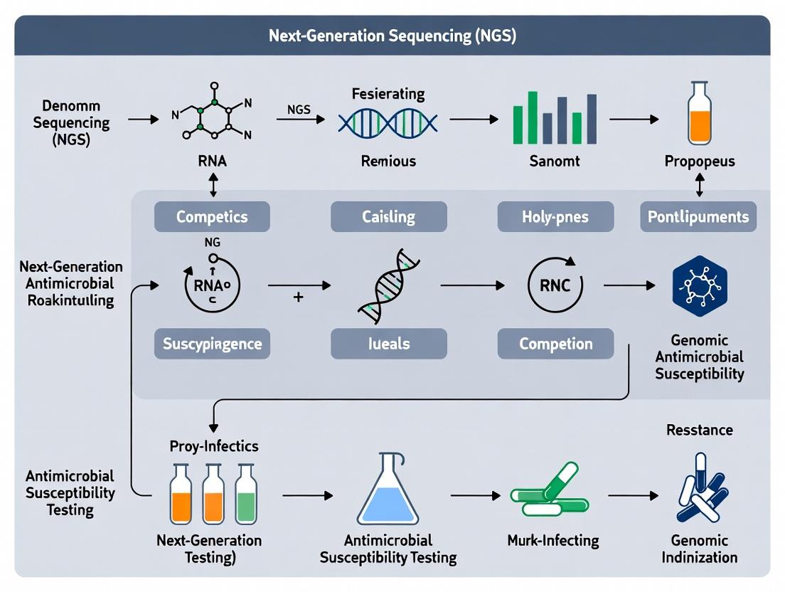
From Reads to Resistance: A Comprehensive Guide to NGS Workflows for Genomic Antimicrobial Susceptibility Testing (AST)
This article provides a detailed roadmap for implementing Next-Generation Sequencing (NGS) for Genomic Antimicrobial Susceptibility Testing (gAST) in research and drug development.
From Reads to Resistance: A Comprehensive Guide to NGS Workflows for Genomic Antimicrobial Susceptibility Testing (AST)
Abstract
This article provides a detailed roadmap for implementing Next-Generation Sequencing (NGS) for Genomic Antimicrobial Susceptibility Testing (gAST) in research and drug development. We explore the scientific rationale behind predicting resistance from genomic data, outline step-by-step workflows from sample preparation to bioinformatic analysis, address common technical challenges, and critically evaluate performance against phenotypic methods. Designed for researchers and industry professionals, this guide synthesizes current best practices and emerging standards to accelerate the development and validation of rapid, precise resistance profiling tools.
Why Sequence for Susceptibility? The Rationale and Revolution of Genomic AST
Application Notes: Integrating NGS for Genomic Antimicrobial Susceptibility Testing (AST)
The slow turnaround time of culture-based phenotypic AST is a critical bottleneck in the antimicrobial resistance (AMR) crisis, often delaying effective therapy by 48-72 hours. Next-generation sequencing (NGS) offers a paradigm shift by enabling genomic AST (gAST), which predicts resistance from microbial DNA sequences within a single day. This approach directly addresses phenotypic delays by detecting known resistance determinants (genes, mutations) and uncovering novel mechanisms through surveillance.
Table 1: Comparison of Phenotypic AST vs. NGS-based gAST Workflows
| Parameter | Traditional Phenotypic AST | NGS-based Genomic AST (gAST) |
|---|---|---|
| Primary Output | Minimum Inhibitory Concentration (MIC) | Detection of resistance genes & predictive mutations |
| Typical Turnaround Time | 48-72 hours post-culture | 6-24 hours post-positive culture or direct from specimen |
| Key Advantage | Functional, phenotypic result | Speed, comprehensiveness, & epidemiological insights |
| Key Limitation | Time delay; blind to novel mechanisms | Inference-based; requires validated genotype-phenotype databases |
| Throughput | Low to medium (isolate-by-isolate) | High (multiplexed, batch processing) |
| Cost per Isolate | Low | Medium to High, but decreasing |
Detailed Protocol: Targeted NGS Panel for Resistance Gene Detection in Enterobacterales
Objective: To prepare sequencing-ready libraries from bacterial DNA for the detection and characterization of AMR genes in Gram-negative Enterobacterales using an amplicon-based targeted NGS panel.
Materials & Equipment:
- Bacterial genomic DNA (extracted from a pure culture, ≥ 2 ng/µL)
- Research Reagent Solutions Toolkit:
Reagent/Material Function Targeted AMR Panel Primer Pool Amplifies specific regions of pre-defined resistance genes & chromosomal targets. High-Fidelity DNA Polymerase Ensures accurate amplification of target amplicons for sequencing. Library Preparation Beads (SPRI) For size selection and purification of amplicon libraries. Dual-Index Barcode Adapters Uniquely tags each sample for multiplexed sequencing. Library Quantification Kit (qPCR-based) Accurately measures concentration of adapter-ligated fragments for pooling. NGS Sequencing Kit v3 (600-cycle) Provides chemistry for sequencing on a mid-output flow cell.
- Thermal cycler, microcentrifuge, magnetic stand, Qubit fluorometer, real-time PCR system, and compatible NGS sequencer.
Procedure:
- PCR Amplification: In a 50 µL reaction, combine DNA with the primer pool and high-fidelity master mix. Cycle: 98°C for 30s; 25 cycles of (98°C for 10s, 60°C for 30s, 72°C for 30s); 72°C for 5 min.
- Amplicon Purification: Clean up PCR product using SPRI beads at a 0.8x ratio. Elute in 25 µL nuclease-free water.
- Indexing PCR: Add dual-index barcodes via a limited-cycle (8 cycles) PCR. Purify final library with SPRI beads at a 0.9x ratio.
- Library QC & Quantification: Assess library fragment size using a bioanalyzer/tapestation. Perform absolute quantification via qPCR using a library quantification kit.
- Pooling & Normalization: Dilute and pool libraries in equimolar ratios based on qPCR data.
- Sequencing: Denature and dilute the pooled library according to sequencer specifications. Load onto the sequencer flow cell. Use a 2x150 bp paired-end sequencing run.
Visualization of Workflows
Title: NGS gAST vs Phenotypic AST Workflow Comparison
Title: Bioinformatic Pipeline for gAST
Application Notes: Establishing Genotype-Phenotype Correlations for Antimicrobial Resistance
Within a Next-Generation Sequencing (NGS)-based Genomic Antimicrobial Susceptibility Testing (AST) workflow, the core principle of linking specific genetic determinants (genotype) to a predicted resistance profile (phenotype) is foundational. This linkage relies on curated knowledge bases that catalog known resistance mechanisms. The primary application is to translate raw genomic variant data into a clinically actionable AST prediction. Key considerations include:
- Mechanism-Based Interpretation: Predictions are not based solely on gene presence/absence. They depend on identifying specific, known mutations (e.g., single nucleotide polymorphisms (SNPs), insertions/deletions) in target genes (e.g., rpoB for rifampicin, gyrA for fluoroquinolones) that are experimentally proven to confer resistance.
- Thresholds for Expression: For some drugs, resistance requires a combination of mutations or a specific mutation "score" (e.g., multiple penicillin-binding protein alterations in Streptococcus pneumoniae). Bioinformatics pipelines must integrate these complex rules.
- Distinguishing Colonization from Infection: The detection of a resistance gene does not inherently define the infection-causing strain, highlighting the need for pure culture or sufficient pathogen reads in direct-from-specimen sequencing.
Table 1: Key Genotype-to-Phenotype Correlations in Bacterial AST
| Pathogen | Antimicrobial Class | Target Gene(s) | Key Resistance-Conferring Mutation(s)/Mechanism | Typical Phenotypic Effect (MIC Increase) |
|---|---|---|---|---|
| Mycobacterium tuberculosis | Rifampicins | rpoB | Missense mutations in RRDR (e.g., S450L) | High-level resistance (MIC >1 mg/L) |
| Escherichia coli | Fluoroquinolones | gyrA, parC | S83L, D87N in gyrA; S80I in parC | Stepwise increase; dual mutations lead to high-level resistance |
| Staphylococcus aureus | β-lactams | mecA / mecC | Acquisition of alternative PBP2a encoded by mecA | Conferred resistance to all β-lactams except ceftaroline/ceftobiprole |
| Pseudomonas aeruginosa | Aminoglycosides | Multiple | Acquisition of modifying enzymes (e.g., aac(6')-Ib, aph(3')-IIb) | Variable, from moderate to high-level resistance |
| Klebsiella pneumoniae | Carbapenems | blaKPC, blaNDM, blaOXA-48-like | Plasmid-borne carbapenemase gene acquisition | High-level resistance (MICs often >8 mg/L) |
Experimental Protocols
Protocol 1: Targeted Amplicon Sequencing for rpoB RRDR Mutation Detection in M. tuberculosis
- Objective: Confirm rifampicin resistance by sequencing the Rifampicin Resistance Determining Region (RRDR) of the rpoB gene.
- Materials: Extracted M. tuberculosis genomic DNA, primers for rpoB RRDR amplification, high-fidelity PCR master mix, NGS library preparation kit, sequencing platform (e.g., Illumina MiSeq).
- Method:
- PCR Amplification: Amplify the ~500bp RRDR region using validated primers. Include a no-template control.
- Amplicon Purification: Clean PCR products using magnetic beads to remove primers and dNTPs.
- Library Preparation: Tag amplicons with dual-index barcodes and sequencing adapters using a limited-cycle PCR.
- Pooling & Quantification: Quantify libraries by qPCR, pool equimolar amounts, and denature.
- Sequencing: Load pooled library onto a MiSeq flow cell for 2x250bp paired-end sequencing.
- Bioinformatics: Demultiplex reads, map to rpoB reference (H37Rv), and call variants. Report any non-synonymous mutation within codons 426-452.
Protocol 2: Whole-Genome Sequencing (WGS) and Bioinformatic Pipeline for Comprehensive Resistance Prediction
- Objective: Predict comprehensive AST profile from bacterial isolate WGS data.
- Materials: Pure culture bacterial isolate, DNA extraction kit, DNA shearing system (e.g., ultrasonicator), WGS library prep kit, sequencing platform (e.g., Illumina NextSeq).
- Method:
- DNA Extraction & QC: Extract high-molecular-weight genomic DNA. Quantify using fluorometry.
- Library Preparation: Fragment DNA, end-repair, A-tail, and ligate indexed adapters. Size-select and PCR-amplify the library.
- Sequencing: Sequence to achieve a minimum of 50x coverage (e.g., 2x150bp on NextSeq).
- Bioinformatics Analysis:
- Quality Control: Assess read quality (FastQC), trim adapters (Trimmomatic).
- Assembly & Annotation: De novo assemble reads (SPAdes) and/or map to reference (BWA, SAMtools). Annotate using PROKKA.
- Resistance Gene Detection: Screen against curated databases (e.g., CARD, ResFinder, NCBI AMRFinderPlus) using ABRicate.
- Variant Calling: For chromosomal targets (e.g., gyrA, parC), call SNPs using Snippy/Bcftools against a susceptible reference genome.
- Interpretation: Integrate results using a rule-based system (e.g., point mutations in gyrA/parC + known ESBL gene = predict fluoroquinolone & 3rd-gen cephalosporin resistance).
The Scientist's Toolkit: Key Research Reagent Solutions
| Item | Function in NGS-AST Workflow |
|---|---|
| High-Fidelity Polymerase (e.g., Q5, Phusion) | Ensures accurate amplification of target genes (like rpoB) prior to sequencing, minimizing PCR-induced errors. |
| Magnetic Bead-based Cleanup Kits (e.g., AMPure XP) | For consistent purification and size-selection of DNA fragments post-amplification and post-ligation during library prep. |
| Dual-Indexed UMI Adapter Kits | Allows multiplexing of samples and incorporation of Unique Molecular Identifiers (UMIs) to correct for sequencing errors and PCR duplicates. |
| Hybridization Capture Probes (e.g., for respiratory panel) | Enables targeted enrichment of pathogen DNA (and associated resistance genes) from complex samples (e.g., sputum) for direct sequencing. |
| Quantitative PCR (qPCR) Library Quantification Kit | Provides accurate molar concentration of final NGS libraries for optimal pooling and cluster density on the flow cell. |
| Curated AMR Database (e.g., CARD, ResFinder) | Essential bioinformatics resource linking known resistance genes/mutations to associated antibiotics and resistance levels. |
Diagrams
NGS-AST Workflow: From Sample to Prediction
Mechanism of Target-Based Resistance
Application Notes
Within the thesis framework of Next-Generation Sequencing (NGS) for Genomic Antimicrobial Susceptibility Testing (AST) workflows, the integration of genomic data into public health and pharmaceutical pipelines is transformative. The primary applications are operationalized as follows:
1. Genomic Surveillance for Antimicrobial Resistance (AMR): Continuous, systematic collection and analysis of WGS data from clinical, agricultural, and environmental isolates to track the emergence, distribution, and temporal trends of AMR genes and mutations. This provides a real-time map of resistance landscape, informing empirical therapy and infection prevention policies.
2. High-Resolution Outbreak Investigation: Utilization of whole-genome sequencing (WGS) to achieve strain-level discrimination. Single Nucleotide Polymorphism (SNP) analysis or core-genome Multilocus Sequence Typing (cgMLST) enables precise tracing of transmission pathways, distinguishing between outbreak-related cases and sporadic infections, and identifying potential point sources.
3. Guiding Novel Drug Discovery and Development: In silico mining of bacterial pangenomes and resistomes to identify novel, conserved targets essential for viability or resistance. Functional genomics (e.g., CRISPRi screening) validates targets. NGS also tracks in vitro and in vivo evolution of resistance against lead compounds, guiding medicinal chemistry efforts to overcome resistance.
Table 1: Quantitative Impact of NGS-Based Applications
| Application | Key Metric | Typical Data/Outcome | Impact |
|---|---|---|---|
| Surveillance | Prevalence of key resistance genes | mcr-1 prevalence in E. coli: <1% in EU (2022), 5-15% in some Asian regions (2023) | Informs national formularies and treatment guidelines |
| Outbreak Investigation | Genetic relatedness threshold | ≤5 SNPs for recent, direct transmission in M. tuberculosis | Enables precise containment measures; reduces nosocomial rates by ~20% |
| Drug Discovery | Target essentiality & conservation | 10-15% of essential genes are highly conserved across Enterobacteriaceae | Prioritizes targets with low risk of natural resistance and broad-spectrum potential |
Experimental Protocols
Protocol 1: NGS-Based Outbreak Investigation Pipeline
Objective: To confirm and delineate a suspected nosocomial outbreak using WGS.
Materials: Bacterial isolates (case and background controls), DNA extraction kit, Qubit fluorometer, Illumina DNA Prep kit, MiSeq sequencer, bioinformatics servers.
Procedure:
- Isolate Selection: Select all epidemiologically suspected isolates. Include 5-10 contemporaneous but epidemiologically unrelated isolates of the same species as background controls.
- Genomic DNA Extraction: Use a standardized mechanical lysis and column-based kit. Elute in 50 µL nuclease-free water. Assess concentration (Qubit) and purity (A260/A280 ~1.8-2.0).
- Library Preparation & Sequencing: Use the Illumina DNA Prep kit for tagmentation-based library prep. Normalize libraries to 4 nM and pool. Sequence on a MiSeq system using a 2x150 bp v3 reagent kit, targeting >50x coverage.
- Bioinformatics Analysis:
- Quality Control: Use FastQC. Trim adapters and low-quality bases with Trimmomatic.
- Assembly: De novo assemble reads using SPAdes. Assess assembly quality with QUAST.
- Core Genome Alignment: Identify core-genome SNPs using Snippy against a reference genome (e.g., E. coli K-12 MG1655).
- Phylogenetic Analysis: Build a maximum-likelihood tree from the core SNP alignment using RAxML. Visualize with FigTree.
- Interpretation: Isolates clustered within ≤5 SNPs are considered part of the same transmission chain. Integrate with epidemiological data to confirm the outbreak.
Protocol 2: In vitro Resistance Evolution Experiment for Drug Discovery
Objective: To predict and characterize resistance mechanisms against a novel antibiotic candidate.
Materials: Novel antibiotic compound, cation-adjusted Mueller Hinton broth (CAMHB), 96-well microtiter plates, shaking incubator.
Procedure:
- Serial Passage: Prepare CAMHB with the compound at 0.25x, 0.5x, 1x, and 2x the MIC. Inoculate each with ~5x10^5 CFU/mL of the target pathogen. Incubate for 18-24h at 37°C.
- Selection: Sub-culture the well showing growth at the highest antibiotic concentration into fresh medium with incrementally increased compound concentrations (e.g., 2x, 4x, 8x the original MIC).
- Harvesting: Repeat step 2 for 20-30 passages. Harvest evolved isolates showing ≥8-fold increase in MIC.
- Whole-Genome Sequencing: Sequence the ancestral and evolved isolates (see Protocol 1, steps 2-4).
- Variant Analysis: Map reads of evolved isolates to the ancestral genome using BWA. Call variants (SNPs, indels) using GATK. Annotate variants to identify mutations in genes related to drug target, efflux pumps, or cell wall biosynthesis.
- Validation: Clone mutated genes into a clean genetic background to confirm their role in resistance.
Visualizations
NGS Workflow for Key AMR Applications
Genomic Outbreak Analysis Protocol
The Scientist's Toolkit: Research Reagent Solutions
| Item | Function in NGS-AST Workflow |
|---|---|
| Magnetic Bead-Based DNA Cleanup Kits (e.g., AMPure XP) | Size-selects and purifies fragmented DNA post-tagmentation or PCR, critical for high-quality library prep. |
| Fragmentase/Nextera Transposase Enzymes | Simultaneously fragments and tags genomic DNA with sequencing adapters in a single, rapid reaction. |
| Unique Dual Index (UDI) Oligos | Provides unique barcodes for both ends of each DNA fragment, enabling accurate sample multiplexing and eliminating index hopping errors. |
| Whole-Cell Lysis & Stabilization Buffers | Allows safe transport and storage of samples at room temperature, inactivating pathogens while preserving DNA for sequencing. |
| Synthetic Spike-in Control DNA | Contains known resistance genes at defined concentrations; added to samples to monitor sequencing efficiency, sensitivity, and limit of detection. |
| qPCR Library Quantification Kits (e.g., with SYBR Green) | Accurately measures the concentration of adapter-ligated DNA fragments, ensuring optimal loading on the sequencer. |
| Validated, Curated AMR Gene Databases (e.g., CARD, ResFinder, AMRFinderPlus) | Bioinformatics repositories used with tools like ABRicate or ARIBA to annotate resistance determinants from WGS data. |
Application Notes
The integration of Next-Generation Sequencing (NGS) into genomic antimicrobial susceptibility testing (AST) workflows represents a paradigm shift, moving beyond traditional culture-based and targeted molecular methods. This approach leverages the core advantages of NGS—comprehensiveness, speed, and the ability to detect novel mutations—to predict phenotypic resistance directly from genomic data. The following notes detail the application of these advantages within a research context aimed at developing robust clinical workflows.
Comprehensiveness: Whole-genome sequencing (WGS) provides an unbiased survey of all resistance determinants in a single assay. Unlike PCR panels, which target a predefined set of known genes, NGS can simultaneously identify:
- Known antimicrobial resistance genes (ARGs) from curated databases (e.g., CARD, ResFinder).
- Chromosomal point mutations in housekeeping genes (e.g., rpoB for rifampicin, gyrA/parC for fluoroquinolones).
- Gene overexpression mechanisms via promoter mutations.
- Insertions/deletions affecting regulatory regions. This comprehensive snapshot is critical for understanding complex, multi-drug resistant (MDR) phenotypes and for outbreak surveillance where strain-tracking (via core-genome MLST) is performed concurrently.
Speed: While traditional culture-based AST requires 24-48 hours post-isolation, NGS-based predictive AST can generate results in a single day. The key accelerant is the direct sequencing from primary samples or positive blood cultures, bypassing the need for sub-culture and pure isolate growth. Advances in library preparation (e.g., transposase-based "tagmentation") and sequencing chemistry (e.g., Illumina NovaSeq X, Oxford Nanopore Technologies PromethIon) have reduced hands-on time and increased throughput. Rapid sequencing platforms like Oxford Nanopore can provide ARG profiles in as little as 1-4 hours, enabling near-real-time resistance prediction.
Detection of Novel Mutations: This is a unique and powerful advantage for research and surveillance. NGS enables the discovery of previously uncharacterized resistance mechanisms by correlating genomic variants with phenotypic resistance profiles in collections of clinical isolates. Comparative genomics of susceptible vs. resistant isolates can reveal:
- Non-synonymous SNPs in genes not previously linked to resistance.
- Gene amplifications.
- Structural variations (inversions, translocations) activating resistance genes. These findings continuously expand the databases used for prediction, improving the accuracy of future assays and informing basic research on drug-target interactions.
The following table summarizes key performance metrics of NGS-AST compared to traditional methods:
| Metric | Traditional Culture AST | Targeted PCR Panel | NGS-Based Predictive AST |
|---|---|---|---|
| Turnaround Time (Post-Isolation) | 18-48 hours | 2-6 hours | 6-24 hours (from isolate) |
| Number of Simultaneous Targets | Limited by panel design | 10-100 known targets | All genes in genome (1000s of potential targets) |
| Novel Variant Discovery | No | No | Yes |
| Strain Typing Correlation | Requires separate test | No | Yes, integrated |
| Primary Sample Feasibility | Low (requires growth) | Moderate (requires known target) | High (metagenomic) |
| Cost per Isolate (Reagent Approx.) | $10-$50 | $50-$150 | $50-$200 (decreasing) |
Experimental Protocols
Protocol 1: Comprehensive ARG & Mutation Detection from Bacterial Isolates
Objective: To extract genomic DNA, perform WGS, and bioinformatically identify known and novel antimicrobial resistance determinants.
Materials: (See "Scientist's Toolkit" for details)
- Pure bacterial culture (>1 McFarland standard).
- Genomic DNA extraction kit (e.g., DNeasy Blood & Tissue Kit).
- DNA quantification instrument (Qubit fluorometer).
- Library Prep Kit (e.g., Illumina DNA Prep).
- Sequencing platform (e.g., Illumina NextSeq 2000).
- High-performance computing cluster.
Methodology:
- DNA Extraction: Follow manufacturer's protocol for Gram-positive or Gram-negative bacteria. Include optional lysozyme/lysostaphin step for tough Gram-positive cells. Elute in 50-100 µL of EB buffer.
- QC & Quantification: Assess DNA purity (A260/A280 ~1.8-2.0) via spectrophotometry. Precisely quantify double-stranded DNA using a fluorometric method (e.g., Qubit dsDNA HS Assay). Minimum requirement: 20 ng/µL in 50 µL.
- Library Preparation: Using 50 ng of input gDNA, perform tagmentation, adapter ligation, and PCR amplification (8-10 cycles) per the Illumina DNA Prep protocol. Clean up with SPB beads.
- Library QC: Assess fragment size distribution using a Bioanalyzer or TapeStation (expected peak: ~550 bp). Quantify final library via qPCR (KAPA Library Quant Kit) for accurate pooling.
- Sequencing: Pool libraries and sequence on a NextSeq 2000 P2 flow cell (100bp paired-end), targeting >50x coverage for most bacterial genomes (~2-5 M reads/isolate).
- Bioinformatic Analysis:
- Quality Control: Use FastQC and Trimmomatic to assess and trim adapter/low-quality bases.
- Assembly & Annotation: De novo assemble reads using SPAdes. Annotate contigs with Prokka.
- ARG Detection: Screen assembled genome against the Comprehensive Antibiotic Resistance Database (CARD) using RGI (Resistance Gene Identifier) in "perfect and strict" mode. Simultaneously, run ARIBA against ResFinder and PointFinder databases to detect known genes and chromosomal mutations.
- Variant Calling for Novel Mutations: Map quality-trimmed reads to a reference genome (e.g., E. coli MG1655) using BWA-MEM. Call variants with BCftools mpileup/call. Filter variants (depth >10, QUAL >30) and annotate using SnpEff.
Protocol 2: Direct Metagenomic Sequencing from Positive Blood Cultures for Rapid AST Prediction
Objective: To rapidly predict resistance from clinical samples without culture isolation, emphasizing speed and comprehensiveness.
Materials:
- Positive blood culture bottle (BacT/ALERT, BACTEC).
- Host DNA depletion kit (e.g., MolYsis Basic5).
- Rapid DNA extraction kit (e.g., QIAamp DNA Micro Kit).
- Rapid library prep kit (e.g., Oxford Nanopore Rapid Barcoding Kit 96).
- Oxford Nanopore MinION or GridION sequencer.
- Real-time analysis compute device (e.g., MinIT, GPU-enabled laptop).
Methodology:
- Sample Processing: Aseptically withdraw 1-2 mL from the positive blood culture bottle.
- Host & Background Depletion: Use the MolYsis protocol: lyse human blood cells with a proprietary buffer, degrade released human DNA with DNase, then lyse bacterial cells to release microbial DNA. Centrifuge to pellet debris.
- DNA Clean-up: Purify the supernatant containing bacterial DNA using the QIAamp Micro Kit. Elute in 25 µL.
- Rapid Library Prep: Dilute DNA to 100 ng in 10 µL. Use the Rapid Barcoding Kit: add rapid barcode, incubate at 30°C for 2 min and 80°C for 2 min, then add rapid adapter and load onto the flow cell within 10 minutes.
- Sequencing & Real-Time Analysis: Load the library onto a MinION R10.4.1 flow cell and start a 24-hour run. Initiate real-time basecalling (Guppy).
- Live ARG Profiling: Stream basecalled reads (FASTQ) to the EPI2ME "What's In My Pot?" (WIMP) workflow for taxonomic classification. Simultaneously, pipe reads to the ARGpore pipeline, which aligns reads in real-time to the CARD database using Minimap2. Generate a dynamic report of detected resistance genes and their relative abundance. Aim for ~50,000 reads for preliminary resistance prediction, typically achievable within 1-2 hours of sequencing.
The Scientist's Toolkit
| Research Reagent / Material | Function in NGS-AST Workflow |
|---|---|
| DNeasy Blood & Tissue Kit (QIAGEN) | Silica-membrane based purification of high-quality, inhibitor-free genomic DNA from bacterial isolates. |
| MolYsis Basic5 (Molzym) | Selectively lyses eukaryotic cells and degrades their DNA, enriching prokaryotic DNA from mixed samples like blood. |
| Illumina DNA Prep Tagmentation Kit | Enzymatically fragments DNA and adds Illumina sequencing adapters in a single, streamlined protocol for library construction. |
| Oxford Nanopore Rapid Barcoding Kit 96 | Ultra-fast (5-10 min) library prep using a transposase-based barcoding approach, critical for same-day turnaround. |
| Qubit dsDNA HS Assay Kit (Thermo Fisher) | Highly specific fluorescent quantification of double-stranded DNA, critical for accurate library input normalization. |
| KAPA Library Quantification Kit (Roche) | qPCR-based absolute quantification of amplifiable library fragments for precise pooling and optimal sequencing cluster density. |
| R10.4.1 Flow Cell (Oxford Nanopore) | Nanopore flow cell with a revised protein pore architecture that provides dramatically improved raw accuracy (>99%) for SNP detection. |
| Comprehensive Antibiotic Resistance Database (CARD) | A curated, ontology-driven resource containing ARG sequences, SNPs, and associated metadata, essential for bioinformatic prediction. |
Diagrams
NGS-AST Research Workflow
Comprehensive Resistance Detection Logic
1. Introduction Within the research framework of Next-Generation Sequencing (NGS) for genomic Antimicrobial Susceptibility Testing (AST), a critical challenge is the predictive gap for complex or entirely undiscovered resistance mechanisms. While NGS excels at identifying known resistance determinants, its predictive power is limited by phenotypic plasticity, epistatic interactions, and novel genetic contexts. This application note details protocols and considerations for addressing these limitations.
2. Key Limitations in Predictive Genomic AST Table 1: Classes of Resistance Difficult to Predict from Genomic Data Alone
| Limitation Class | Description | Impact on Predictive AST |
|---|---|---|
| Undiscovered Genes/SNPs | Novel resistance determinants not present in reference databases. | False susceptible calls; incomplete resistance profiling. |
| Gene Expression & Regulation | Resistance conferred by variable expression (e.g., efflux pump upregulation, porin downregulation) without coding sequence mutation. | Discordance between genotype (no mutation) and phenotype (resistant). |
| Epistasis & Genetic Context | The phenotypic effect of a mutation depends on the presence/absence of other genetic variants (e.g., compensatory mutations). | Variable MIC outcomes from identical resistance alleles in different strains. |
| Cryptic Resistance | Resistance genes that are silent under standard lab conditions but can be induced in host or under specific stresses. | Underestimation of resistance potential. |
| Complex Multi-Gene Traits | Resistance requiring the cumulative effect of many small-effect loci (e.g., low-level, adaptive resistance). | Polygenic scores often lack the precision for clinical prediction. |
3. Experimental Protocols for Investigating Predictive Gaps
Protocol 3.1: Phenotype-Genotype Correlation for Anomalous Isolates Objective: To identify genetic basis for resistance in isolates where WGS fails to predict observed phenotype. Materials: Bacterial isolate with discrepant genotype-phenotype, LB broth & agar, appropriate antibiotics, DNA extraction kit, PCR reagents, NGS library prep kit, sequencer. Procedure:
- Confirm Phenotype: Perform repeat MIC assay (e.g., broth microdilution per CLSI/EUCAST) for the antibiotic in question.
- Deep Sequencing: Extract genomic DNA. Prepare and sequence paired-end libraries (2x150bp) on an Illumina platform to high coverage (>100x).
- Comprehensive Genomic Analysis: a. De novo Assembly: Assemble reads using SPAdes or Unicycler. Assess quality with QUAST. b. Resistance Gene Screening: Analyze against curated databases (CARD, ResFinder, NDARO) using ABRicate. c. Variant Analysis: Map reads to a reference genome (e.g., E. coli MG1655). Call SNPs/indels with Snippy. Annotate variants. d. Context Analysis: Examine genomic region surrounding any candidate resistance gene for promoters, insertional sequences, or gene truncations using Artemis or BRIG.
- Functional Validation: Clone candidate genes/regions into a susceptible background via plasmid vector or allelic exchange. Re-test MIC of transformants.
Protocol 3.2: Functional Metagenomics for Unculturable/Undiscovered Resistome Objective: To capture novel resistance genes from complex microbial samples (e.g., gut microbiome, environmental). Materials: Environmental or fecal sample, metagenomic DNA extraction kit, copy-control fosmid or cosmid vector (e.g., pCC1FOS), E. coli EPI300 host, LB agar with antibiotic and copy-control inducer. Procedure:
- Extract Metagenomic DNA: Isolate high-molecular-weight DNA from sample.
- Library Construction: Partially digest DNA, size-select fragments (30-40 kb). Ligate into fosmid vector. Package using phage packaging extract.
- Transformation & Selection: Transfect E. coli EPI300. Plate on LB agar containing chloramphenicol (vector marker) and the antibiotic of interest (e.g., meropenem). Include control plate with inducer (e.g., arabinose) for copy-number amplification.
- Sequence Resistance-Conferring Clones: Isolate fosmid DNA from resistant colonies. Perform long-read sequencing (PacBio/Oxford Nanopore) of the insert.
- Bioinformatic Analysis: Annotate open reading frames. Compare to known protein databases (BLASTP, HMMER) to identify homology to known resistance families or novel protein families.
4. Visualizing the Analysis Workflow for Complex Resistance
Title: Analysis Path for Genotype-Phenotype Discrepancy
5. The Scientist's Toolkit: Key Research Reagent Solutions Table 2: Essential Materials for Investigating Complex Resistance
| Item | Function in Research |
|---|---|
| Copy-Control Fosmid Vectors (e.g., pCC1FOS) | Maintains large (30-45 kb) environmental DNA inserts at single copy to avoid toxicity, inducible to high copy for expression screening. |
| EPI300 E. coli Strain | RecA- host for fosmid propagation, engineered for high transformation efficiency and induced copy number control. |
| Broad-Host-Range Cloning Vectors (e.g., pUCP24) | Allows expression of candidate genes in diverse Gram-negative bacterial backgrounds for functional validation. |
| CRISPR-Cas9 Allelic Exchange Systems | Enables precise deletion or insertion of putative regulatory elements (promoters, SNPs) in the native genomic context. |
| Polar Transposon Mutagenesis Kit (e.g., Tn5) | For genome-wide identification of genes contributing to low-level or adaptive resistance phenotypes. |
| Real-Time PCR Assays for efflux pump/porin genes | Quantifies expression changes of regulatory networks that are not encoded in the primary DNA sequence. |
| Curated AMR Database (e.g., CARD with RGI) | Provides comprehensive reference of known resistance mechanisms for genotype screening and homology detection. |
Blueprint for Success: A Step-by-Step NGS gAST Laboratory and Bioinformatics Pipeline
Within the research framework of a Next-Generation Sequencing (NGS) workflow for genomic Antimicrobial Susceptibility Testing (gAST), sample preparation and DNA extraction constitute the critical foundational step. The quality and integrity of the nucleic acid template directly dictate the accuracy of subsequent sequencing, variant calling, and resistance genotype prediction. This protocol outlines detailed considerations and methodologies to ensure the recovery of high-quality, inhibitor-free microbial DNA from complex clinical specimens, suitable for whole-genome sequencing (WGS)-based AST.
Key Considerations for Sample Preparation
Sample Type and Input
The choice of protocol is heavily influenced by the sample matrix, which impacts pathogen biomass, host DNA contamination, and the presence of PCR inhibitors.
| Sample Type | Typical Pathogen Load (CFU/mL) | Major Challenges | Recommended Minimum Input for gAST |
|---|---|---|---|
| Pure Bacterial Colony | 10^8 - 10^9 | Minimal; primarily lysis efficiency | 1-5 colonies |
| Positive Blood Culture Broth | 10^7 - 10^9 | Host blood cells, charcoal, resin beads | 0.5 - 1 mL broth |
| Sputum | Variable (10^6 - 10^9) | Viscous mucin, host cells, diverse flora | 0.5 - 1 mL (post-digestion) |
| Urine | Variable (10^3 - 10^7) | Low biomass, urea, salts | 1-10 mL (after centrifugation) |
| Swab (e.g., wound) | Variable | Low biomass, swab material inhibitors | Swab eluted in 1 mL buffer |
Host DNA Depletion
For samples with significant human cell contamination (e.g., sputum, blood culture), host DNA depletion is essential to increase the microbial sequencing depth.
Protocol: Selective Lysis for Blood Culture Samples
- Reagents: Lysis Buffer (0.1% Saponin, 0.5% Triton X-100 in TE buffer), DNase I (optional for host DNA digestion).
- Procedure:
- Transfer 1 mL of positive blood culture broth to a microcentrifuge tube.
- Add 2 mL of selective lysis buffer. Vortex for 10 seconds.
- Incubate at room temperature for 10 minutes to lyse human blood cells.
- Centrifuge at 12,000 x g for 5 minutes to pellet intact microbial cells.
- Carefully discard supernatant. Wash pellet with 1 mL of sterile phosphate-buffered saline (PBS).
- Proceed to microbial DNA extraction.
Inhibitor Removal
Clinical samples contain substances that inhibit downstream enzymatic reactions (PCR, sequencing).
| Common Inhibitor | Source | Mitigation Strategy |
|---|---|---|
| Hemoglobin/Heme | Blood | Use inhibitor-removal columns; add bovine serum albumin (BSA) to PCR. |
| Humic Acids | Sputum, tissue | Modified CTAB extraction; commercial clean-up kits. |
| Urea & Salts | Urine | Extensive washing with PBS or TE buffer. |
| Melanin | Swabs | Pre-treatment with polyvinylpyrrolidone (PVP). |
Detailed DNA Extraction Protocols
Protocol A: Magnetic Bead-Based Extraction for Pure Cultures and Processed Samples
This scalable method yields high-purity DNA suitable for library preparation.
Materials (Research Reagent Solutions):
- Lysis Buffer (Guanidine Hydrochloride-based): Disrupts cell membranes and denatures proteins.
- Proteinase K (20 mg/mL): Digests nucleases and structural proteins.
- Magnetic Silica Beads: Bind DNA under high-salt conditions.
- Wash Buffer 1 (High Salt): Removes contaminants while retaining DNA on beads.
- Wash Buffer 2 (Ethanol-based): Removes residual salts and organics.
- Nuclease-free Water (Elution Buffer): Low-ionic-strength solution to elute pure DNA.
Procedure:
- Cell Lysis: Resuspend pelleted microbial cells in 200 µL of lysis buffer. Add 20 µL of Proteinase K. Mix thoroughly.
- Incubation: Incubate at 56°C for 30 minutes with intermittent vortexing. For Gram-positive bacteria, add 20 µL of lysozyme (50 mg/mL) and pre-incubate at 37°C for 30 minutes prior to step 1.
- Binding: Add 200 µL of binding buffer (provided in kit) and 50 µL of magnetic bead suspension. Mix and incubate at room temperature for 10 minutes.
- Capture: Place tube on a magnetic rack for 2 minutes. Carefully discard supernatant.
- Washing: Remove from magnet. Add 500 µL Wash Buffer 1. Resuspend beads fully. Capture on magnet. Discard supernatant. Repeat with 500 µL Wash Buffer 2. Air-dry beads for 5-10 minutes.
- Elution: Remove from magnet. Add 50-100 µL nuclease-free water. Resuspend beads and incubate at 55°C for 5 minutes. Capture beads and transfer eluted DNA to a clean tube.
- Quality Control: Quantify using a fluorometric method (e.g., Qubit). Assess purity via A260/A280 (target: 1.8-2.0) and A260/A230 (target: >2.0). Check integrity by agarose gel electrophoresis.
Protocol B: Column-Based Extraction with Inhibitor Removal for Challinical Samples
Ideal for sputum, stool, or tissue where inhibitors are prevalent.
Procedure:
- Pre-treatment: For sputum, add equal volume of 1% DTT (Dithiothreitol) and incubate at 37°C for 15 minutes to liquefy.
- Lysis: Transfer 200 µL of processed sample to a tube with 200 µL of lysis buffer and Proteinase K. Vortex vigorously.
- Inhibitor Removal: Add 100 µL of inhibitor removal resin. Vortex for 30 seconds. Centrifuge at 12,000 x g for 2 minutes.
- Column Binding: Transfer supernatant to a silica spin column. Centrifuge at 10,000 x g for 1 minute.
- Washing: Add 500 µL of wash buffer 1. Centrifuge. Discard flow-through. Add 500 µL of wash buffer 2 (ethanol-based). Centrifuge. Dry column with an additional spin.
- Elution: Place column in a clean tube. Apply 50-100 µL of pre-heated (70°C) elution buffer to the membrane. Incubate for 2 minutes. Centrifuge to elute.
The Scientist's Toolkit: Key Reagent Solutions
| Item | Function in gAST Workflow |
|---|---|
| Lysis Buffer (Guanidine HCl/Detergent) | Chaotropic agent that disrupts cell membranes, inactivates nucleases, and promotes DNA binding to silica. |
| Proteinase K | Broad-spectrum serine protease that degrades proteins and aids in the removal of histone contaminants. |
| Lysozyme (for Gram-positives) | Enzyme that hydrolyzes peptidoglycan in the bacterial cell wall, enabling access of lysis buffers. |
| Magnetic Silica Beads | Paramagnetic particles providing a solid-phase for DNA purification, enabling automation and high yield. |
| Inhibitor Removal Technology (e.g., resins) | Selectively binds humic acids, polyphenols, and other common PCR inhibitors from complex samples. |
| DNase I (RNase-free) | Used in host depletion protocols to digest free human genomic DNA after selective lysis of eukaryotic cells. |
| Dithiothreitol (DTT) | Reducing agent that breaks disulfide bonds in mucin, liquefying sputum for efficient pathogen recovery. |
| Fluorometric DNA Quantification Kit | Enables accurate, dye-based quantification of double-stranded DNA, unaffected by RNA or contaminants. |
Quality Control Metrics and Thresholds
| QC Parameter | Method | Target for gAST-WGS | Impact of Failure |
|---|---|---|---|
| DNA Yield | Fluorometry (Qubit) | >10 ng (Minimum for library prep) | Insufficient library complexity. |
| Purity (A260/A280) | Spectrophotometry (NanoDrop) | 1.8 - 2.0 | Protein/phenol contamination inhibits enzymes. |
| Purity (A260/A230) | Spectrophotometry (NanoDrop) | >2.0 | Salt/carbohydrate carryover inhibits PCR. |
| Integrity | Agarose Gel / Fragment Analyzer | Clear high-molecular-weight band (>20 kb) | Fragmented DNA leads to poor library assembly. |
| Inhibitor Presence | qPCR Inhibition Assay (Spike-in) | Cq shift < 2 cycles | Failed amplification during library enrichment. |
Diagrams
Diagram 1: gAST Sample Prep Decision Workflow
Diagram 2: DNA Extraction Core Steps
Diagram 3: Inhibitor Impact & Mitigation Pathway
Within the broader thesis on Next-Generation Sequencing (NGS) for genomic Antimicrobial Susceptibility Testing (gAST), library preparation is the critical step that determines the scope and resolution of genetic data available for predicting antimicrobial resistance (AMR). The choice between Whole-Genome Sequencing (WGS) and Targeted Amplicon Sequencing (TAS) dictates the balance between comprehensive discovery of resistance mechanisms and sensitive, cost-effective detection of known variants. This decision directly impacts the downstream analysis's ability to correlate genotype with phenotype in clinical and research settings.
Comparative Analysis: WGS vs. TAS for gAST
The selection between WGS and TAS is guided by specific research goals, available resources, and the required depth of analysis. The following table summarizes the key comparative parameters.
Table 1: Comparison of WGS and TAS for gAST Applications
| Parameter | Whole-Genome Sequencing (WGS) | Targeted Amplicon Sequencing (TAS) |
|---|---|---|
| Primary Goal | Unbiased, comprehensive profiling of entire genome. | Highly sensitive detection of known AMR loci/alleles. |
| Target Region | Entire microbial genome (typically 2-10 Mbp for bacteria). | Specific, pre-defined AMR genes, promoters, or SNPs (e.g., 10-200 bp amplicons). |
| Library Prep Time | ~4-8 hours (varies by kit). | ~3-6 hours (including initial PCR). |
| Typical Input DNA | 1-100 ng (high quality). | As low as 1 pg - 10 ng (can tolerate some degradation). |
| Multiplexing Capacity | High (96+ samples via dual indexing). | Very High (100s-1000s of samples via sample-specific primers). |
| Sequencing Depth Required | 50x - 100x coverage for reliable variant calling. | >500x - 10,000x for low-frequency variant detection. |
| Key Advantage for gAST | Discovery of novel resistance mutations, plasmids, and horizontal gene transfer events; strain typing. | Extreme sensitivity for minority populations (heteroresistance); low cost per sample for high-throughput. |
| Main Limitation for gAST | Higher cost per sample; data analysis complexity; lower sensitivity for rare variants unless deeply sequenced. | Limited to known targets; cannot detect novel resistance mechanisms outside amplicon regions. |
| Best Suited For | Research into unknown resistance mechanisms, outbreak surveillance, comprehensive isolate characterization. | High-throughput screening of clinical isolates for a defined panel of AMR markers, detecting heteroresistance. |
Table 2: Quantitative Cost & Data Output Comparison (Per Sample Estimates)
| Component | Whole-Genome Sequencing | Targeted Amplicon Sequencing |
|---|---|---|
| Library Prep Reagent Cost | $50 - $150 | $10 - $30 |
| Sequencing Cost (to achieve recommended depth) | $100 - $300 (30-50x on NovaSeq/HiSeq) | $5 - $20 (10,000x on MiSeq) |
| Average Data Output (per sample) | 1 - 5 Gbp | 0.1 - 0.5 Mbp (per target) |
| Bioinformatics Data Storage Need | High (GBs per sample) | Low (MBs per sample) |
Detailed Experimental Protocols
Protocol 3.1: Illumina DNA Prep for Whole-Genome Sequencing (gAST Workflow)
Based on Illumina DNA Prep (formerly Nextera Flex) methodology for bacterial genomes.
Materials: Illumina DNA Prep Kit, IDT for Illumina DNA/RNA UD Indexes, AMPure XP Beads, 80% Ethanol, Qubit dsDNA HS Assay Kit, magnetic stand, thermal cycler. Principle: Utilizes tagmentation to simultaneously fragment and tag genomic DNA with adapter sequences.
- Input DNA Normalization: Dilute high-quality, high-molecular-weight genomic DNA to 20 ng/µL in 10 mM Tris-HCl, pH 8.5. Use 20 ng (1 µL) as input.
- Tagmentation: Combine 1 µL DNA with 5 µL Tagmentation Mix and 4 µL Tagmentation Buffer. Incubate at 55°C for 10 minutes. Immediately add 5 µL Neutralization Buffer and mix. Incubate at room temperature for 5 minutes.
- PCR Amplification & Indexing: Add 15 µL of PCR Mix and 5 µL of a unique, dual-unique (UD) index pair (i5 and i7) to the neutralized tagmentation reaction. Perform PCR: 68°C for 3 min; 98°C for 45 sec; then 12-14 cycles of [98°C for 15 sec, 60°C for 30 sec, 68°C for 60 sec]; final hold at 4°C.
- Clean-up: Add 30 µL of AMPure XP beads (0.6X ratio) to the 30 µL PCR reaction. Purify following standard bead-based protocol. Elute in 25 µL Resuspension Buffer.
- Library QC: Quantify using Qubit dsDNA HS Assay. Assess size distribution (expected peak ~550 bp) via TapeStation or Bioanalyzer High Sensitivity DNA chip.
- Pooling & Sequencing: Normalize libraries based on molarity and pool. Sequence on an Illumina platform (e.g., NextSeq 2000, NovaSeq) to achieve a minimum of 50x average coverage.
Protocol 3.2: Two-Step PCR Amplicon Sequencing for Targeted AMR Detection
Protocol for high-plex detection of known AMR gene variants.
Materials: Primer pools for AMR targets (e.g., ResFinder, CARD database-derived), high-fidelity DNA polymerase (e.g., Q5 Hot Start), dNTPs, AMPure XP Beads, Illumina PCR Indexing Kit (e.g., Nextera XT Index Kit v2), thermal cycler. Principle: Initial PCR enriches specific AMR targets; second PCR adds sample-specific indices and full sequencing adapters.
- Primary (Target) PCR: Design multiplexed primer pools covering critical regions of AMR genes (e.g., blaKPC, mecA, gyrA QRDR). Set up 25 µL reactions: 1X PCR Buffer, 200 µM dNTPs, 0.5 µM primer pool, 0.02 U/µL polymerase, and 1-10 ng genomic DNA. Thermocycling: Initial denaturation 98°C, 30s; 25 cycles of [98°C, 10s; 60-65°C (annealing), 30s; 72°C, 30s]; final extension 72°C, 2 min.
- Primary PCR Clean-up: Pool all primary amplicons from a single sample. Perform a 0.8X SPRI bead clean-up. Elute in 20 µL nuclease-free water.
- Secondary (Indexing) PCR: Use 5 µL of cleaned primary amplicon as template. Perform a limited-cycle (8 cycles) PCR using Illumina index primers (i5 and i7) to add full adapter sequences.
- Final Library Clean-up: Perform a 0.9X SPRI bead clean-up. Elute in 25 µL buffer.
- QC & Normalization: Quantify libraries (Qubit). Verify amplicon sizes (TapeStation). Normalize libraries by molarity.
- Pooling & Sequencing: Pool equal volumes of normalized libraries. Sequence on a MiSeq or iSeq system using a 300-cycle kit (2x150 bp) to achieve ultra-deep coverage (>5,000x per target).
Visualizations
Diagram 1: gAST Workflow Decision Logic for Library Prep
Diagram 2: Comparative Workflow: WGS vs. TAS Library Prep
The Scientist's Toolkit: Key Research Reagent Solutions
Table 3: Essential Materials for NGS Library Preparation in gAST Research
| Item / Solution | Primary Function in gAST Workflow | Example Product(s) |
|---|---|---|
| High-Fidelity DNA Polymerase | Accurate amplification of AMR gene targets in TAS; critical for minimizing PCR errors that could mimic resistance SNPs. | Q5 Hot Start (NEB), KAPA HiFi HotStart ReadyMix (Roche) |
| Tagmentase / Fragmentation Enzyme | Fragments genomic DNA and adds sequencing adapters simultaneously in WGS library preps; ensures unbiased representation. | Illumina Tagmentase (in DNA Prep kits), Nextera Transposase |
| SPRI (Solid Phase Reversible Immobilization) Beads | Size-selective clean-up of DNA fragments post-amplification and adapter ligation; used for purification and size selection in both WGS and TAS. | AMPure XP Beads (Beckman Coulter), SPRIselect (Beckman Coulter) |
| Dual-Indexed UD Index Primers | Allows unique combinatorial indexing of each sample for high-level multiplexing; essential for pooling dozens to hundreds of gAST samples. | IDT for Illumina Nextera UD Indexes, Illumina CD Indexes |
| NGS Library Quantification Kit | Accurate quantification of final library concentration (in nM) for precise pooling and optimal cluster density on the flow cell. | KAPA Library Quantification Kit (Roche), qPCR-based assays |
| Bioanalyzer/TapeStation DNA Kits | Qualitative and semi-quantitative assessment of library fragment size distribution, critical for calculating molarity and checking for adapter dimers. | Agilent High Sensitivity DNA Kit (Bioanalyzer), D1000 ScreenTape (TapeStation) |
| Custom Ampliseq or Primers | Pre-designed primer pools targeting specific AMR gene panels (e.g., for Mycobacterium tuberculosis resistance); enables standardized, reproducible TAS. | Thermo Fisher Ampliseq panels, Custom oligo pools from IDT/Twist |
Within the genomic antimicrobial susceptibility testing (AST) workflow, selecting the appropriate sequencing platform is critical. This step determines the balance between accuracy, read length, cost, and turnaround time, directly impacting the feasibility of rapid, culture-independent AST. This Application Note compares the dominant short-read (Illumina) and long-read (Oxford Nanopore Technologies, ONT) platforms, providing protocols for their implementation in a bacterial whole-genome sequencing (WGS) workflow aimed at predicting resistance genotypes.
Platform Comparison & Quantitative Data
The following tables summarize key performance metrics and suitability for genomic AST.
Table 1: Technical Specifications and Comparative Throughput (Current as of 2024)
| Parameter | Illumina (NovaSeq X Series) | Oxford Nanopore (PromethION 2 Solo) |
|---|---|---|
| Core Technology | Reversible dye-terminator sequencing-by-synthesis | Protein nanopore-based electronic sensing |
| Read Type | Short-read (paired-end) | Long-read (single-pass, continuous) |
| Typical Read Length | 2x150 bp | 10-100+ kb (N50 often >20 kb) |
| Max Output per Flow Cell/Run | 8-16 Tb (NovaSeq X Plus) | 200-300 Gb (PromethION P2 Solo) |
| Accuracy (Raw Read) | >99.9% (Q30+) | ~97-99% (Q20-Q30); improved with duplex |
| Run Time (Standard) | 13-44 hours | 12-72 hours (configurable) |
| Time to First Base | ~6-24 hours | ~10 minutes - 1 hour |
| Capital Cost (Instrument) | Very High | Moderate |
| Cost per Gb (Consumables) | Low ($5-$10) | Moderate-High ($15-$30) |
| Key Strength for AST | High accuracy for SNP/SNV detection, established variant pipelines | Structural variant detection, plasmid assembly, rapid turnaround. |
Table 2: Suitability for Genomic Antimicrobial Susceptibility Testing Applications
| AST Application | Recommended Platform | Rationale |
|---|---|---|
| Comprehensive Resistance Gene Cataloging | Illumina | High accuracy ensures reliable detection of known resistance SNPs and gene alleles. |
| Plasmid & Mobile Genetic Element (MGE) Analysis | Oxford Nanopore | Long reads span repetitive regions and resolve complete plasmid structures, tracking horizontal transfer. |
| Metagenomic Direct-from-Specimen AST | Oxford Nanopore | Rapid time-to-first base enables same-day analysis; long reads improve binning and assembly. |
| High-Throughput Surveillance & Outbreak Typing | Illumina | Superior throughput and lower per-sample cost for processing hundreds of bacterial isolates. |
| Novel Resistance Mechanism Discovery | Hybrid (Both) | Illumina provides accuracy for SNPs; ONT provides context for complex rearrangements and novel insertions. |
Experimental Protocols
Protocol 3.1: Bacterial WGS for AST Using Illumina NovaSeq
Objective: Generate high-accuracy, short-read data from a bacterial isolate for resistance variant calling. Reagents: QIAamp DNA Mini Kit (Qiagen), Qubit dsDNA HS Assay Kit, Illumina DNA Prep kit, IDT for Illumina DNA/RNA UD Indexes, NovaSeq X Series Reagents. Equipment: Thermocycler, Qubit fluorometer, magnetic stand, Agilent TapeStation, NovaSeq X.
- DNA Extraction: Lyse bacterial pellet using enzymatic/mechanical lysis. Purify genomic DNA using the QIAamp kit. Elute in 50 µL TE buffer.
- QC & Quantification: Measure DNA concentration with Qubit HS assay. Assess integrity via TapeStation (DIN >7.0). Dilute to 50 ng/µL in 10mM Tris-HCl.
- Library Preparation (Illumina DNA Prep): a. Tagmentation: Combine 50 ng DNA with ATM and Tagment DNA Buffer. Incubate at 55°C for 10 min. Neutralize with NT Buffer. b. PCR Amplification & Indexing: Add DNA Prep Master Mix and unique dual index primers (UDI). PCR: 68°C for 3 min; 98°C for 45s; then 12 cycles of [98°C for 15s, 60°C for 30s]; final hold at 68°C for 1 min. c. Clean-up: Add Sample Purification Beads, wash twice with 80% EtOH. Elute in 25 µL Resuspension Buffer.
- Library QC: Quantify with Qubit. Analyze fragment size distribution on TapeStation (expected peak: ~450 bp).
- Pooling & Denaturation: Pool equimolar amounts of indexed libraries. Denature with 0.2N NaOH. Dilute to final loading concentration of 200 pM.
- Sequencing: Load onto NovaSeq X flow cell. Use 2x150 bp paired-end chemistry. Run time: ~44 hours.
- Data Analysis (BaseSpace): Use DRAGEN AMR pipeline for simultaneous alignment, variant calling, and resistance gene/SNP identification against CARD/NCBI AMR databases.
Protocol 3.2: Rapid WGS for AST Using ONT PromethION
Objective: Generate long-read data for rapid resistance profiling and plasmid reconstruction. Reagents: Quick-DNA HMW MagBead Kit (Zymo), Qubit dsDNA HS/Broad Range Assay, SQK-LSK114 Ligation Sequencing Kit, Flow Cell Priming Kit, PromethION R10.4.1 flow cell. Equipment: Thermomixer, Hula mixer, magnetic stand, PromethION 2 Solo.
- High Molecular Weight (HMW) DNA Extraction: Resuspend bacterial pellet in MagBinding Bead slurry. Lyse with Proteinase K and GSB buffer. Bind DNA to beads, wash twice. Elute HMW DNA gently in 50 µL EB buffer.
- QC & Quantification: Use Qubit Broad Range for concentration. Assess fragment size via pulsed-field gel electrophoresis or FEMTO Pulse system (>20 kb desired).
- Library Preparation (LSK114, Rapid): a. DNA Repair & End-Prep: Combine 1 µg DNA, NEBNext FFPE DNA Repair Buffer, and Ultra II End-prep enzyme mix. Incubate at 20°C for 5 min, then 65°C for 5 min. Clean with AMPure XP beads (0.4x ratio). b. Native Barcode Ligation: Add Rapid Adapter (RAP) and a unique Native Barcode (EXP-NBDxxx) to eluted DNA. Add Blunt/TA Ligase Master Mix. Incubate at room temperature for 10 min. Clean with AMPure XP beads (0.4x ratio). c. Adapter Ligation: Combine barcoded DNA with Adapter Mix II (AMII) and NEBNext Quick T4 DNA Ligase. Incubate at room temperature for 10 min. d. Clean-up & Elution: Add SQK-LSK114 Bead Binding Buffer, bind to beads, wash twice. Elute in 15 µL Elution Buffer.
- Flow Cell Priming & Loading: Prime PromethION R10.4.1 flow cell with Flush Buffer (FB) and Flush Tether (FT). Load prepared library onto the spot-on port.
- Sequencing: Start a 72-hour sequencing run via MinKNOW software. For rapid AST, analyze data in real-time after 1-2 hours of sequencing.
- Real-Time Analysis (EPI2ME): Use the EPI2ME "What's in my pot?" or ARMA workflow for live species ID and resistance gene detection. For hybrid assembly, basecall with super-accurate (sup) model and assemble with Flye, polishing with Medaka.
Visualization: Sequencing Workflow Decision Pathway
Title: Sequencing Platform Decision Pathway for Genomic AST
The Scientist's Toolkit: Key Research Reagent Solutions
Table 3: Essential Materials for Sequencing-Based AST Workflows
| Item (Supplier) | Function in Workflow | Key Consideration for AST |
|---|---|---|
| QIAamp DNA Microbiome Kit (Qiagen) | Co-extracts host and microbial DNA; critical for direct-from-specimen metagenomics. | Minimizes human DNA background, enriching for bacterial pathogen signal. |
| Nextera XT DNA Library Prep Kit (Illumina) | Rapid, tagmentation-based library prep for low-input isolates. | Fast (90 min) but best for pure isolates; not ideal for complex samples. |
| SQK-RBK114.24 (ONT) | Rapid barcoding kit for multiplexing 24 isolates on one ONT flow cell. | Enables cost-effective, high-throughput long-read sequencing of isolate panels. |
| Qubit dsDNA HS Assay Kit (Thermo Fisher) | Fluorometric quantification of dilute DNA samples. | Essential for accurate input mass pre-library prep; more specific for DNA than spectrophotometry. |
| AMPure XP Beads (Beckman Coulter) | Solid-phase reversible immobilization (SPRI) magnetic beads. | Used for size selection and clean-up in most NGS protocols; ratio determines cutoff. |
| PhiX Control v3 (Illumina) | Sequencing control for run monitoring, focusing, and phasing/pre-phasing calculations. | Crucial for low-diversity libraries (e.g., bacterial genomes) on Illumina platforms. |
| Sequencing Control Kit (ONT, SQK-ACC114) | External positive control (ERC) DNA for monitoring pore performance. | Verifies flow cell functionality before loading precious clinical samples. |
| CARD & NCBI AMR Databases | Curated repositories of resistance genes, variants, and associated phenotypes. | Essential bioinformatics resources for genotype-to-phenotype prediction in analysis pipelines. |
Within a Next-Generation Sequencing (NGS) workflow for Genomic Antimicrobial Susceptibility Testing (AST), the bioinformatics core is the critical bridge that translates raw sequencing data into actionable predictions of antimicrobial resistance (AMR). This phase involves computational processing to identify genetic determinants—Antimicrobial Resistance Genes (ARGs)—and sometimes associated mutations, from microbial genomes. The accuracy and comprehensiveness of this step directly influence the reliability of the phenotypic resistance prediction.
Application Notes: Core Analytical Steps & Considerations
Primary Data Assessment & Quality Control
Initial quality metrics determine downstream analysis success. Current benchmarks (2024) suggest the following thresholds for Illumina short-read data:
Table 1: Key Quality Control Metrics for NGS Data in AMR Analysis
| Metric | Recommended Threshold | Purpose & Rationale |
|---|---|---|
| Q-Score (Phred) | ≥30 (Q30) for >80% of bases | Ensures base call accuracy >99.9%, minimizing false variant calls. |
| Total Reads | ≥50x intended genome coverage | Provides sufficient depth for reliable ARG detection and variant calling. |
| Adapter Content | <5% | High adapter content indicates poor library prep and can interfere with alignment. |
| Per Base Sequence Content | A/T and G/C ratios within 10% after first 10-15 bases | Abnormalities may indicate overrepresented sequences or contamination. |
Protocol 2.1.1: FastQC & MultiQC for Aggregate QC
- Run FastQC on all raw FASTQ files:
fastqc *.fastq -o ./qc_results/. - Consolidate reports using MultiQC:
multiqc ./qc_results/. - Review the
multiqc_report.html. Flag samples failing >2 core metrics (Table 1) for exclusion or reprocessing.
Preprocessing: Trimming & Adapter Removal
Low-quality bases and adapter sequences must be removed to improve mapping accuracy.
Protocol 2.2.1: Trimming with fastp
- Execute fastp with default quality and length filtering, plus adapter auto-detection:
- Post-trimming, verify improved metrics by repeating FastQC on the output files.
Read Alignment & Taxonomic Profiling
For metagenomic samples or pure cultures, aligning reads to reference databases is a primary ARG detection method.
Protocol 2.3.1: Alignment to a Comprehensive AMR Database using KMA KMA (k-mer alignment) offers rapid and accurate mapping to resistance gene databases.
- Index a curated ARG database (e.g., CARD, ResFinder):
Align trimmed reads:
The output file
sample_vs_card.rescontains aligned genes, coverage, and template depth.
ARG Detection & Annotation
This step identifies specific ARG variants and their potential phenotypic correlates.
Table 2: Comparison of Primary ARG Detection Approaches (2024)
| Method | Type | Key Database | Output | Best Use Case |
|---|---|---|---|---|
| Alignment-Based (KMA, BWA) | Reads/Contigs aligned to ARG DB | CARD, ResFinder, MEGARes | Gene identity, coverage, %identity | Targeted, known ARG detection. |
| Hidden Markov Model (HMM) | Protein sequence search | Resfams, PFAM | Protein family membership | Detecting divergent or remote ARG homologs. |
| De Novo Assembly + Screening | Assemble genome, then screen | ARG-ANNOT, NCBI AMRFinderPlus | ARG in genomic context, linkage | Complete genome analysis, plasmid detection. |
Protocol 2.4.1: Comprehensive ARG Detection Pipeline using ABRicate ABRicate wrappers multiple databases for consolidated screening.
- Install ABRicate and associated databases.
- Run screening against multiple databases (using assembled contigs or raw reads):
- Aggregate results:
abricate --summary *.tsv > summary_report.csv. Filter results based on thresholds (e.g., ≥90% coverage, ≥95% identity).
Interpretation & Reporting
The final step translates ARG presence into a structured AST prediction.
Protocol 2.5.1: Generating a Clinical/Research Report
- Curate Findings: Filter ARG hits based on established clinical breakpoints or literature-based rules (e.g., specific blaKPC variants imply carbapenem resistance).
- Contextualize: For assembled data, check ARG location (chromosome vs. plasmid) using tools like mlplasmids or PlasmidFinder.
- Generate Report: Create a table with columns: Antibiotic Class, Detected ARG, Coverage/Identity, Predicted Phenotype (R/S), Confidence Level (High/Medium/Low).
The Scientist's Toolkit: Research Reagent Solutions
Table 3: Essential Bioinformatics Resources for AMR Analysis
| Item | Function/Description | Example/Provider |
|---|---|---|
| Curated ARG Database | Reference sequences for known resistance genes. | Comprehensive Antibiotic Resistance Database (CARD). |
| Resistance Gene Identifier (RGI) | Software for predicting resistome from protein or nucleotide data using CARD. | https://card.mcmaster.ca/analyze/rgi |
| AMRFinderPlus | NCBI's tool for identifying AMR genes, stress response, and virulence factors. | https://github.com/ncbi/amr |
| ResFinder | Database & tool for detection of acquired ARGs and chromosomal point mutations. | https://cge.food.dtu.dk/services/ResFinder/ |
| K-mer Alignment (KMA) Tool | Fast and accurate alignment for read/contig classification against ARG DBs. | https://bitbucket.org/genomicepidemiology/kma/src/master/ |
| Trimming Tool (fastp) | All-in-one FASTQ preprocessor for adapter/quality trimming and reporting. | https://github.com/OpenGene/fastp |
| Quality Control Suite (MultiQC) | Aggregates results from bioinformatics analyses across many samples into a single report. | https://multiqc.info/ |
| De Novo Assembler (SPAdes) | Genome assembler for isolating complete ARGs and understanding genomic context. | https://github.com/ablab/spades |
Visualized Workflows
Title: Bioinformatics Pipeline from Raw Reads to ARG Report
Title: Logic for Translating ARG Data to AST Prediction
This protocol details the bioinformatic prediction of antimicrobial resistance (AMR) from assembled microbial genomes or metagenomic sequences. It is a critical component of a Next-Generation Sequencing (NGS) workflow for genomic Antimicrobial Susceptibility Testing (AST), designed to translate genetic data into actionable predictions of phenotypic resistance. By integrating curated public databases with customizable panels, researchers can balance comprehensive screening against specific, hypothesis-driven analysis.
Core Public Databases: Comparison and Application
The following table summarizes the key characteristics of three major public AMR gene databases, essential for selecting the appropriate tool for a given study.
Table 1: Comparative Analysis of Major Public AMR Gene Databases
| Database | Primary Curation Focus | Gene Nomenclature | Update Frequency | Key Feature for Prediction |
|---|---|---|---|---|
| CARD (Comprehensive Antibiotic Resistance Database) | Antibiotic Resistance Ontology (ARO) terms; intrinsic & acquired resistance mechanisms. | Strict ARO accession numbers and names. | Quarterly | Includes Resistance Gene Identifier (RGI) tool with model-based detection of perfect, strict, and loose hits. |
| ResFinder (at Center for Genomic Epidemiology) | Acquired antimicrobial resistance genes in bacterial pathogens. | Gene family names (e.g., blaCTX-M-1). | Regularly updated (no fixed schedule). | Includes point mutation detection for specific species (e.g., M. tuberculosis, H. pylori). |
| ARG-ANNOT (Antibiotic Resistance Gene-ANNOTation) | Acquired resistance genes from literature, including rare/variant sequences. | Gene names and variant types. | Periodically, as new variants are published. | Known for high sensitivity in detecting divergent resistance gene sequences. |
Detailed Experimental Protocol
3.1. Protocol: Standardized AMR Gene Detection from Assembled Genomes
Objective: To identify known AMR genes and mutations in a bacterial whole-genome assembly using multiple database approaches.
Materials & Input:
- Input Data: High-quality assembled genome in FASTA format.
- Software: Abricate (v1.0.1), AMRFinderPlus (v3.12.8), or RGI (v6.0.0).
- Databases: Locally installed copies of CARD, ResFinder, and ARG-ANNOT (downloaded within the last 3 months).
- Computing Environment: Unix-based command-line environment (Linux/macOS) with Perl/Python.
Procedure:
- Database Preparation:
- Ensure all databases are downloaded and formatted for the chosen tool. For Abricate:
abricate --setupdb. - Note the download date and version of each database for reproducibility.
- Ensure all databases are downloaded and formatted for the chosen tool. For Abricate:
Parallel Gene Detection:
- Run the detection tool against each database independently.
- Example using Abricate:
Result Consolidation & Interpretation:
- Merge results based on genomic coordinates or gene identity.
- Resolve conflicts (e.g., the same region hit in multiple databases) by cross-referencing ARO terms (CARD) and gene family names.
- Filter hits based on quality metrics: % Coverage of reference gene > 90% and % nucleotide identity > 80% are standard thresholds for confident assignment of acquired genes.
Mutation Analysis (if applicable):
- For specific pathogens (e.g., M. tuberculosis), use dedicated tools like TB-Profiler (which integrates ResFinder) or run AMRFinderPlus with its protein variant model to detect resistance-conferring point mutations in core genes (e.g., rpoB for rifampicin).
3.2. Protocol: Designing and Applying a Custom AMR Panel
Objective: To create a focused sequence database for targeted screening of specific resistance mechanisms relevant to a research project or clinical panel.
Materials:
- Sequence Curation Source: Literature, in-house isolate data, or subset of public databases.
- Software: BLAST+ (v2.13.0), SeqKit (v2.3.0), any NGS alignment tool (Bowtie2, BWA).
- File Format: FASTA for sequences, TSV for metadata.
Procedure:
- Panel Definition:
- Define the scope (e.g., "ESBL and Carbapenemase Genes in Enterobacteriaceae," "Macrolide Resistance Determinants in Streptococcus spp.").
- Extract relevant nucleotide or protein sequences from primary databases or literature. Include canonical sequences and known major variants.
Database Construction:
- Compile sequences into a single FASTA file. Annotate each entry with a consistent identifier, gene name, and variant.
- Index the database for the chosen alignment tool (e.g.,
bowtie2-build custom_panel.fasta custom_panel_index).
Deployment and Analysis:
- Align raw reads or assembled contigs against the custom panel.
Example using Bowtie2 for read mapping:
Calculate depth of coverage and breadth of coverage for each panel gene. A gene is considered "present" if >90% of its length is covered at a depth ≥10x.
Visualization of the AMR Prediction Workflow
Workflow for AMR Gene Prediction
Table 2: Key Resources for AMR Prediction Analysis
| Item / Resource | Provider / Example | Function in Workflow |
|---|---|---|
| CARD Database & RGI | McMaster University | Provides a standardized ontology (ARO) and tool for predicting resistance mechanisms based on curated models. |
| ResFinder Suite | Center for Genomic Epidemiology (CGE) | Specialized toolset for identifying acquired AMR genes and key chromosomal mutations in bacterial pathogens. |
| AMRFinderPlus | NCBI | Integrates protein family and variant models to detect both acquired genes and resistance-conferring mutations. |
| Abricate Tool | Seemann Lab, GitHub | A lightweight, wrapper tool for running multiple AMR databases (CARD, ResFinder, etc.) seamlessly. |
| BLAST+ Executables | NCBI | Foundational tool for creating custom BLAST databases and performing sequence similarity searches for panel creation. |
| Unix Command-Line Environment | Linux distribution or macOS Terminal | Essential operating environment for running bioinformatics tools and scripting automated analysis pipelines. |
| Curated Reference Genome(s) | NCBI RefSeq, PATRIC | High-quality genome(s) of the target species used for alignment context and mutation detection. |
Within the Next-Generation Sequencing (NGS) for Genomic Antimicrobial Susceptivity Testing (AST) workflow, the final analytical step transforms raw genomic data into clinically and microbiologically actionable reports. This phase integrates computational predictions of resistance genotypes with phenotypic correlation databases to generate a susceptibility profile that guides therapeutic decision-making. The interpretative framework must balance the sensitivity of variant detection with the predictive value for phenotypic resistance, a core challenge addressed in broader NGS-AST thesis research.
Core Data Integration and Interpretation Framework
The actionable profile is generated by synthesizing data from multiple bioinformatics modules.
Table 1: Key Input Data for Susceptibility Profile Generation
| Data Input Type | Description | Typical Source/Algorithm |
|---|---|---|
| Identified AMR Determinants | List of acquired resistance genes and chromosomal point mutations. | Alignment to curated databases (e.g., CARD, ResFinder, PointFinder). |
| Genotype-Phenotype Correlation | Likelihood of resistance phenotype (S/I/R) for a given genotype. | Expert rules or statistical models (e.g., logistic regression) trained on genotype-phenotype databases. |
| Variant Characteristics | Variant allele frequency, read depth, genomic context. | Variant calling output (e.g., from GATK, FreeBayes). |
| Quality Metrics | Coverage uniformity, Q-score, contamination checks. | QC modules within the pipeline. |
| Epidemiological Data | Local resistance prevalence, outbreak strain data. | External surveillance databases (e.g., ECDC, CDC). |
Table 2: Example Interpretative Categories for Genotypic AST
| Interpretative Category | Definition | Reporting Implication |
|---|---|---|
| Confirmed Resistance | High-confidence genotype with strong, established phenotypic correlation. | Report as "Resistant" with supporting evidence. |
| Presumptive Resistance | Genotype with moderate correlation or emerging evidence. | Report as "Likely Resistant" with a confidence score. |
| Heteroresistance | Detection of resistant variant at low allele frequency (e.g., 5-20%). | Flag for review; may indicate emerging resistance. |
| Susceptible, Wild-Type | No known resistance determinants identified. | Report as "Suspectible" with note on limitations of known database. |
| Indeterminate | Variant of unknown significance (VUS) or insufficient data quality. | Recommend confirmatory phenotypic testing. |
Detailed Protocol: Generating an Actionable Susceptibility Profile
Protocol 1: Integration and Interpretation Workflow
Objective: To integrate bioinformatics outputs and apply interpretative rules for final report generation.
Materials:
- Input Files: CSV/JSON files containing: (1) AMR gene calls, (2) chromosomal mutation calls, (3) quality metrics.
- Software: Custom Python/R script or commercial interpretation software (e.g., AREScloud, ARDaP).
- Reference Database: Curated genotype-phenotype correlation database (e.g., WHO AGAR list, EUCAST breakpoints).
Procedure:
- Data Collation: Load all input files from the previous pipeline steps (read QC, alignment, variant calling, AMR gene detection) into the interpretation engine.
- Quality Assurance Check: Apply pre-defined thresholds (e.g., minimum 30x coverage over target region, Q-score >30). Samples failing QC are flagged.
- Rule-Based Interpretation: For each detected determinant, query the correlation database.
- Apply expert rules: e.g., "Detection of mecA or mecC confers categorical resistance to all β-lactams except ceftaroline in S. aureus."
- Apply statistical model scores if using a machine learning-based interpreter.
- Resolve Conflicts: If multiple determinants predict conflicting phenotypes (e.g., one gene suggests resistance, another suggests susceptibility to the same drug), apply a hierarchy (e.g., resistance trumps susceptibility) or a weighted scoring system.
- Generate Preliminary Profile: Compile a per-isolate, per-antibiotic list of predictions with associated confidence levels (High, Medium, Low).
- Clinical Contextualization: Append relevant epidemiological comments (e.g., "mcr-1 detected: Consider alternative to colistin").
- Report Formatting: Output in standardized formats (PDF, HL7) suitable for the laboratory information system (LIS) or electronic health record (EHR).
Protocol 2: Validation Against Phenotypic AST
Objective: To validate and calibrate the genotypic susceptibility profile using reference phenotypic methods (e.g., broth microdilution).
Materials:
- Bacterial isolate with NGS-derived genotype.
- Cation-adjusted Mueller-Hinton broth (CAMHB).
- Antibiotic stock solutions.
- 96-well microtiter plates.
- Automated plate reader.
Procedure:
- Strain Preparation: Subculture the isolate and prepare a 0.5 McFarland standard suspension in saline.
- Plate Preparation: Dispense CAMHB into wells. Create two-fold serial dilutions of each antibiotic in the corresponding wells of the microtiter plate.
- Inoculation: Dilute the bacterial suspension and inoculate each well to a final concentration of ~5 x 10^5 CFU/mL.
- Incubation: Incubate plate at 35±2°C for 16-20 hours.
- Minimum Inhibitory Concentration (MIC) Determination: Read the MIC as the lowest antibiotic concentration that completely inhibits visible growth.
- Correlation Analysis: Compare the phenotypic MIC (and derived S/I/R category using CLSI/EUCAST breakpoints) with the genotypic prediction. Calculate essential agreement (EA), categorical agreement (CA), and rates of very major errors (VME) and major errors (ME).
- Model Refinement: Use discrepant results to refine the genotypic interpretation rules in the database.
The Scientist's Toolkit: Research Reagent Solutions
Table 3: Essential Materials for NGS-AST Reporting & Validation
| Item | Function in Workflow | Example Product/Provider |
|---|---|---|
| Curated AMR Database | Provides the reference sequences and phenotype correlations for interpretation. | Comprehensive Antibiotic Resistance Database (CARD), NCBI Pathogen Detection. |
| Genotype-Phenotype Correlation Software | Applies expert rules or statistical models to predict resistance. | ARDaP, ARIBA with integrated rules. |
| Quality Control (QC) Software Suite | Assesses NGS run and sample-level metrics to ensure data integrity for reporting. | FastQC, MultiQC. |
| Reference Antimicrobials for MIC Testing | Used in the gold-standard phenotypic assay to validate genotypic predictions. | Sigma-Aldrick antibiotic standards, TREK Diagnostic Sensititre panels. |
| Standardized Reporting Template | Ensures consistent, clear, and actionable format for final profiles. | Custom templates based on CLSI M100 or EUCAST guidelines. |
| Bioinformatics Pipeline Manager | Orchestrates the workflow from raw data to preliminary report. | Nextflow, Snakemake, Galaxy. |
Visualizations
Diagram 1: NGS-AST Interpretation Workflow (80 chars)
Diagram 2: Rule-Based Logic for Profile Generation (75 chars)
Overcoming Hurdles: Critical Challenges and Optimization Strategies in gAST Workflows
Addressing Low-Biomass Samples and Host DNA Contamination
Within the critical framework of Next-Generation Sequencing (NGS) for genomic antimicrobial susceptibility testing (AST) workflows, the accurate detection of microbial genomic content is paramount. Two persistent and interrelated challenges are the analysis of low-biomass samples, where pathogen nucleic acid is scarce, and host DNA contamination, where overwhelming human genetic material obscures microbial signals. This application note details protocols and solutions for mitigating these issues to ensure reliable, sensitive, and specific detection of antimicrobial resistance (AMR) markers from complex clinical samples.
Table 1: Comparison of Host DNA Depletion and Microbial Enrichment Techniques
| Technique | Principle | Avg. Host DNA Reduction | Avg. Microbial Yield Retention | Best Suited For |
|---|---|---|---|---|
| Selective Lysis | Differential lysis of human/mammalian cells followed by centrifugation. | ~80-95% | Variable (30-80%) | Sputum, BAL, cultures. |
| Nuclease Treatment (sDNAse) | Degrades short, fragmented host DNA (e.g., from apoptotic cells). | ~90-99% | High (>90%) | Plasma, CSF, low-biomass liquid biopsies. |
| Probe-Based Hybridization | Sequence-specific probes capture/host DNA for removal. | >99.9% | High (>85%) | Any sample with high host burden (e.g., tissue). |
| Methylation-Based Capture | Immunoprecipitation of methylated (host) vs. unmethylated (microbial) DNA. | ~95-99% | Moderate-High (70-90%) | Blood, tissue, stool. |
| Selective rRNA Depletion | Probes remove host ribosomal RNA, enriching microbial mRNA. | N/A (RNA focus) | Microbial RNA enriched | Metatranscriptomics of active communities. |
Table 2: Impact of Library Prep Kits on Low-Biomass/High-Host Background Samples
| Kit Type | Input DNA Flexibility | Duplicate Rate Management | Recommended Input for High-Host Samples | Suitability for Metagenomic AST |
|---|---|---|---|---|
| Standard Illumina | Moderate (1ng-100ng) | Low | Not optimal | Low |
| Ultra-Low Input / Whole Genome Amplification | Very High (fg-pg) | Very High | Caution: Amplifies host & contaminant | Moderate (with controls) |
| Ligation-Free, Transposase-Based | High (pg-ng) | Moderate | Good with prior depletion | High |
| Duplex-Sequencing Compatible | Low-Moderate (ng) | Extremely Low | Excellent (reduces errors) | High (for variant calling) |
Detailed Experimental Protocols
Protocol 1: Saponin-Based Selective Lysis for Sputum Samples Prior to DNA Extraction
Objective: To reduce human cellular biomass while preserving bacterial cells for downstream DNA extraction and NGS-based AST.
Materials:
- Sputum sample.
- Saponin solution (0.1-1% w/v in PBS, sterile-filtered).
- PBS (Phosphate Buffered Saline).
- Proteinase K.
- Lysozyme (for Gram-positive lysis).
- Standard microbial DNA extraction kit (e.g., QIAamp DNA Microbiome Kit).
Methodology:
- Homogenization: Mix sputum with an equal volume of Saponin solution (e.g., 500µL each). Vortex thoroughly.
- Incubation: Incubate at room temperature for 15-20 minutes with gentle agitation. Saponin lyses eukaryotic (host) cell membranes.
- Centrifugation: Centrifuge at 500 x g for 10 minutes. This pellets intact bacterial cells while host cell debris remains in suspension.
- Wash: Carefully discard the supernatant. Resuspend the pellet in 1 mL of PBS. Centrifuge again at 500 x g for 10 minutes. Discard supernatant.
- Enzymatic Lysis: Resuspend the final bacterial pellet in kit-specific lysis buffer. Add Lysozyme (final conc. 20 mg/mL) and Proteinase K (as per kit). Incubate at 56°C for 30-60 min.
- DNA Extraction: Complete the DNA extraction following the manufacturer's protocol, including steps to remove residual contaminants.
- QC: Quantify DNA using a fluorometric method (e.g., Qubit dsDNA HS Assay). Assess host depletion via qPCR targeting a single-copy human gene (e.g., RNase P) versus a universal bacterial 16S rRNA gene.
Protocol 2: Probe-Based Host DNA Depletion Using Commercial Kits
Objective: To remove >99% of human DNA from extracted total nucleic acids, enriching for microbial sequences.
Materials:
- Extracted total DNA from sample (e.g., tissue, blood).
- Commercial host depletion kit (e.g., NEBNext Microbiome DNA Enrichment Kit, QIAseq Host Depletion Kit).
- Magnetic stand.
- PCR thermocycler.
Methodology:
- DNA Shearing and End-Prep: If required, fragment input DNA (50-500 ng in 5-50µL) to desired size (e.g., ~300 bp) and perform end-repair/dA-tailing per kit instructions.
- Hybridization: Combine prepared DNA with biotinylated host-DNA-specific oligonucleotide probes. Denature at 95°C for 5 min and hybridize at a defined temperature (e.g., 60°C) for 5-60 min. Probes hybridize to human DNA sequences.
- Capture and Removal: Add streptavidin-coated magnetic beads to the hybridization mix. Incubate to allow bead-probe-human DNA complex formation. Place on a magnetic stand for 2-5 min. Carefully transfer the supernatant, which contains enriched microbial DNA, to a new tube.
- Clean-Up: Purify the enriched microbial DNA using SPRI beads or a provided column.
- QC and Library Prep: Quantify the enriched DNA. Proceed directly to NGS library preparation, preferably using a kit designed for low-input or degraded DNA. Validate depletion efficiency via qPCR as in Protocol 1.
Visualizations
Title: Integrated Workflow for Host DNA Reduction
Title: Impact on Genomic Antimicrobial Susceptibility Testing
The Scientist's Toolkit: Research Reagent Solutions
Table 3: Essential Materials for Addressing Low-Biomass and Contamination
| Item / Reagent | Function / Purpose | Key Consideration for gAST |
|---|---|---|
| QIAamp DNA Microbiome Kit | Simultaneously extracts microbial DNA while degrading >99% of contaminating host DNA via an enzymatic cocktail. | Preserves broad-range microbial integrity for AMR gene detection. |
| NEBNext Microbiome DNA Enrichment Kit | Uses human DNA-specific probes to deplete host sequences from extracted DNA. | High depletion efficiency increases sensitivity for rare resistance variants. |
| Molzym MolYsis kits | Selective lysis series for different sample types (blood, tissue, saliva). Removes host DNA pre-extraction. | Minimizes background for direct-from-sample culture-free NGS. |
| Nuclease-resistant DNA Spikes (e.g., SeraCare SeraSeq) | Quantified synthetic microbial DNA controls added to sample pre-processing. | Distinguishes true low biomass from technical loss; monitors limit of detection. |
| Duplex Sequencing Adapter Kits | Tags each DNA strand uniquely, enabling error correction to <1 error per 10^7 bp. | Critical for identifying low-frequency resistance mutations in mixed populations. |
| RNase H-based rRNA Depletion Probes | Removes abundant host ribosomal RNA, enriching for microbial mRNA in transcriptomic studies. | Enables functional AST by detecting expressed resistance mechanisms. |
| Ultra-Low Input Library Prep Kits (e.g., Nextera XT) | Enables library construction from picogram amounts of DNA. | Required after aggressive host depletion which yields minimal microbial DNA. |
Ensuring Adequate Sequencing Depth and Coverage for Reliable Variant Calling
The integration of Next-Generation Sequencing (NGS) into genomic antimicrobial susceptibility testing (AST) workflows promises a paradigm shift from phenotypic to genotypic resistance prediction. The core thesis of this broader research is that a robust, clinically actionable NGS-AST workflow requires the accurate detection of all relevant antimicrobial resistance (AMR) determinants, including single nucleotide polymorphisms (SNPs), insertions/deletions (indels), and gene amplifications. The reliability of this variant calling is fundamentally dependent on achieving adequate sequencing depth and uniform coverage across target regions. Inadequate depth leads to false negatives, while poor coverage uniformity can miss critical variants in low-coverage areas, directly compromising AST accuracy and leading to potential therapeutic failure.
Key Concepts: Depth, Coverage, and Their Impact on Variant Calling
- Sequencing Depth (Read Depth): The average number of reads aligning to a specific genomic position. Higher depth increases confidence in base calling, especially for heterozygous variants.
- Coverage (Breadth): The percentage of the target region (e.g., AMR gene panel, bacterial genome) sequenced at a minimum depth (e.g., 1x, 10x). Uniform coverage ensures no region is systematically under-sampled.
- Variant Calling Confidence: Directly correlated with depth at the variant position. Common thresholds for reliable SNP calling in mixed samples (e.g., resistant subpopulations) are significantly higher than for homogeneous samples.
Table 1: Recommended Minimum Sequencing Depth for Variant Calling in NGS-AST
| Application Context | Recommended Minimum Depth | Rationale & Key Considerations |
|---|---|---|
| Homogeneous Culture (Pure Isolate) | 50x - 100x | For confident calling of homozygous variants in clonal samples. |
| Detection of Heteroresistance (Mixed Populations) | 500x - 2000x | To identify minor alleles present at low frequencies (e.g., 1-5%) which may confer resistance. |
| Metagenomic Direct-from-Specimen | 100x - 200x per genome equiv.* | Highly variable; depends on host DNA depletion and pathogen load. Focuses on species-level detection and major variants. |
| Comprehensive AMR/VR Panels | 200x - 500x | Ensures coverage of all known resistance determinants, even those with lower capture efficiency. |
Note: Estimates based on theoretical models for complex samples.
Experimental Protocol: Determining Optimal Depth for Heteroresistance Detection
Aim: To establish the minimum sequencing depth required to detect a known resistance-conferring SNP present at 1% allele frequency in a bacterial culture mixture.
Materials:
- Two isogenic bacterial strains: Susceptible (Wild-type) and Resistant (with known SNP, e.g., gyrA p.S83L).
- Spectrophotometer or cell counter for precise quantification.
- DNA extraction kit (e.g., QIAGEN DNeasy Blood & Tissue Kit).
- Library prep kit for Illumina (e.g., Nextera XT DNA Library Preparation Kit).
- Sequencing platform (e.g., Illumina MiSeq, NextSeq).
- Bioinformatic tools: BWA (alignment), SAMtools (processing), GATK or VarScan2 (variant calling).
Procedure:
- Create Validation Mixtures: Mix genomic DNA from the resistant and susceptible strains at precise ratios to simulate 1%, 5%, and 10% resistant allele frequencies.
- Library Preparation & Sequencing: Prepare sequencing libraries for each mixture and the pure controls. Pool libraries and sequence on an Illumina MiSeq system using a v3 600-cycle kit. Target an average depth of >2000x to allow for robust down-sampling analysis.
- Bioinformatic Down-sampling: Use
seqtkor Picard tools to randomly subsample the aligned BAM files to lower average depths (e.g., 50x, 100x, 200x, 500x, 1000x). - Variant Calling & Analysis: Call variants at each down-sampled depth using a sensitive caller (e.g., VarScan2 with
--min-var-freq 0.005). Record the detection/non-detection of the known SNP and its measured allele frequency. - Threshold Determination: Identify the depth at which the 1% variant is called consistently (e.g., in 95% of technical replicates) with a measured frequency within 20% of the expected value.
Diagram: NGS-AST Workflow with QC Checkpoints
Diagram Title: NGS-AST Workflow with QC Checkpoints
The Scientist's Toolkit: Essential Research Reagent Solutions
Table 2: Key Reagents & Kits for NGS-AST Depth Optimization
| Item | Function & Role in Ensuring Depth/Coverage |
|---|---|
| High-Fidelity DNA Polymerase (e.g., Q5, KAPA HiFi) | Minimizes PCR errors during library amplification, ensuring reads are accurate before sequencing, reducing false positive variant calls. |
| Hybridization Capture Probes (e.g., Twist Pan-Bacterial AMR Panel) | Ensures uniform enrichment of target AMR genes and regulatory regions, improving coverage breadth and reducing dropouts. |
| PCR Duplicate Removal Beads (e.g., AMPure XP with size selection) | Allows for precise size selection of libraries and reduces over-representation of identical fragments, providing a more accurate estimate of true depth. |
| Quantitative QC Kits (e.g., Agilent TapeStation, Qubit dsDNA HS Assay) | Accurate library quantification is critical for balanced multiplexing, preventing sample under- or over-sequencing which leads to variable depth. |
| PhiX Control v3 (Illumina) | Serves as a run quality control and aids in base calling calibration, especially for low-diversity libraries (e.g., amplicon panels), improving base quality scores. |
| Reference Genomes & AMR Databases (e.g., CARD, NCBI AMRFinderPlus) | Essential for accurate alignment and annotation of called variants. Poor reference choice leads to misalignment, lowering effective coverage. |
Protocol: Assessing Coverage Uniformity for an AMR Panel
Aim: To evaluate the uniformity of coverage across a targeted hybridization capture panel containing 500 AMR genes.
Materials:
- Sequenced data (FASTQ) from a panel-based NGS-AST run.
- Reference file of target regions in BED format.
- Software: Mosdepth, R with ggplot2 package.
Procedure:
- Alignment: Align FASTQ reads to the appropriate reference genome using BWA-MEM. Output a BAM file.
- Calculate Depth: Run Mosdepth on the BAM file, providing the target BED file:
mosdepth -n -b <targets.bed> <output_prefix> <sample.bam>. - Generate Metrics: Mosdepth outputs
.mosdepth.global.dist.txtand per-region summary files. - Analyze Uniformity:
- Calculate the percentage of target bases covered at ≥100x (or your minimum threshold).
- Compute the "fold-80 penalty" or the mean coverage divided by the coverage at the 80th percentile of sorted target bases. A value close to 1 indicates high uniformity.
- Visualization: Use the per-base depth output to plot coverage across each target gene, identifying systematic low-coverage regions that may require probe redesign.
Diagram: Factors Influencing Sequencing Depth & Coverage
Diagram Title: Factors Affecting Depth and Coverage
Within the NGS-AST research workflow, establishing and validating sample-specific thresholds for sequencing depth and coverage uniformity is non-negotiable. The protocols and guidelines presented here provide a framework for researchers to empirically determine these parameters, ensuring that downstream genotypic resistance predictions are built on a foundation of reliable variant data. As the field moves towards standardized clinical implementation, these quality metrics will form essential components of any accreditation standard for genomic AST.
Application Notes: Critical Considerations for NGS-based AST Workflows
Within genomic antimicrobial susceptibility testing (AST) research, next-generation sequencing (NGS) promises rapid, comprehensive pathogen profiling. However, bioinformatic analysis introduces significant risks of false conclusions, directly impacting diagnostic accuracy and therapeutic decisions. This document details key pitfalls and protocols to mitigate them.
Pitfall 1: False Positives in Resistance Gene Detection False positives arise from sequence homology, contamination, or database errors. A primary source is the misannotation of conserved housekeeping genes or non-functional gene fragments as resistance determinants.
Table 1: Common Sources of False Positives in NGS-AST
| Source | Example | Impact on AST Prediction |
|---|---|---|
| Intrinsic Genes | E. coli acrB efflux pump homolog | May be mis-called as acquired resistance gene. |
| Silent Mutations | Synonymous SNP in gyrA | Incorrect prediction of fluoroquinolone resistance. |
| Cross-Contamination | Index hopping in multiplexed runs | False attribution of resistance to a sample. |
| Database Over-calling | Inclusion of non-confirmed sequences | Prediction of resistance without phenotypic correlation. |
Experimental Protocol 1: Orthogonal Confirmation of Putative Resistance Variants Objective: To validate bioinformatically called resistance SNPs or genes via an independent method. Materials: DNA from original sample, PCR reagents, Sanger sequencing reagents/bioinformatics tools. Procedure:
- Design primers flanking the genomic region containing the putative resistance SNP or gene identified via NGS pipeline.
- Perform PCR amplification using the original sample DNA. Include a negative control (no template).
- Purify PCR amplicons and subject to Sanger sequencing.
- Align Sanger sequences to the reference gene using a tool like BLASTN or Geneious.
- Confirm the presence/absence of the specific variant. Discrepancies indicate a potential NGS false positive (e.g., from misalignment).
Pitfall 2: False Negatives Due to Coverage Gaps and Curation Gaps False negatives occur when true resistance elements are missed. Causes include low sequencing depth, poor genome assembly in repetitive regions, and the absence of novel or rare resistance mechanisms from reference databases.
Table 2: Factors Leading to False Negatives
| Factor | Quantitative Benchmark for Risk | Mitigation Strategy |
|---|---|---|
| Sequencing Depth | <30x mean coverage for WGS | Aim for >100x depth for reliable variant calling. |
| Database Completeness | Absence of novel gene variant (e.g., new blaCTX-M allele) | Use multiple, curated DBs; perform homology searches. |
| Assembly Quality | Contig break within a resistance gene | Use both assembly and read-based mapping approaches. |
| Expression-level Resistance | Silent gene or unexpressed promoter | Integrate transcriptomic (RNA-seq) data where possible. |
Experimental Protocol 2: De Novo Assembly and BLAST-Based Screening for Novel Elements Objective: To detect resistance genes not present in primary curated databases. Materials: Quality-filtered NGS reads (FASTQ), high-performance computing cluster. Procedure:
- Perform de novo genome assembly using SPAdes or Unicycler.
- Annotate assembled contigs using Prokka or RAST.
- Extract all predicted protein sequences (FASTA format).
- Perform BLASTP search against a comprehensive, non-redundant protein database (e.g., NCBI nr).
- Manually inspect top hits with high similarity but low identity (<95%) to known resistance genes. Functional characterization (cloning, expression, MIC testing) is required for confirmation.
The Scientist's Toolkit: Research Reagent Solutions for Robust NGS-AST
Table 3: Essential Materials for Mitigating Bioinformatics Pitfalls
| Item / Reagent | Function in Workflow | Rationale |
|---|---|---|
| Strain-specific Positive Control DNA | In-run control for known resistance variants. | Identifies wet-lab and bioinformatic dropouts (false negatives). |
| PhiX Control v3 Library | Sequencing process control. | Monitors error rates, identifies cluster recognition issues. |
| Commercial Mock Microbial Communities (e.g., ZymoBIOMICS) | Control for contamination and cross-talk. | Benchmarks false positive rate from index hopping or contamination. |
| Multiple Curated DBs (e.g., CARD, ResFinder, NDARO, BV-BRC) | Parallel bioinformatic screening. | Highlights discrepancies and curation gaps between databases. |
| Dedicated Analysis Workstation with containerized pipelines (Docker/Singularity) | Reproducible, version-controlled analysis. | Eliminates environment-specific software errors affecting results. |
Visualizations
Title: NGS-AST Bioinformatics Pipeline & Pitfall Points
Title: How Database Curation Gaps Cause False AST Results
Application Notes for NGS-based Antimicrobial Susceptibility Testing (AST)
The integration of Next-Generation Sequencing (NGS) into genomic Antimicrobial Susceptibility Testing (AST) workflows presents a paradigm shift from phenotypic methods. The primary challenge lies in optimizing the critical triad of Turnaround Time (TAT), cost, and accuracy to enable clinically actionable results. This document outlines application notes and protocols to achieve this balance within a research context focused on developing robust NGS-AST pipelines.
Quantitative Comparison of NGS-AST Workflow Components
Table 1: Comparison of Key NGS Platform Options for AST Research
| Platform | Approx. Run Time (Library Prep to Data) | Approx. Cost per Gb (Reagents) | Read Length (bp) | Key Suitability for AST |
|---|---|---|---|---|
| Illumina MiSeq | 24-55 hours | $90-$120 | 2x300 | High accuracy for SNP/indel detection in resistance genes. |
| Illumina NextSeq 550 | 12-30 hours | $40-$65 | 2x150 | Higher throughput for multiplexing samples. |
| Oxford Nanopore MinION | 1-72 hours (flow cell dependent) | ~$70-$90 | Variable, up to >1Mb | Ultra-fast turnaround for real-time analysis; higher error rate. |
| PacBio HiFi | 4-30 hours | ~$80-$100 | 10-25 kb | Excellent for resolving complex resistance loci and plasmids. |
Table 2: Impact of Bioinformatics Pipeline Choices on TAT & Accuracy
| Pipeline Step | Fast Method (Lower Accuracy) | Balanced Method | High-Accuracy Method (Slower) |
|---|---|---|---|
| Read QC/Trimming | Fastp (min) | Trimmomatic (min) | rigorous BBDuk (min) |
| Alignment (to resistome) | KMA/kallisto (min) | BWA-MEM (min-hr) | Minimap2/PB align (hr) |
| Variant Calling | LoFreq (hr) | GATK Best Practices (hr) | DeepVariant (hr-day) |
| Estimated Total Compute TAT | 1-3 hours | 4-8 hours | 12-24 hours+ |
| Relative Accuracy | Lower | High | Highest |
Experimental Protocols
Protocol 1: Rapid Library Prep for Bacterial WGS from Pure Culture
Objective: Generate sequencing-ready libraries from bacterial colonies in under 4 hours. Materials: Lysozyme, Proteinase K, RNase A, Magnetic beads for cleanup, Tagmentation enzyme mix (e.g., Nextera XT), PCR master mix with dual-index barcodes. Procedure:
- Cell Lysis (30 min): Resuspend 1-5 colonies in 50µl TE buffer with 10µl lysozyme (20mg/ml). Incubate 15 min at 37°C. Add 5µl Proteinase K and 5µl RNase A, incubate 15 min at 56°C.
- DNA Cleanup (30 min): Bind lysate to 1.8X magnetic beads. Wash twice with 80% ethanol. Elute DNA in 20µl nuclease-free water.
- Tagmentation & Amplification (2 hours): Combine 5µl DNA with 10µl tagmentation buffer and 5µl enzyme. Incubate 10 min at 55°C. Neutralize with 5µl Neutralization Buffer.
- Indexing PCR (1.5 hours): Add 15µl PCR master mix and 5µl dual-index primers to neutralized tagment. Cycle: 72°C/3min; 95°C/30sec; [95°C/10sec, 55°C/30sec, 72°C/1min] x 12-15 cycles; 72°C/5min.
- Library Cleanup & Normalization (30 min): Clean PCR product with 1X magnetic beads. Quantify via fluorometry and pool at equimolar ratios.
Protocol 2: Targeted Hybridization Capture for Resistance Gene Enrichment
Objective: Enrich for known AMR genes to increase sensitivity and reduce sequencing depth requirements, cutting cost and TAT. Materials: Biotinylated RNA baits (e.g., SureSelectXT), Streptavidin-coated magnetic beads, Hybridization buffer, Wash buffers. Procedure:
- Library Preparation (As per Protocol 1): Prepare standard WGS library.
- Hybridization (4-16 hours): Denature library at 95°C for 5 min. Mix with hybridization buffer and biotinylated bait pool. Incubate at 65°C with agitation.
- Capture (1 hour): Add streptavidin beads to hybridization mix. Incubate at room temperature. Wash beads stringently with pre-warmed buffers to remove non-specific binding.
- Amplification of Captured Library (2 hours): Perform a limited-cycle (10-12) PCR to amplify the enriched library. Clean up with magnetic beads.
- Validation: Check enrichment via qPCR for a target (e.g., mecA) and non-target (e.g., 16S) gene.
Visualization
Title: NGS-AST Workflow TAT Decision Pathway
Title: The Core TAT-Cost-Accuracy Balance in NGS-AST
The Scientist's Toolkit: Research Reagent Solutions
Table 3: Essential Reagents for NGS-AST Workflow Optimization
| Item | Function & Relevance to TAT/Cost/Accuracy |
|---|---|
| Magnetic Bead-based Cleanup Kits (e.g., SPRIselect) | Enable rapid, automatable purification of DNA and libraries, reducing manual TAT and cost vs. column-based methods. |
| Tagmentation-based Library Prep Kits (e.g., Nextera XT) | Significantly reduce library construction time to <4 hours vs. traditional ligation-based methods (>6 hours). |
| Biotinylated Hybridization Capture Probes | Target AMR genes specifically, reducing required sequencing depth (cost) and enabling detection of low-abundance targets (accuracy). |
| PCR-Free Library Prep Kits | Eliminate PCR bias and errors, improving accuracy for variant calling, but require more input DNA (can affect TAT if growth is needed). |
| Multiplexed Indexing Primers (96+ unique combos) | Allow high-level sample multiplexing, drastically reducing per-sample sequencing cost and increasing throughput. |
| Rapid Sequencing Kits (e.g., Illumina Rapid Run, Nanopore Rapid Barcoding) | Engineered for faster sequencing cycles, directly shortening the longest TAT component in the workflow. |
| Internal Control DNA (Phage/ Synthetic) | Spiked into samples to monitor extraction efficiency, library prep, and sequencing uniformity, ensuring accuracy. |
| Cloud Computing Credits | Provide scalable, on-demand bioinformatics processing power, optimizing compute TAT without capital hardware cost. |
Handling Mixed Infections and Heteroresistance with NGS Data
Within the broader thesis on Next-Generation Sequencing (NGS) for genomic Antimicrobial Susceptibility Testing (AST) workflows, addressing mixed infections and heteroresistance is a critical frontier. Mixed infections involve the presence of multiple distinct pathogen strains or species in a single sample, complicating resistance profiling. Heteroresistance describes a phenomenon where a seemingly clonal bacterial population contains subpopulations with differing resistance levels, often below standard detection thresholds. NGS, particularly whole-genome sequencing (WGS) and targeted deep sequencing, provides the resolution needed to detect and characterize these complexities, enabling more accurate AST predictions and treatment decisions.
Table 1: Prevalence and Detection Limits of Mixed Infections and Heteroresistance
| Pathogen/Context | Reported Prevalence of Heteroresistance/Mixed Infections | Typical NGS Detection Limit (Variant Allele Frequency, VAF) | Key Implicated Resistance Mechanisms |
|---|---|---|---|
| Mycobacterium tuberculosis | Heteroresistance: 10-20% in clinical isolates | ~1-5% VAF (deep sequencing) | rpoB (rifampin), katG (isoniazid), pncA (pyrazinamide) |
| Staphylococcus aureus (MRSA) | Heteroresistance to vancomycin (hVISA): 1-15% | 1-10% VAF | Cell wall thickening, vraSR/vraT operon |
| Gram-negative bacilli (e.g., Pseudomonas, Acinetobacter) | Mixed infections common in chronic wounds/CF; Heteroresistance to colistin: up to 30% | ~0.1-1% VAF (ultra-deep sequencing) | mcr genes, pmrAB mutations, ampC amplification |
| Candida auris | Heteroresistance to azoles, echinocandins reported | ~5% VAF | ERG11, FKS1 hotspot mutations |
| Sepsis (polymicrobial) | Mixed bacterial-fungal infections: ~12% of sepsis cases | Species-level: <0.1% abundance (shotgun metagenomics) | Diverse species-specific mechanisms |
Detailed Protocols
Protocol 3.1: Targeted Amplicon Deep Sequencing for Heteroresistance Detection
Objective: Detect low-frequency resistance-conferring variants (1-5% VAF) in a specific bacterial gene from a culture isolate.
Materials:
- Isolated genomic DNA.
- Pathogen-specific primers flanking known resistance hotspots (e.g., rpoB RRDR for TB, mecA for S. aureus).
- High-fidelity PCR master mix.
- Library prep kit for Illumina (e.g., Nextera XT).
- Illumina MiSeq or iSeq platform (≥10,000x depth target).
Procedure:
- Amplification: Perform PCR in triplicate using high-fidelity polymerase. Pool replicates to reduce PCR bias.
- Library Preparation: Fragment and tag amplicons using a bead-based tagmentation kit. Use dual indexing to minimize index hopping.
- Sequencing: Load library onto a mid-output flow cell. Sequence with 2x150bp or 2x250bp chemistry to ensure full coverage of amplicon.
- Bioinformatic Analysis: a. Demultiplex: Separate reads by sample index. b. Quality Trim: Use Trimmomatic or Fastp. c. Align: Map reads to a reference gene sequence using BWA-MEM or Bowtie2. d. Variant Calling: Use LoFreq or VarScan2 with stringent parameters (baseQ≥30, mappingQ≥30) to call low-frequency SNPs/indels. Manually inspect BAM files in IGV for validation.
Protocol 3.2: Metagenomic Shotgun Sequencing for Mixed Infection Profiling
Objective: Identify all microbial species and their relative abundances, and profile resistance genes in a complex clinical sample (e.g., sputum, tissue biopsy).
Materials:
- Clinical sample with minimal host contamination (pre-enriched if necessary).
- Host depletion kit (e.g., NEBNext Microbiome DNA Enrichment Kit).
- DNA extraction kit for tough microbiological samples (e.g., QIAamp PowerFecal Pro).
- Fragmentation system (sonicator or enzymatic).
- Illumina-compatible shotgun library prep kit (e.g., KAPA HyperPrep).
- Illumina NovaSeq or NextSeq (20-50 million reads/sample).
Procedure:
- Sample Processing: Homogenize sample. Use host depletion if sample is tissue or blood.
- DNA Extraction: Extract total DNA. Assess quality and quantity (Qubit, Bioanalyzer).
- Library Prep: Fragment DNA to ~350bp. Perform end-repair, A-tailing, and adapter ligation. Perform limited-cycle PCR (≤10 cycles).
- Sequencing: Pool libraries and sequence.
- Bioinformatic Analysis: a. Quality Control: FastQC and MultiQC. b. Host Read Removal: Align to host genome (e.g., hg38) and discard mapped reads. c. Taxonomic Profiling: Use Kraken2/Bracken with a comprehensive database (e.g., GTDB or RefSeq) for species/strain-level assignment. d. Resistance Gene Profiling: Align reads to a curated resistance database (e.g., CARD, ResFinder) using SRST2 or ABRicate. Quantify gene coverage and abundance.
Visualizations
Diagram 1: NGS Workflow for Complex Resistance Detection
Diagram 2: Heteroresistance Mechanism & Detection Logic
The Scientist's Toolkit
Table 2: Essential Research Reagent Solutions for NGS-Based Resistance Studies
| Item | Function & Application | Example Product/Kit |
|---|---|---|
| High-Fidelity PCR Master Mix | Amplifies target resistance regions with minimal error for accurate variant calling. Essential for amplicon deep sequencing. | Q5 High-Fidelity (NEB), KAPA HiFi HotStart ReadyMix |
| Microbial DNA Enrichment Kit | Depletes abundant host nucleic acids from human-derived samples (e.g., sputum, tissue), enriching for pathogen DNA for shotgun metagenomics. | NEBNext Microbiome DNA Enrichment Kit, QIAseq FastSelect |
| Methylation-Aware Library Prep Kit | Preserves base modification data (e.g., 6mA) during sequencing, which can be linked to resistance gene regulation in some bacteria. | PacBio SMRTbell prep kits, Oxford Nanopore Ligation Sequencing Kits |
| Ultra-Deep Sequencing Panel | Targeted sequencing panels designed to cover hundreds of resistance genes and associated regulatory regions with uniform, high coverage. | Illumina AmpliSeq for Antibiotic Resistance Genes, ARGpanel |
| Metagenomic Standard | Defined, mock microbial community with known composition and abundance. Used to validate and benchmark wet-lab and bioinformatic workflows for mixed infection analysis. | ZymoBIOMICS Microbial Community Standard |
| Automated AST Correlation Software | Bioinformatic pipeline that integrates called variants, resistance genes, and species ID to predict phenotypic susceptibility profiles from NGS data. | ARIBA, PointFinder, Mykrobe Predictor |
Quality Control (QC) Checkpoints for Each Stage of the gAST Pipeline
Within the broader research on Next-Generation Sequencing (NGS) workflows for genomic Antimicrobial Susceptibility Testing (gAST), rigorous Quality Control is paramount. This document outlines the essential QC checkpoints across the gAST pipeline, providing detailed application notes and protocols to ensure data integrity, reproducibility, and accurate prediction of antimicrobial resistance (AMR) from bacterial genomes.
Sample Preparation & Nucleic Acid Extraction
QC Checkpoints & Metrics
Quantitative and qualitative assessments are required prior to library construction.
Table 1: QC Metrics for Extracted Genomic DNA
| QC Parameter | Target Specification | Assessment Method | Action Threshold |
|---|---|---|---|
| Concentration | > 0.2 ng/µL (for WGS) | Fluorometric (Qubit) | Below target: Re-extract or concentrate |
| Purity (A260/A280) | 1.8 - 2.0 | Spectrophotometry (NanoDrop) | <1.7: Protein contamination; >2.0: Possible solvent/chaotrope carryover |
| Fragment Size | > 10 kb (intact genomic DNA) | Agarose Gel Electrophoresis or FEMTO Pulse | Significant smearing <5kb: Degraded sample; re-extract |
| Inhibitor Presence | Negative | qPCR with exogenous control (e.g., Pseudomonas aeruginosa phage) | Cq delay >2 cycles: Purify with cleanup kit |
Protocol 1.1: Assessment of DNA Purity and Integrity
Materials: Extracted gDNA, 0.8% agarose gel, 1X TAE buffer, DNA ladder (1 kb - 10 kb), GelRed nucleic acid stain, spectrophotometer. Method:
- Spectrophotometry: Load 1-2 µL of sample onto a NanoDrop pedestal. Record A260/A280 and A260/A230 ratios.
- Gel Electrophoresis: a. Prepare a 0.8% agarose gel in 1X TAE with GelRed. b. Mix 100 ng of DNA with 6X loading dye. c. Run gel at 5 V/cm for 45 minutes. d. Visualize under blue light. A tight, high-molecular-weight band indicates intact DNA.
Library Preparation
QC Checkpoints & Metrics
Library preparation must be monitored for appropriate fragment size distribution and yield.
Table 2: QC Metrics for NGS Libraries
| QC Parameter | Target Specification | Assessment Method | Action Threshold |
|---|---|---|---|
| Library Concentration | > 1 nM | Fluorometric (Qubit, dsDNA HS Assay) | Below 1 nM: Re-pool or re-amplify |
| Average Fragment Size | Platform-specific (e.g., 550 bp for Illumina) | Microcapillary Electrophoresis (Bioanalyzer/TapeStation) | Deviation > ±15% from target: Re-size select |
| Adapter Dimer Presence | < 5% of total signal | Microcapillary Electrophoresis | >10%: Perform bead-based cleanup |
Protocol 2.1: Library Quantification and Size Profiling Using a Bioanalyzer
Materials: High Sensitivity DNA Kit (Agilent), library sample, heat block. Method:
- Prepare the gel-dye mix and load onto the chip primed in the station.
- Add 5 µL of marker to specified wells. Add 1 µL of ladder to the designated well.
- For samples, mix 1 µL of library with 5 µL of marker.
- Load 1 µL of each sample mixture into a sample well.
- Run the chip on the Agilent 2100 Bioanalyzer. Analyze the electropherogram for peak size (bp) and molarity (nM).
Sequencing Run
QC Checkpoints & Metrics
Real-time and post-run metrics are critical for assessing sequencing performance.
Table 3: Key Sequencing Run QC Metrics (Illumina Platform)
| QC Parameter | Target Specification | Monitoring Tool | Action Threshold |
|---|---|---|---|
| Cluster Density (clusters/mm²) | Platform optimal range (e.g., NovaSeq: 200-300K) | Illumina Sequencing Analysis Viewer (SAV) | ±10% outside optimal range |
| Q30 Score (%) | > 80% for bases passing filter | SAV / InterOp | < 70%: Investigate reagent/flow cell issues |
| % PhiX Alignment | 1-10% (for low-diversity libraries) | SAV / BaseSpace | Drastic deviation: Indicates library or sequencing issues |
| Error Rate | < 0.5% | SAV | Sustained increase: May signal cycle chemistry failure |
Bioinformatic Analysis
QC Checkpoints & Metrics
Post-sequencing bioinformatic QC ensures data suitability for AMR gene detection.
Table 4: Bioinformatic QC Metrics for gAST
| QC Parameter | Target Specification | Tool | Action Threshold |
|---|---|---|---|
| Reads Passing Filter | > 1M reads for bacterial WGS | FastQC / MultiQC | < 500k reads: Insufficient coverage |
| Mean Coverage Depth | > 50x (minimum 30x) | SAMtools depth | < 30x: Sequence deeper or re-prep library |
| Genome Coverage Breadth | > 95% at 1x depth | SAMtools / bedtools | < 90%: Gaps may miss key resistance loci |
| Contamination Estimate | < 5% from other species | Kraken2 / Bracken | > 10%: Decontaminate or re-isolate sample |
Protocol 4.1: Pre-Assembly QC with FastQC and MultiQC
Materials: Raw FASTQ files, Linux server with conda. Method:
- Install tools:
conda install -c bioconda fastqc multiqc. - Run FastQC on all files:
fastqc *.fastq.gz -t 8. - Aggregate reports:
multiqc . -o multiqc_report. - Review key metrics in
multiqc_report.html: Per base sequence quality, adapter content, sequence duplication levels.
Diagram 1: gAST Pipeline with QC Checkpoints
Diagram 2: AMR Prediction Logic After QC
The Scientist's Toolkit: Key Research Reagent Solutions
Table 5: Essential Materials for gAST Pipeline QC
| Item | Supplier Examples | Function in gAST QC |
|---|---|---|
| Qubit dsDNA HS Assay Kit | Thermo Fisher Scientific | Accurate, dye-based quantification of low-concentration DNA and libraries, critical for yield QC. |
| Agilent High Sensitivity DNA Kit | Agilent Technologies | Microcapillary electrophoresis for precise library fragment size distribution analysis. |
| Illumina PhiX Control v3 | Illumina | Sequencing run control for error rate calibration and low-diversity library normalization. |
| Nextera XT DNA Library Prep Kit | Illumina | Standardized library preparation kit enabling consistent fragment size and adapter ligation. |
| MagPure Cell-Free DNA Buffers | Magen | Bead-based cleanup systems for removing adapter dimers and size selection. |
| FastQC Software | Babraham Bioinformatics | Initial quality check of raw sequencing reads for per-base quality, adapter contamination. |
| Kraken2/Bracken Database | Ben Langmead Lab / CCBC | Pre-compiled genomic database for rapid taxonomic identification and contamination screening. |
Benchmarking gAST: Validation Frameworks and Comparative Analysis with Phenotypic Methods
Within Next-Generation Sequencing (NGS) workflows for genomic Antimicrobial Susceptibility Testing (AST), accurate performance evaluation is critical for clinical translation and research validation. This document defines and details the core metrics—Essential Agreement (EA), Categorical Agreement (CA), and associated error rates—used to benchmark NGS-based genotypic predictions against phenotypic AST reference methods (e.g., broth microdilution). These metrics form the statistical backbone for assessing a workflow's accuracy, reliability, and potential for guiding antimicrobial therapy.
Definitions of Core Metrics
Essential Agreement (EA): The percentage of isolates where the NGS-derived minimum inhibitory concentration (MIC) or equivalent quantitative prediction is within ±1 two-fold dilution of the reference phenotypic MIC. EA measures quantitative precision.
Categorical Agreement (CA): The percentage of isolates where the interpretive category (Susceptible (S), Intermediate (I), or Resistant (R)) from the NGS prediction agrees with the category derived from the reference phenotypic MIC and established clinical breakpoints (e.g., EUCAST, CLSI). CA measures clinical interpretive accuracy.
Error Rates: Discrepancies between NGS predictions and reference phenotypes are classified as:
- Very Major Error (VME): False Susceptibility. NGS predicts S, but phenotype is R. (Most serious error)
- Major Error (ME): False Resistance. NGS predicts R, but phenotype is S.
- Minor Error (mE): NGS prediction or phenotype is I, and the other is S or R.
Table 1: Acceptable Performance Thresholds for NGS-based AST
Performance goals are adapted from FDA and ISO guidelines for AST device validation.
| Metric | Acceptable Threshold (for a given organism-drug combination) | Rationale |
|---|---|---|
| Essential Agreement (EA) | ≥ 90% | Ensures quantitative MIC predictions are within an acceptable technical range of reference method. |
| Categorical Agreement (CA) | ≥ 90% | Ensures clinical interpretation is correct for the majority of isolates. |
| Very Major Error (VME) Rate | ≤ 3% | Minimizes the critical risk of falsely predicting susceptibility. |
| Major Error (ME) Rate | ≤ 3% | Minimizes the risk of falsely predicting resistance, which may lead to unnecessary use of broader-spectrum agents. |
| Minor Error (mE) Rate | ≤ 10% | Controls for discrepancies involving the intermediate category. |
Table 2: Example Performance Data from a Simulated NGS-AST Study
Hypothetical data for 150 *E. coli isolates tested against ciprofloxacin.*
| Comparison Metric | Number of Isolates | Percentage | Pass/Fail (vs. Threshold) |
|---|---|---|---|
| Total Isolates | 150 | 100% | N/A |
| Essential Agreement (EA) | 138 | 92.0% | Pass (≥90%) |
| Categorical Agreement (CA) | 141 | 94.0% | Pass (≥90%) |
| Very Major Errors (VME) | 2 | 1.4%* | Pass (≤3%) |
| Major Errors (ME) | 3 | 2.7% | Pass (≤3%) |
| Minor Errors (mE) | 4 | 2.7%* | Pass (≤10%) |
VME% = (VME / Phenotype R isolates) x 100. ME% = (ME / Phenotype S isolates) x 100. *mE% = (mE / Total isolates) x 100.
Experimental Protocol: Validating NGS-AST Predictions Against Broth Microdilution
Objective: To calculate EA, CA, and error rates for NGS-derived AST predictions using broth microdilution (BMD) as the reference phenotypic method.
Materials:
- Bacterial Isolate Panel: A well-characterized panel of 100-200 clinical isolates, encompassing target species and a range of susceptibility profiles (S, I, R) for the antimicrobial(s) of interest.
- Reference Method: Cation-adjusted Mueller-Hinton broth (CAMHB) for BMD, prepared according to CLSI M07.
- Antimicrobial Stock Solutions: Prepared from USP reference standards at appropriate concentrations.
- NGS Workflow: DNA extraction kits, library preparation reagents, sequencing platform (e.g., Illumina, Oxford Nanopore), bioinformatics pipeline for resistance gene/variant detection and MIC prediction.
- Clinical Breakpoints: Current EUCAST or CLSI breakpoint tables.
Procedure:
Phenotypic Reference Testing (BMD): a. Prepare BMD panels according to CLSI M07. Include quality control strains (E. coli ATCC 25922, P. aeruginosa ATCC 27853, etc.). b. Inoculate panels with a 0.5 McFarland standard suspension of each test isolate, diluted to yield ~5 x 10^5 CFU/mL per well. c. Incubate at 35±2°C for 16-20 hours in ambient air. d. Read and record the MIC (μg/mL) as the lowest concentration that completely inhibits visible growth.
NGS-Based Genotypic Prediction: a. Extract genomic DNA from the same batch of each test isolate. b. Prepare sequencing libraries using a validated protocol (e.g., Illumina Nextera XT). c. Sequence to an appropriate depth of coverage (e.g., >50x). d. Process raw reads through a bioinformatics pipeline: quality trimming, alignment to a reference genome or resistance database, and variant calling. e. Input identified resistance determinants (genes, SNPs, indels) into a validated genotype-to-phenotype prediction algorithm or database to generate a predicted MIC and/or an interpretive category (S/I/R).
Data Analysis & Metric Calculation: a. Calculate Essential Agreement (EA): For each isolate-drug pair, determine if the NGS-predicted MIC is within ±1 two-fold dilution of the BMD MIC. Calculate EA as: (Number of isolates within ±1 dilution / Total number of isolates) x 100. b. Assign Interpretive Categories: Convert both BMD MICs and NGS-predicted MICs to S/I/R categories using the same clinical breakpoint table. c. Calculate Categorical Agreement (CA): Count isolates where NGS and BMD categories match exactly. Calculate CA as: (Number of category matches / Total number of isolates) x 100. d. Classify and Calculate Error Rates: i. Very Major Error (VME): Isolate where NGS = S and BMD = R. VME% = (Number of VMEs / Total number of BMD-R isolates) x 100. ii. Major Error (ME): Isolate where NGS = R and BMD = S. ME% = (Number of MEs / Total number of BMD-S isolates) x 100. iii. Minor Error (mE): Isolate where either NGS or BMD = I, and the other = S or R. mE% = (Number of mEs / Total number of isolates) x 100.
Visualizations
Title: Workflow for Calculating AST Performance Metrics
The Scientist's Toolkit: Research Reagent Solutions
Table 3: Essential Materials for NGS-AST Metric Validation Experiments
| Item | Function in Protocol | Example Product/Solution |
|---|---|---|
| Cation-Adjusted Mueller-Hinton Broth (CAMHB) | Standardized medium for broth microdilution, ensuring consistent cation concentrations for accurate MIC determination. | BD BBL CAMHB, Sigma-Aldrich CAMHB. |
| Antimicrobial Reference Powder | Provides the exact, known quantity of drug for preparing in-house BMD panels or verifying commercial panel concentrations. | USP Reference Standards, Sigma-Aldrich antibiotic powders. |
| Commercial Broth Microdilution Panels | Pre-made, QC-passed panels for high-throughput phenotypic AST, serving as the gold standard reference. | Thermo Fisher Sensititre, Beckman Coulter MicroScan MIC Panels. |
| High-Fidelity DNA Extraction Kit | Ensures high-yield, inhibitor-free genomic DNA for optimal NGS library preparation, critical for detecting low-abundance resistance variants. | Qiagen DNeasy Blood & Tissue Kit, MagNA Pure system (Roche). |
| NGS Library Prep Kit | Fragments and attaches platform-specific adapters to DNA for sequencing. Choice affects coverage uniformity and GC bias. | Illumina DNA Prep, Nextera XT, Oxford Nanopore Ligation Sequencing Kit. |
| Resistance Gene Database | Curated catalog of known AMR genes/mutations with associated phenotypes, used to interpret NGS data. | CARD, ResFinder, ARG-ANNOT, EUCAST Breakpoint Tables. |
| Bioinformatics Pipeline Software | Suite of tools for processing raw NGS reads, aligning to references, calling variants, and predicting resistance. | BWA, Bowtie2, SAMtools, GATK, ARIBA, Mykrobe Predictor. |
| Quality Control Strains | Reference strains with known MICs and resistance genotypes for validating both phenotypic and genotypic methods. | E. coli ATCC 25922, S. aureus ATCC 29213, P. aeruginosa ATCC 27853. |
Within the broader thesis research on Next-Generation Sequencing (NGS) for genomic Antimicrobial Susceptibility Testing (AST), establishing a robust validation protocol is paramount. This protocol must ensure that the bioinformatic pipeline and genomic markers used for predicting resistance are accurate, reproducible, and clinically relevant. The selection of appropriate bacterial strains—encompassing well-characterized reference strains and diverse clinical isolates—coupled with statistically sound experimental design, forms the cornerstone of a credible validation framework. This document outlines detailed application notes and protocols for this critical phase.
Core Components of the Validation Set
Reference Strains
Reference strains, obtained from repositories like the American Type Culture Collection (ATCC) or the National Collection of Type Cultures (NCTC), provide a gold-standard baseline. They have known, stable genotypes and phenotypes, crucial for benchmarking the NGS-AST workflow's analytical performance.
Key Functions:
- Control for Sequencing and Bioinformatics: Monitor sequencing accuracy, library preparation efficiency, and bioinformatic tool performance.
- Assay Precision: Evaluate repeatability and reproducibility.
- Limit of Detection: Establish the minimum genomic coverage or variant allele frequency reliably detected.
Clinical Isolates
Clinical isolates represent the real-world heterogeneity of bacterial populations. They introduce genetic diversity, mixed populations, and novel resistance mechanisms not found in reference collections.
Key Functions:
- Clinical Accuracy Assessment: Determine the diagnostic sensitivity and specificity of the NGS-AST workflow against phenotypic AST (the reference standard).
- Capture Diversity: Include isolates with common and rare resistance genotypes, multiple resistance mechanisms, and from diverse geographical locations and host species.
- Identify Limitations: Uncover workflow failures (e.g., due to poor-quality DNA, missing targets in the database, or complex genotypes).
Statistical Considerations for Protocol Design
A validation study must be powered to yield statistically significant results. Key parameters include:
- Primary Endpoints: Typically, the categorical agreement (CA) between NGS-predicted and phenotypic AST results, and the rates of very major errors (VME: false-susceptible), major errors (ME: false-resistant), and minor errors (MiE).
- Statistical Power: The probability of correctly rejecting the null hypothesis (e.g., that the error rate exceeds an acceptable threshold). A power of 80% or 90% is standard.
- Acceptable Error Rates: Based on guidelines from the FDA or ISO 20776-2. For a new method, targets might be VME < 1.5%, ME < 3%, and CA > 90%.
- Sample Size Calculation: The number of isolates required per drug-bug combination depends on the acceptable error rates, desired power (1-β), significance level (α, often 0.05), and the expected prevalence of resistant isolates in the sample set.
Table 1: Example Sample Size Calculation for a Single Drug-Bug Combination
| Parameter | Description | Value for Calculation |
|---|---|---|
| α | Significance Level | 0.05 |
| 1-β | Desired Statistical Power | 0.90 |
| p0 | Acceptable VME Rate | 0.015 (1.5%) |
| p1 | Expected/Unacceptable VME Rate | 0.05 (5%) |
| R | Expected Resistance Rate in Isolate Set | 0.20 (20%) |
| n (Resistant) | Minimum No. of Resistant Isolates Needed | ~200 |
| n (Total) | Minimum Total Isolates (if 20% are resistant) | ~1000 |
Note: Calculation based on a one-sided exact test for a single proportion. Actual numbers vary widely based on assumptions and statistical model.
Detailed Experimental Protocol
Protocol 1: Strain Selection and Characterization
Objective: To assemble a validated strain panel for NGS-AST workflow evaluation. Materials: See "The Scientist's Toolkit" below. Procedure:
- Define Scope: Determine the bacterial species and antimicrobial agents to be validated.
- Acquire Reference Strains: Select 5-10 reference strains spanning key resistance phenotypes (wild-type susceptible, and resistant to specific drug classes).
- Acquire Clinical Isolates: Source a target of 100-500 clinical isolates from partner clinical laboratories. Prioritize isolates with recent, high-quality phenotypic AST results (e.g., broth microdilution).
- Phenotypic Re-testing: Perform confirmatory phenotypic AST on all clinical isolates using a reference method (e.g., CLSI broth microdilution) in parallel with NGS workflow initiation.
- Metadata Curation: Create a database for each isolate: source, date, species ID, phenotypic MICs/interpretations for all target drugs.
Protocol 2: NGS-AST Workflow Execution
Objective: To generate genomic data and resistance predictions for the validation panel. Procedure:
- Genomic DNA Extraction: Use a standardized, validated extraction kit for all isolates. Quantify DNA using fluorometry.
- Whole Genome Sequencing: Perform library preparation (e.g., Illumina DNA Prep) and sequence on a platform like Illumina NextSeq 2000 to a target depth of 100x mean coverage. Include positive (reference strain) and negative (water) controls in each sequencing run.
- Bioinformatic Analysis:
a. Quality Control: Use FastQC/MultiQC to assess raw read quality.
b. Assembly & Typing: De novo assemble reads using SPAdes. Determine MLST using
mlst. c. Resistance Gene Detection: Screen assemblies against curated databases (e.g., NCBI's AMRFinderPlus, CARD) using ABRicate. d. Variant Calling: Map reads of selected species (e.g., M. tuberculosis) to a reference genome using BWA/GATK to identify SNPs in resistance-associated genes (e.g., rpoB for rifampin). - Genotype-to-Phenotype Prediction: Apply predefined rules (e.g., presence of mecA = oxacillin resistant in S. aureus) or machine learning models to generate a categorical prediction (S, I, R) from the genomic data.
Protocol 3: Data Analysis and Performance Calculation
Objective: To compare genomic predictions to phenotypic results and calculate performance metrics. Procedure:
- Create Comparison Table: Align phenotypic results with genotypic predictions for each isolate-drug pair.
- Calculate Metrics:
- Categorical Agreement (CA): (Number of agreements / Total comparisons) x 100.
- Very Major Error (VME) Rate: (False Susceptible / Phenotypic Resistant) x 100.
- Major Error (ME) Rate: (False Resistant / Phenotypic Susceptible) x 100.
- Minor Error (MiE) Rate: (Intermediate vs. S/R or vice versa / Total) x 100.
- Statistical Analysis: Calculate 95% confidence intervals for each metric. Perform hypothesis testing (e.g., Chi-square) to compare error rates to predefined acceptable limits.
Visualization of Workflow and Statistical Design
Title: NGS-AST Validation Protocol Four-Phase Workflow
Title: Statistical Hypothesis Testing Logic for AST Validation
The Scientist's Toolkit: Research Reagent Solutions
Table 2: Essential Materials for NGS-AST Validation Protocol
| Item / Reagent Solution | Function in Protocol | Example Product / Specification |
|---|---|---|
| Reference Strain Panels | Provide genotypic/phenotypic ground truth for benchmarking. | ATCC MRSA Strains Panel, EUCAST QC Strains. |
| Clinical Isolate Biobank | Source of diverse, phenotypically characterized isolates. | Must include metadata (MICs, source, date). |
| High-Fidelity DNA Extraction Kit | Yield pure, high-molecular-weight genomic DNA for WGS. | Qiagen DNeasy Blood & Tissue Kit, MagMAX Microbiome kits. |
| Fluorometric DNA Quantifier | Accurate quantification of low-concentration DNA for library prep. | Qubit dsDNA HS Assay, Quantus Fluorometer. |
| Whole Genome Sequencing Kit | Robust library preparation for Illumina or other NGS platforms. | Illumina DNA Prep, Nextera XT Library Prep Kit. |
| Bioinformatic Database | Curated catalog of resistance genes/mutations for detection. | NCBI AMRFinderPlus, CARD, ResFinder. |
| Reference AST Method | Gold-standard phenotypic method for comparator. | CLSI M07 Broth Microdilution, Sensititre plates. |
| Statistical Software | For sample size calculation and performance analysis. | R (binom.test, epiR), PASS, SAS. |
This application note provides a comparative analysis of next-generation sequencing-based genomic antimicrobial susceptibility testing (NGS-gAST) against traditional phenotypic methods: broth microdilution (BMD, the reference standard), disk diffusion (DD), and automated susceptibility testing systems. Within the broader thesis on developing a robust NGS-gAST workflow, this analysis is crucial for validating genomic predictions against established phenotypic endpoints, defining performance metrics (e.g., categorical agreement, essential agreement), and establishing the use cases for each method in both research and clinical settings.
Table 1: Core Characteristics and Performance Metrics of AST Methods
| Feature | NGS-gAST | Broth Microdilution (Reference) | Disk Diffusion | Automated Systems (e.g., Vitek 2, BD Phoenix) |
|---|---|---|---|---|
| Principle | Detection of known resistance genes/mutations from sequenced DNA. | Direct measurement of microbial growth in serial antibiotic dilutions. | Measurement of inhibition zone diameter around an antibiotic disk. | Automated measurement of growth (optical, turbidimetric, fluorogenic) in panels. |
| Turnaround Time | ~6-24 hours (after culture) + bioinformatics. | 16-24 hours (manual). | 16-24 hours (manual reading). | 4-18 hours. |
| Throughput | Very High (multiplexed, many isolates/genes per run). | Low to moderate. | Low to moderate. | High. |
| Primary Output | Predictive susceptibility based on genotype. | Minimum Inhibitory Concentration (MIC). | Zone diameter (mm). | MIC or categorical result (S/I/R). |
| Key Advantage | Detects mechanisms, predicts resistance ahead of phenotype, high throughput. | Gold standard, provides definitive MIC. | Low cost, flexible, provides clear visual result. | Fast, standardized, low hands-on time. |
| Key Limitation | Limited to known determinants; cannot detect novel mechanisms; cannot assess expression. | Labor-intensive, low throughput. | Subjective reading; only categorical; not for all bug-drug combinations. | Panel-dependent; may require subculture; cost of instrument/panels. |
| Major Error Rate* | ~2-5% (for well-curated databases) | N/A (Reference) | ~3-7% | ~3-5% |
| Essential Agreement (EA) with BMD | 85-98% (varies by organism/drug) | 100% (Self) | N/A (does not produce MIC) | 90-97% |
| Categorical Agreement (CA) with BMD | 90-99% | 100% (Self) | 90-95% | 92-98% |
Major Error (ME) rate: Percentage of isolates called resistant by reference method but susceptible by test method.
Detailed Experimental Protocols
Protocol 1: Broth Microdilution (Reference Method) as per CLSI M07
- Objective: Determine the Minimum Inhibitory Concentration (MIC) of an antibiotic.
- Materials: Cation-adjusted Mueller-Hinton Broth (CAMHB), sterile 96-well microtiter plates, antibiotic stock solutions, bacterial suspension equivalent to 0.5 McFarland standard.
- Procedure:
- Prepare two-fold serial dilutions of the antibiotic in CAMHB across the wells of a microtiter plate (e.g., 128 µg/mL to 0.06 µg/mL).
- Dilute the standardized bacterial suspension to achieve a final inoculum of ~5 x 10⁵ CFU/mL in each well.
- Seal the plate and incubate at 35±2°C for 16-20 hours in ambient air.
- Read the MIC visually as the lowest concentration of antibiotic that completely inhibits visible growth.
- Quality Control: Include control strains (e.g., E. coli ATCC 25922, P. aeruginosa ATCC 27853) with each run.
Protocol 2: NGS-gAST Wet-Lab Workflow
- Objective: Generate sequencing-ready libraries from bacterial isolates for resistance gene detection.
- Materials: DNA extraction kit (e.g., DNeasy UltraClean Microbial Kit), fluorometric quantifier (Qubit), library prep kit (e.g., Illumina Nextera XT), sequencing platform (e.g., Illumina MiSeq).
- Procedure:
- DNA Extraction: Extract high-quality genomic DNA from an overnight pure culture.
- DNA Quantification & Normalization: Quantify DNA using a fluorometric method and normalize to 0.2 ng/µL.
- Tagmented Library Preparation: Using a tagmentation-based kit, fragment DNA and attach sequencing adapters with sample-specific barcodes.
- Library Clean-up & Normalization: Purify libraries using magnetic beads and normalize concentrations.
- Pooling & Sequencing: Pool equal volumes of normalized libraries and sequence using a 2x250 bp paired-end run.
- Bioinformatics Pipeline: (1) Quality control (FastQC), (2) De novo assembly (SPAdes), (3) Resistance gene screening (ABRicate against CARD, ResFinder, NCBI AMRFinderPlus).
Protocol 3: Comparative Validation Study Design
- Objective: Systematically compare NGS-gAST predictions against BMD, DD, and automated system results.
- Materials: A curated collection of bacterial isolates (n=100-200) with diverse resistance phenotypes.
- Procedure:
- Perform BMD, DD, and automated testing on all isolates in parallel, following standard operating procedures.
- Perform NGS-gAST (Protocol 2) on all isolates.
- For each isolate-antibiotic combination, compare the NGS-gAST prediction (S/R) to the phenotypic result.
- Calculate performance metrics: Categorical Agreement (CA), Essential Agreement (EA for MIC methods), Major Error (ME), Very Major Error (VME).
Visualization of the Comparative Analysis Workflow
Diagram 1: Comparative validation workflow for NGS-gAST.
The Scientist's Toolkit: Key Research Reagent Solutions
Table 2: Essential Materials for NGS-gAST Comparative Studies
| Item | Function/Benefit in NGS-gAST Workflow |
|---|---|
| Cation-Adjusted Mueller Hinton Broth (CAMHB) | Standardized medium for reference BMD, ensuring accurate and reproducible MICs. |
| ATCC/DSMZ Quality Control Strains (e.g., E. coli 25922) | Essential for validating the performance of all phenotypic AST methods in the study. |
| High-Fidelity DNA Extraction Kit (e.g., microbial-specific) | Ensures high-molecular-weight, inhibitor-free DNA for optimal sequencing library preparation. |
| Fluorometric DNA Quantification Assay (Qubit) | Provides accurate quantification of low-concentration DNA critical for library normalization. |
| Tagmentation-based Library Prep Kit (e.g., Illumina) | Enables fast, efficient, and highly multiplexed library construction for bacterial genomes. |
| Curated Resistance Gene Database (CARD, ResFinder) | The essential bioinformatics resource for translating genetic content into AST predictions. |
| Automated AST System Panels (e.g., Gram-negative) | Provides a standardized, high-throughput phenotypic comparator used in modern clinical labs. |
| Statistical Software (R, Python with pandas) | Required for calculating performance metrics (CA, EA) and generating comparative visualizations. |
In the context of Next-Generation Sequencing (NGS) for genomic antimicrobial susceptibility testing (AST), breakpoints are critical decision thresholds. Epidemiological cut-off values (ECOFFs) distinguish wild-type from non-wild-type strains, identifying microorganisms with acquired resistance mechanisms. Clinical breakpoints (CBPs), set by bodies like EUCAST and CLSI, predict clinical treatment success or failure. A core research thesis is to establish robust genomic correlates for these phenotypic breakpoints to enable reliable NGS-based AST.
Key Definitions and Quantitative Data
Table 1: Comparison of Epidemiological and Clinical Breakpoints
| Aspect | Epidemiological Cut-off (ECOFF) | Clinical Breakpoint (CBP) |
|---|---|---|
| Primary Purpose | Detect acquired resistance mechanisms; surveillance | Predict clinical outcome of therapy |
| Defining Body | EUCAST, CLSI | EUCAST, CLSI, FDA |
| Basis | MIC distribution of wild-type isolates | Pharmacokinetic/Pharmacodynamic (PK/PD), clinical outcome data |
| Categories | Wild-type vs. Non-wild-type | Susceptible (S), Intermediate (I), Resistant (R) |
| Influence on Genomic AST | Defines genetic basis of resistance | Target for predictive genomic correlates |
Table 2: Current Status of Genomic Correlates for Key Antibiotics (Example Data)
| Antibiotic Class | Key Resistance Gene/Mutation | Correlation with ECOFF | Correlation with CBP | Validation Level |
|---|---|---|---|---|
| Fluoroquinolones | gyrA (S83L), parC (S80I) | Strong | Moderate-High (high-dose) | Clinical isolate studies |
| β-lactams (E. coli) | blaCTX-M variants | Strong for ECOFF | Variable; depends on MIC | Well-established |
| Aminoglycosides | aac(6')-Ib-cr | Strong | Strong for specific agents | Established |
| Colistin | mcr-1 to mcr-10 | Strong | Strong (EUCAST) | Surveillance setting |
| Vancomycin (Enterococcus) | vanA operon | Strong | Strong | Diagnostic standard |
Experimental Protocols
Protocol 1: Establishing a Genomic Correlate for an ECOFF
Objective: To link a genetic variant to a non-wild-type MIC phenotype. Materials: See "The Scientist's Toolkit" below. Method:
- Strain Collection: Assemble a diverse, globally-sourced collection of ≥100 isolates for the target species.
- Phenotypic Testing: Determine MICs for the target antibiotic using a reference broth microdilution method (ISO 20776-1).
- ECOFF Determination: Apply EUCAST ECOFF Finder method to the MIC distribution to define the wild-type cutoff.
- Whole Genome Sequencing: Sequence all isolates to high coverage (≥50x) using an Illumina platform. Perform hybrid assembly for reference genomes.
- Genotype-Phenotype Association:
- Map reads to a reference genome or pangenome.
- Call variants (SNPs, indels) and detect acquired genes via AMR databases (CARD, ResFinder).
- Use statistical association (e.g., linear regression for MIC, Fisher's exact test for ECOFF binary) to link genetic variants to elevated MICs exceeding the ECOFF.
- Validation: Confirm causality, if possible, via cloning and heterologous expression of the candidate gene/variant in a susceptible background, followed by MIC testing.
Protocol 2: Validating a Genomic Correlate Against Clinical Breakpoints
Objective: To assess the predictive performance of a genetic marker for clinical S/I/R categorization. Materials: As above, plus patient outcome data if available. Method:
- Cohort with CBPs: Use a characterized isolate set with associated clinical breakpoint categorizations (S, I, R).
- Genotyping: Perform targeted NGS (amplicon or panel-based) or extract data from WGS for the candidate genomic correlate(s).
- Predictive Model: Define a genotypic rule (e.g., presence of blaKPC → predicts "R" to meropenem).
- Performance Analysis:
- Create a confusion matrix comparing genotypic prediction to phenotypic CBP result.
- Calculate metrics: Sensitivity, Specificity, Positive Predictive Value (PPV), Negative Predictive Value (NPV), and essential agreement.
- Multivariate Analysis: For complex resistance, develop models incorporating multiple variants to improve correlation with the precise MIC and CBP.
Diagrams
Title: Breakpoint Determination and Genomic Correlate Workflow (100 chars)
Title: Genomic AST Breakpoint Validation Pathway (98 chars)
The Scientist's Toolkit
Table 3: Essential Research Reagent Solutions for NGS-based AST Breakpoint Studies
| Item | Function/Description | Example Vendor/Product |
|---|---|---|
| Reference AST Panels | Provides gold-standard MICs for phenotype-genotype correlation. | Sensititre BROTH MIC panels, UMIC plates. |
| NGS Library Prep Kits | Prepares genomic DNA for sequencing on various platforms. | Illumina Nextera XT, QIAseq FX DNA Library Kit. |
| Hybridization Capture Panels | For targeted sequencing of known AMR genes/regions. | Twist Comprehensive AMR Panel, Illumina AmpliSeq AMR Panel. |
| Bioinformatics Pipelines | For variant calling, gene detection, and association analysis. | CARD RGI, ResFinder, ARIBA, Snippy. |
| QC Reference Strains | Controls for sequencing and phenotypic AST (e.g., known MIC). | ATCC/CDC/NRCMS control strains. |
| Cloning & Expression Kits | For functional validation of candidate resistance variants. | Gibson Assembly Master Mix, pUC19 vector, electrocompetent cells. |
| Statistical Software | For ECOFF calculation (ECOFF Finder) and association statistics. | R (with ecoffinder package), Python SciPy. |
| Curated AMR Databases | Essential for annotating detected resistance determinants. | CARD, ResFinder, EUCAST Breakpoint Tables. |
Application Note: NGS-Based AMR Gene Detection inMycobacterium tuberculosis
Background: The rapid and accurate prediction of antimicrobial resistance (AMR) in M. tuberculosis (Mtb) is critical for effective treatment and containment. Next-Generation Sequencing (NGS) offers a comprehensive solution by identifying resistance-conferring mutations across the entire genome.
Case Study Summary: A 2024 study evaluated a targeted NGS panel for predicting drug resistance in Mtb clinical isolates. The panel targeted full gene sequences of rpoB, katG, inhA, embB, pncA, gyrA, gyrB, rrs, and eis promoter region.
Quantitative Performance Data:
Table 1: Diagnostic Performance of NGS AMR Prediction vs. Phenotypic DST
| Drug | Gene Target | Sensitivity (%) | Specificity (%) | Concordance (%) | Turnaround Time (Days) |
|---|---|---|---|---|---|
| Rifampicin | rpoB | 98.7 | 96.2 | 97.8 | 3-5 |
| Isoniazid | katG, inhA promoter | 94.5 | 99.1 | 97.5 | 3-5 |
| Fluoroquinolones | gyrA, gyrB | 92.1 | 98.3 | 96.4 | 3-5 |
| Amikacin | rrs | 89.8 | 100 | 96.1 | 3-5 |
| Ethambutol | embB | 81.4 | 95.6 | 90.3 | 3-5 |
Protocol: Targeted NGS for Mtb AMR Prediction
I. DNA Extraction and QC
- Sample: Heat-inactivated Mtb culture (MGIT or solid).
- Reagent: Use a bead-beating lysis kit optimized for mycobacteria (e.g., QIAGEN QIAamp DNA Mini Kit with enhanced lysis).
- Procedure: Lyse cells with bead-beating for 2 minutes. Follow kit protocol for binding, washing, and elution in 50 µL nuclease-free water.
- QC: Quantify DNA using Qubit dsDNA HS Assay. Acceptable yield: >1 ng/µL. Check fragmentation on TapeStation (DV200 > 30%).
II. Library Preparation (Amplicon-Based)
- Panel: Use a commercially available or custom-designed amplicon panel covering critical resistance loci.
- PCR 1 - Target Amplification:
- Mix: 10-20 ng DNA, multiplex primer pool, PCR master mix.
- Cycle: 95°C for 5 min; 30 cycles of (95°C for 15s, 60°C for 5 min); 4°C hold.
- Clean-up: Purify amplicons with AMPure XP beads (0.8x ratio).
- PCR 2 - Indexing:
- Attach dual indices and sequencing adapters using a limited-cycle (8-10 cycles) PCR.
- Final Clean-up: Purify with AMPure XP beads (0.8x ratio). Elute in 25 µL.
III. Sequencing & Analysis
- Sequencing Platform: Illumina MiSeq or iSeq. Use 2x150 bp paired-end run.
- Bioinformatics Pipeline:
- Demultiplexing: Generate FASTQ files.
- Trimming & Alignment: Trim adapters with Trimmomatic. Align to H37Rv reference genome (NC_000913.3) using BWA-MEM.
- Variant Calling: Use GATK HaplotypeCaller for SNP/indel detection. Minimum depth: 50x; allele frequency threshold: 5% for heteroresistance.
- Interpretation: Compare variants to curated databases (e.g., WHO mutation catalogue, PhyResSE, TB-Profiler) to predict resistance profile.
The Scientist's Toolkit: Key Reagents for NGS AMR Workflow
| Item | Function | Example Product |
|---|---|---|
| Mycobacterial DNA Extraction Kit | Efficient lysis of tough cell wall and high-yield, inhibitor-free DNA extraction. | QIAGEN QIAamp DNA Microbiome Kit |
| High-Fidelity PCR Master Mix | Accurate amplification of target regions with minimal error introduction. | Thermo Fisher Platinum SuperFi II |
| Targeted Amplicon Panel | Multiplex PCR primer set for specific capture of AMR-associated genes. | Illumina AmpliSeq for Mycobacteria |
| SPRI Beads | Size-selective purification of DNA fragments and cleanup of PCR reactions. | Beckman Coulter AMPure XP |
| Sequencing Kit | Provides chemistry for cluster generation and sequencing-by-synthesis. | Illumina MiSeq Reagent Kit v3 (150-cycle) |
| Positive Control DNA | Genomically characterized Mtb DNA with known resistance mutations for run QC. | ATCC 35818 (H37Rv) & specific mutant strains |
NGS Workflow for Mtb AMR Testing
Application Note: Utilizing NGS in Pre-Clinical Development of Novel Anti-Fungal Agents
Background: In pre-clinical drug development, understanding the genomic basis of resistance emergence is vital. NGS enables high-resolution tracking of population dynamics and resistance mutation acquisition in fungal pathogens during in vitro evolution experiments.
Case Study Summary: A 2023-2024 study investigated the evolutionary pathways of Candida auris when exposed to sub-inhibitory concentrations of a novel antifungal drug candidate (OPH-001, a glucan synthase inhibitor). Population genomics via whole-genome sequencing was performed at serial time points.
Quantitative Evolution Data:
Table 2: Emergence of Genomic Variants in C. auris under OPH-001 Pressure
| Time Point (Days) | Drug Concentration (xMIC) | Non-Synonymous SNVs (avg.) | Copy Number Variations (avg.) | Dominant Resistance Pathway |
|---|---|---|---|---|
| 0 (Baseline) | 0 | 0 | 0 | N/A |
| 7 | 0.5 | 3.2 | 0.5 | Cell Wall Remodeling (FKS1) |
| 14 | 1.0 | 8.7 | 1.2 | Efflux Upregulation (CDR1, MDR1) |
| 28 | 2.0 | 15.4 | 3.1 | Aneuploidy (Chr5 Gain) + FKS1 mutation |
Protocol: In vitro Evolution & Population NGS for Antifungal Resistance
I. Serial Passage Experiment
- Strain & Media: Candida auris B11220 in RPMI-1640 + MOPS.
- Drug Preparation: Prepare 2x serial dilutions of the novel antifungal in DMSO/media.
- Passaging: Inoculate 96-well plates with ~1x10^5 CFU/mL in media containing sub-MIC drug. Incubate at 35°C.
- Harvesting: Every 7 days, take culture from the well with the highest concentration showing growth. Use to inoculate fresh drug plates. Aliquot cell pellet (1mL culture) for DNA extraction at each passage. Store at -80°C.
II. Population Genomic DNA Prep & Sequencing
- Extraction: Use a fungal-specific enzymatic/mechanical lysis kit (e.g., Zymo Research YeaStar Genomic Kit).
- Library Prep: Utilize a tagmentation-based library prep for fragmented gDNA (e.g., Illumina DNA Prep). This accommodates diverse fragment sizes.
- Sequencing: Perform whole-genome sequencing on Illumina NextSeq 2000, targeting ~100x coverage per population sample.
III. Population Genomics Analysis
- Alignment: Map reads to C. auris B8441 reference using BWA-MEM.
- Variant Calling: Use Breseq (version 0.38.0) in population mode to identify SNPs, indels, and copy number variants present in the population.
- Frequency Tracking: Track allele frequencies of identified mutations across time points to map evolutionary trajectories.
- Pathway Analysis: Annotate mutations against fungal databases (CGD, FungiDB) to infer affected biological pathways and resistance mechanisms.
Fungal AMR Evolution under Drug Pressure
Regulatory and Standardization Landscape for gAST (CLSI, EUCAST)
The integration of Next-Generation Sequencing (NGS) into genomic Antimicrobial Susceptibility Testing (gAST) necessitates alignment with established antimicrobial susceptibility testing (AST) standards. The Clinical and Laboratory Standards Institute (CLSI) and the European Committee on Antimicrobial Susceptibility Testing (EUCAST) are the two primary global bodies defining phenotypic AST breakpoints and methodologies. While formal NGS-specific standards for clinical gAST are under development, current guidelines focus on using genomic data to infer resistance, relying on the correlation between the presence of known resistance determinants and established phenotypic breakpoints.
Table 1: Key Regulatory and Standardization Bodies for gAST
| Organization | Primary Document/Initiative | Focus & Current Status (2024-2025) | Key Relevance to gAST Workflow |
|---|---|---|---|
| CLSI | M100 Performance Standards for Antimicrobial Susceptibility Testing (Ed. 34, 2024) | Defines phenotypic MIC breakpoints, QC ranges, and testing methods. | Provides the phenotypic breakpoints and quality control standards against which genotypic predictions must be benchmarked. |
| CLSI | MM09 Molecular Methods for Clinical Genetics and Oncology Testing; QMS22 Quality Management for Molecular Diagnostics. | General quality standards for molecular assays. | Framework for establishing analytic validity, quality control, and assurance in NGS workflows. |
| CLSI | Developing Standard: M50 - Analysis and Presentation of Cumulative AST Data. | Under development; includes considerations for genotypic data. | Future guidance on aggregating and interpreting both phenotypic and genotypic AST data. |
| EUCAST | EUCAST Clinical Breakpoints (v 14.0, 2024) | Definitive AST breakpoints for Europe and widely used globally. | Serves as the target for correlating genotypic resistance predictions. |
| EUCAST | EUCAST Next Generation Sequencing Working Group | Active group developing guidelines for using NGS in AST. | Developing the "EUCAST NGS guideline for detection of resistance mechanisms" (expected 2025). |
| EUCAST | Published Guideline: EUCAST guidelines for detection of resistance mechanisms (v 4.0, 2023). | Covers specific PCR and targeted methods. | Predecessor document informing the development of broader NGS guidelines. |
Core Application Notes for gAST Protocol Development
Application Note 1: Establishing Genotype-Phenotype Correlation
The clinical utility of gAST hinges on accurate prediction of phenotype from genotype. This requires a curated, up-to-date database of resistance determinants (genes, SNPs, promoters, efflux pumps) and their established or statistically correlated phenotypic outcomes (S/I/R based on CLSI/EUCAST breakpoints). Key challenges include interpreting novel variants, combinatorial effects of multiple mutations, and gene expression impacts.
Table 2: Quantitative Metrics for gAST Assay Validation (Example)
| Validation Parameter | Target Benchmark (based on phenotypic AST standards) | Measurement in gAST Protocol |
|---|---|---|
| Analytic Sensitivity | >99.5% detection of target variants at ≥5% allele frequency. | Variant detection limit using serial dilutions of characterized DNA samples. |
| Analytic Specificity | >99% for target resistance loci. | Percent agreement with reference sequence for known resistant and susceptible isolates. |
| Repeatability | >95% concordance. | Intra-run reproducibility of genotype call for the same sample. |
| Reproducibility | >90% concordance. | Inter-run, inter-operator, inter-instrument concordance. |
| Predictive Agreement (Essential) | Category Agreement (CA) ≥ 90% with reference phenotype. Major Error (ME) < 3%. Very Major Error (VME) < 3%. | Comparison of gAST-predicted S/I/R vs. broth microdilution (reference method) results for a challenge panel of isolates. |
Application Note 2: Quality Control and Bioinformatics Pipeline Standardization
The NGS wet-lab and bioinformatics process must be controlled using defined metrics. This includes controls for DNA extraction, library preparation, sequencing, variant calling, and database interpretation.
Detailed Experimental Protocols
Protocol 1: Reference Method for Phenotypic AST Correlation (Broth Microdilution)
This protocol is the gold standard against which gAST predictions must be validated, as per CLSI M07 and EUCAST guidelines.
Objective: To determine the Minimum Inhibitory Concentration (MIC) of antimicrobial agents against bacterial isolates for subsequent correlation with genotypic data.
Materials (Research Reagent Solutions):
- Cation-Adjusted Mueller-Hinton Broth (CAMHB): Standardized growth medium for AST.
- Frozen or Lyophilized MIC Panels: Pre-dispensed antibiotics in serial two-fold dilutions in 96-well microtiter plates.
- Turbidity Standard (0.5 McFarland): For standardizing inoculum density.
- Sterile Saline (0.85-0.9% NaCl): For diluting bacterial suspensions.
- Quality Control Strains: E. coli ATCC 25922, P. aeruginosa ATCC 27853, S. aureus ATCC 29213.
- Multichannel Pipettes and Sterile Reservoirs: For efficient inoculation.
Procedure:
- Inoculum Preparation: From a fresh overnight agar plate, suspend colonies in saline to match the 0.5 McFarland turbidity standard (~1-2 x 10^8 CFU/mL).
- Inoculum Dilution: Dilute the suspension 1:150 in sterile CAMHB to achieve a final inoculum of ~5 x 10^5 CFU/mL.
- Panel Inoculation: Using a multichannel pipette, transfer 50-100 µL of the adjusted inoculum into each well of the MIC panel (excluding the sterility control well).
- Incubation: Seal the panel and incubate at 35±2°C for 16-20 hours in ambient air.
- Reading and Interpretation: Examine each well for visible growth. The MIC is the lowest concentration of antibiotic that completely inhibits growth. Interpret S/I/R using current CLSI M100 or EUCAST breakpoint tables.
- Quality Control: Concurrently test QC strains. Results must fall within published acceptable MIC ranges.
Protocol 2: NGS-based gAST Workflow for Bacterial Isolates
Objective: To generate and analyze whole-genome sequencing (WGS) data from a bacterial isolate to predict antimicrobial susceptibility.
Materials (Research Reagent Solutions):
- DNA Extraction Kit (for Gram-positive/-negative bacteria): For high-molecular-weight, pure genomic DNA.
- DNA Quantitation Kit (fluorometric): e.g., Qubit dsDNA HS Assay.
- NGS Library Preparation Kit (Illumina-compatible): For fragmented DNA end-repair, adapter ligation, and PCR amplification.
- Size Selection Beads: e.g., SPRIselect beads, for library fragment size purification.
- Hybridization Capture Probes (Optional, for targeted enrichment): Panels targeting resistance-associated genomic regions.
- Sequencing Control (e.g., PhiX): For run quality monitoring.
- Bioinformatics Server/Cloud Environment: With installed pipeline tools (see below).
Procedure: Part A: Wet-Lab Sequencing
- Genomic DNA Extraction: Extract DNA from a pure bacterial culture. Assess purity (A260/A280 ~1.8-2.0) and quantity (>1 ng/µL required).
- Library Preparation: Fragment DNA, perform end-repair & A-tailing, and ligate indexed adapters. Amplify the library with limited-cycle PCR.
- Library QC: Quantify final library concentration by qPCR (for molarity) and analyze fragment size distribution (e.g., TapeStation).
- Sequencing: Pool libraries at equimolar concentrations. Sequence on an Illumina platform (e.g., MiSeq, NextSeq) to a minimum coverage of 50-100x (target dependent). Include 1-5% PhiX control.
Part B: Bioinformatics Analysis
- Primary Analysis: Demultiplex reads. Assess quality with FastQC. Trim adapters and low-quality bases using Trimmomatic or fastp.
- Variant Detection Workflow (Two Parallel Approaches): a. Reference-Based Mapping: - Map reads to a reference genome (e.g., E. coli K-12) using BWA-MEM. - Sort and index BAM files with SAMtools. - Call variants (SNPs, indels) using GATK or FreeBayes. - Call consensus sequence. b. De Novo Assembly: - Assemble reads into contigs using SPAdes or Shovill. - Assess assembly quality (contig number, N50).
- Resistance Gene Identification: Analyze the consensus sequence/contigs using dedicated tools:
- ABRicate (w/ databases: CARD, ResFinder, NCBI AMRFinderPlus) to identify acquired resistance genes.
- PointFinder or Mykrobe to identify specific chromosomal mutations (e.g., in gyrA, rpoB).
- Interpretation and Reporting: Compile the list of detected resistance determinants. Predict phenotype (S/I/R) using a rules-based system derived from CLSI/EUCAST guidelines and curated literature.
Diagram Title: NGS gAST workflow and validation pathway
The Scientist's Toolkit: Key Reagents & Materials for NGS gAST
| Item | Function in gAST Workflow |
|---|---|
| Cation-Adjusted Mueller-Hinton Broth | Standardized medium for reference phenotypic MIC testing. |
| High-Fidelity DNA Polymerase | For accurate, unbiased amplification during NGS library prep. |
| Dual-Indexed Adapter Kits | Allows multiplexing of many samples in one sequencing run. |
| SPRIselect Beads | For precise size selection and cleanup of DNA fragments. |
| PhiX Control v3 | Provides a balanced nucleotide library for sequencing run QC. |
| Resistance Gene Databases (CARD, ResFinder) | Curated knowledge bases linking genetic variants to resistance. |
| Bioinformatics Tools (FastQC, SPAdes, ABRicate) | Open-source software for quality control, assembly, and gene detection. |
Conclusion
NGS-based gAST represents a paradigm shift from observing phenotypic consequences to directly identifying genetic determinants of resistance, offering unprecedented speed and depth for research and drug development. A successful workflow hinges on a robust integration of optimized wet-lab protocols, rigorous bioinformatics, and comprehensive validation against gold-standard methods. While challenges in standardization, interpretation of novel mutations, and cost remain, the trajectory points toward increasingly automated, integrated, and clinically actionable systems. For the biomedical field, widespread adoption will accelerate antimicrobial stewardship, enhance surveillance of emerging threats, and provide powerful tools for identifying novel targets and stratifying patients in clinical trials for new antimicrobial agents. The future lies in refining predictive algorithms, establishing universal genomic breakpoints, and seamlessly linking genomic data to patient outcomes.