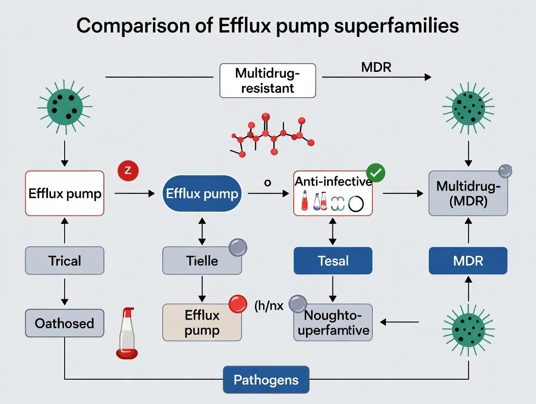
Efflux Pump Superfamilies: A Comparative Analysis of Mechanisms and Therapeutic Targets in MDR Pathogens
This article provides a comprehensive analysis of the major efflux pump superfamilies—RND, MFS, MATE, SMR, and ABC—that drive multidrug resistance (MDR) in critical Gram-negative and Gram-positive pathogens.
Efflux Pump Superfamilies: A Comparative Analysis of Mechanisms and Therapeutic Targets in MDR Pathogens
Abstract
This article provides a comprehensive analysis of the major efflux pump superfamilies—RND, MFS, MATE, SMR, and ABC—that drive multidrug resistance (MDR) in critical Gram-negative and Gram-positive pathogens. Aimed at researchers and drug development professionals, it explores foundational structures and mechanisms, details current methodological approaches for studying pump activity and inhibition, addresses key challenges in assay design and compound discovery, and directly compares the clinical impact and druggability of each superfamily. The synthesis offers a strategic framework for prioritizing targets and developing novel efflux pump inhibitors (EPIs) to restore antibiotic efficacy.
The Molecular Architecture of Resistance: Defining Efflux Pump Superfamilies and Their Core Mechanisms
Multidrug efflux pumps are integral membrane proteins that actively extrude a wide array of antimicrobial agents, contributing significantly to multidrug resistance (MDR) in bacterial pathogens. This guide compares the five primary superfamilies of efflux pumps, their substrate profiles, and experimental approaches for their study, framed within the broader thesis of comparing efflux pump superfamilies in MDR pathogens research.
Comparison of Efflux Pump Superfamilies in MDR Pathogens
The table below summarizes the key characteristics, representative pumps, and substrate profiles of the five major superfamilies.
Table 1: Comparative Overview of Efflux Pump Superfamilies
| Superfamily | Representative Pump (Organism) | Energy Source | Typical Substrates (Drug Classes) | Key Structural Features |
|---|---|---|---|---|
| ATP-Binding Cassette (ABC) | MsrA (S. aureus) | ATP Hydrolysis | Macrolides, streptogramins B, lincosamides | Two transmembrane domains (TMDs), two nucleotide-binding domains (NBDs) |
| Major Facilitator Superfamily (MFS) | MdfA (E. coli), NorA (S. aureus) | Proton Motive Force (PMF) | Fluoroquinolones, β-lactams, chloramphenicol, tetracyclines | 12 or 14 TMDs, single polypeptide. |
| Resistance-Nodulation-Division (RND) | AcrB (E. coli), MexB (P. aeruginosa) | Proton Motive Force (PMF) | Broadest spectrum: β-lactams, fluoroquinolones, macrolides, dyes, detergents | Three-component complex: inner membrane, periplasmic adaptor, outer membrane protein. |
| Small Multidrug Resistance (SMR) | EmrE (E. coli) | Proton Motive Force (PMF) | Quaternary ammonium compounds, dyes, some biocides | Small size (~110 aa), four TMDs, functions as oligomer. |
| Multidrug and Toxic Compound Extrusion (MATE) | NorM (V. parahaemolyticus) | Sodium or Proton Motive Force | Fluoroquinolones, cationic dyes, aminoglycosides | 12 TMDs, functions as dimer or trimer. |
Table 2: Quantitative Comparison of Substrate Extrusion Efficiency
| Superfamily | Pump (Organism) | Substrate (Model Drug) | Experimental MIC Increase (Fold)* | Relative Efflux Rate (nmol/min/mg protein)* | Primary Reference Strain |
|---|---|---|---|---|---|
| RND | AcrAB-TolC (E. coli) | Erythromycin | 32 - 64 | 85 - 120 | AG100A vs. AG100 (ΔacrAB) |
| MFS | NorA (S. aureus) | Ciprofloxacin | 4 - 8 | 15 - 25 | SA-1199 vs. SA-1199B (ΔnorA) |
| MATE | NorM (V. parahaemolyticus) | Norfloxacin | 8 - 16 | 30 - 50 | WT vs. ΔnorM mutant |
| ABC | MsrA (S. aureus) | Erythromycin | 16 - 32 | 40 - 60 (ATP-dependent) | RN4220/pUL5054 vs. control |
| SMR | EmrE (E. coli) | Ethidium Bromide | 2 - 4 | 8 - 12 | BW25113 vs. JW5503 (ΔemrE) |
*Representative ranges from published literature; actual values depend on specific experimental conditions.
Experimental Protocols for Efflux Pump Analysis
Protocol 1: Minimum Inhibitory Concentration (MIC) Reduction Assay
Purpose: To determine the contribution of an efflux pump to resistance against a specific antibiotic. Method:
- Prepare two sets of serial two-fold dilutions of the antibiotic in Mueller-Hinton Broth.
- To one set, add a sub-inhibitory concentration (typically ¼ MIC) of a known efflux pump inhibitor (EPI) like Phe-Arg-β-naphthylamide (PAβN) for Gram-negatives or reserpine for Gram-positives.
- Inoculate each well with ~5 x 10^5 CFU/mL of the bacterial strain.
- Incubate at 35°C for 16-20 hours.
- The MIC is the lowest concentration that inhibits visible growth. A ≥4-fold decrease in MIC in the presence of the EPI indicates significant efflux activity.
Protocol 2: Ethidium Bromide Accumulation/Efflux Assay (Fluorometric)
Purpose: To directly measure the real-time activity of efflux pumps using a fluorescent substrate. Method:
- Grow bacterial cells to mid-log phase (OD600 ~0.4). Harvest and wash twice with PBS or assay buffer (pH 7.0).
- Resuspend cells to an OD600 of 0.2 in buffer containing glucose (0.2%) as an energy source.
- Load the cells with Ethidium Bromide (EtBr, final conc. 2-5 µg/mL) in the presence of an EPI (e.g., 50 µg/mL PAβN) to inhibit efflux and allow dye accumulation. Incubate 30-60 min.
- Centrifuge, wash, and resuspend in fresh buffer with glucose.
- Transfer suspension to a quartz cuvette in a fluorometer (excitation 530 nm, emission 600 nm). Record baseline fluorescence for 60 sec.
- Add glucose (final 0.2%) to energize the cells and initiate active efflux. Monitor the decrease in fluorescence (dye extrusion) for 5-10 minutes.
- Add a protonophore (e.g., CCCP, final 50 µM) to collapse PMF and confirm energy-dependent efflux.
- The initial rate of fluorescence decrease after glucose addition is proportional to efflux pump activity.
Research Reagent Solutions: The Scientist's Toolkit
Table 3: Essential Reagents for Efflux Pump Research
| Reagent/Solution | Function & Application | Example Product/Catalog # |
|---|---|---|
| Phe-Arg-β-naphthylamide (PAβN) | Broad-spectrum EPI for RND pumps in Gram-negative bacteria. Used in MIC reduction and accumulation assays. | Sigma-Aldrich, P4157 |
| Reserpine | EPI for MFS pumps in Gram-positive bacteria (e.g., NorA). Also inhibits some ABC transporters. | Sigma-Aldrich, R0875 |
| Carbonyl Cyanide m-Chlorophenyl Hydrazone (CCCP) | Protonophore that dissipates the proton motive force, used to confirm energy-dependent efflux. | Thermo Fisher Scientific, 50-230-7264 |
| Ethidium Bromide | Fluorescent substrate for many MDR pumps. Standard dye for accumulation/efflux assays. | Thermo Fisher Scientific, 15585011 |
| Hoechst 33342 | DNA-binding dye, specific substrate for pumps like AcrAB-TolC. Used in competitive efflux assays. | Thermo Fisher Scientific, H3570 |
| Nile Red | Lipophilic fluorescent dye extruded by RND pumps. Useful for studying hydrophobic compound efflux. | Sigma-Aldrich, 72485 |
| Crystal Violet | Dye used in basic assay to screen for efflux-deficient mutants (increased accumulation). | Sigma-Aldrich, C3886 |
| Müller-Hinton Broth (cation-adjusted) | Standardized medium for antimicrobial susceptibility testing (MIC assays). | BD Diagnostics, 212322 |
Visualization of Efflux Mechanisms and Assays
Title: RND Tripartite Efflux Pump Mechanism
Title: Fluorescent Dye Efflux Assay Protocol Workflow
Within the critical research landscape of multidrug-resistant (MDR) pathogens, efflux pumps represent a primary defense mechanism, mediating the expulsion of antimicrobial agents and contributing to treatment failure. This comparison guide objectively analyzes the performance characteristics of five major transporter superfamilies—Resistance-Nodulation-Division (RND), Major Facilitator Superfamily (MFS), Multidrug and Toxic Compound Extrusion (MATE), Small Multidrug Resistance (SMR), and ATP-Binding Cassette (ABC)—based on experimental data from recent studies.
Comparative Performance Data
Table 1: Structural and Functional Comparison of Efflux Pump Superfamilies
| Superfamily | Topology (Transmembrane Segments) | Energy Coupling | Typical Substrate Range | Proton: Drug Stoichiometry (Typical) | Representative Pathogen & Pump |
|---|---|---|---|---|---|
| RND | 12 TMS (per subunit) | Proton Motive Force (H+) | Very Broad: β-lactams, macrolides, tetracyclines, fluoroquinolones, dyes, detergents | 1H+:1Drug (Variable) | P. aeruginosa (MexB), E. coli (AcrB) |
| MFS | 12 or 14 TMS | Proton Motive Force (H+) | Broad: Tetracyclines, chloramphenicol, fluoroquinolones, β-lactams | 1H+:1Drug (or 2H+) | S. aureus (NorA), E. coli (TetA) |
| MATE | 12 TMS | Na+ or H+ motive force | Fluoroquinolones, aminoglycosides, ethidium, norfloxacin | 1Na+/2H+:1Drug | E. coli (NorM), V. parahaemolyticus (VecM) |
| SMR | 4 TMS (homodimer/oligomer) | Proton Motive Force (H+) | Narrow: Quaternary ammonium compounds, dyes, some fluoroquinolones | 2H+:1Drug | E. coli (EmrE), S. aureus (QacC) |
| ABC | 12 TMS (2x6 TMS domains) | ATP Hydrolysis | Broad: Macrolides, aminoglycosides, peptides, ions | 1ATP:1Drug (Variable) | E. faecalis (LmrA), S. pneumoniae (PatAB) |
Table 2: Experimental Performance Metrics from Recent Efflux Inhibition Assays (Model: E. coli)
| Superfamily (Target Pump) | Baseline MIC (μg/mL) Ciprofloxacin | MIC with Carbonyl Cyanide m-chlorophenyl hydrazone (CCCP) (Efflux Disruptor) | Fold Reduction in MIC | Common Inhibitor (Experimental) |
|---|---|---|---|---|
| RND (AcrAB-TolC) | 0.03 | 0.004 | 7.5 | Phenylalanine-arginine β-naphthylamide (PAβN) |
| MFS (TetA) | 8.0 | 2.0 | 4.0 | - |
| MATE (NorM) | 0.25 | 0.06 | 4.0 | - |
| SMR (EmrE) | 0.125 | 0.125 | 1.0 (No change) | - |
| ABC (LmrA) | 0.5 | 0.5 | 1.0 (No change) | Verapamil |
Detailed Experimental Protocols
Protocol for Minimum Inhibitory Concentration (MIC) Reduction Assay with Efflux Pump Inhibitors (EPIs)
Objective: To determine the contribution of a specific efflux pump superfamily to antibiotic resistance by measuring the decrease in MIC in the presence of a disruptor (e.g., CCCP) or a specific inhibitor.
Materials: Bacterial culture (e.g., E. coli K-12 and its isogenic efflux pump knockout mutants), Cation-Adjusted Mueller Hinton Broth (CAMHB), antibiotic stock (e.g., ciprofloxacin), EPI stock (e.g., CCCP, PAβN), 96-well microtiter plates.
Methodology:
- Prepare serial two-fold dilutions of the antibiotic in CAMHB across a 96-well plate.
- In one set of rows, supplement each well with a sub-inhibitory concentration of the EPI (e.g., 10-50 μM PAβN or 5-20 μM CCCP). The control set contains no EPI.
- Inoculate each well with a standardized bacterial suspension (5 × 10^5 CFU/mL final concentration).
- Incubate the plate at 37°C for 18-24 hours.
- The MIC is defined as the lowest concentration of antibiotic that completely inhibits visible growth.
- The fold reduction is calculated as: MIC (without EPI) / MIC (with EPI).
Protocol for Ethidium Bromide Accumulation Assay (Fluorometric)
Objective: To directly measure efflux pump activity by quantifying the intracellular accumulation of a fluorescent substrate (e.g., ethidium bromide) in real-time.
Materials: Bacterial cells in exponential growth phase, phosphate-buffered saline (PBS) with glucose (0.4% w/v), ethidium bromide (EtBr) stock solution, EPIs, microplate reader with fluorescence capabilities (excitation ~530 nm, emission ~600 nm).
Methodology:
- Harvest and wash bacterial cells, resuspending them in PBS-glucose to an OD600 of ~0.5.
- Aliquot cell suspension into a black-walled, clear-bottom 96-well plate.
- Pre-incubate with or without EPI for 10 minutes.
- Rapidly add EtBr to a final concentration (e.g., 5-10 μM) and immediately begin kinetic fluorescence readings every 1-2 minutes.
- After a steady-state accumulation phase (10-20 min), add an energy disruptor like CCCP (final 50 μM). This collapses the proton motive force, halting PMF-driven efflux and causing a sharp increase in fluorescence as EtBr accumulates.
- Data is normalized and expressed as relative fluorescence units (RFU) over time. The initial slope or the final fluorescence plateau post-CCCP indicates baseline efflux capacity.
Visualization: Efflux Pump Characterization Workflow
Title: Workflow for Validating Efflux Pump Function in MDR Pathogens
The Scientist's Toolkit: Key Research Reagent Solutions
Table 3: Essential Materials for Efflux Pump Research
| Item | Function in Research | Example/Note |
|---|---|---|
| Proton Motive Force (PMF) Disruptors | Collapses H+ gradient; distinguishes PMF-driven (RND, MFS, MATE, SMR) from ATP-driven (ABC) pumps. | Carbonyl cyanide m-chlorophenyl hydrazone (CCCP), 2,4-Dinitrophenol (DNP) |
| Broad-Spectrum EPIs | Competitively inhibits multiple pumps, especially RND; used for phenotypic screening. | Phenylalanine-arginine β-naphthylamide (PAβN / MC-207,110) |
| Fluorescent Efflux Substrates | Directly visualize and quantify transport activity in real-time accumulation/efflux assays. | Ethidium Bromide (EtBr), Hoechst 33342, Rhodamine 6G |
| ATPase Inhibitors | Specifically targets ABC transporters by inhibiting ATP hydrolysis. | Sodium orthovanadate (Na3VO4) |
| Isogenic Knockout Mutant Strains | Gold standard for attributing resistance phenotype to a specific pump gene. | e.g., E. coli ΔacrB, P. aeruginosa ΔmexB |
| Membrane Permeabilizers | Controls for intrinsic permeability; ensures substrate reaches cytoplasmic pump. | Polymyxin B nonapeptide, EDTA (for Gram-negatives) |
Within the critical research field of multidrug-resistant (MDR) pathogens, the comparison of efflux pump superfamilies is paramount. This guide provides an objective comparison of three fundamental structural and mechanistic blueprints: the number of Transmembrane Domains (TMDs), reliance on Proton Motive Force (PMF), and direct ATP dependence. These features define the major superfamilies—RND, MFS, MATE, ABC, and SMR—that mediate antibiotic efflux in Gram-negative and Gram-positive bacteria.
Table 1: Core Architectural and Energetic Features of Major Efflux Pump Superfamilies
| Efflux Pump Superfamily | Typical # of TMDs (Per Subunit) | Primary Energy Coupling Mechanism | ATP Hydrolysis Directly Required? | Representative Organism & Protein | Typical Substrate Profile |
|---|---|---|---|---|---|
| Resistance-Nodulation-Division (RND) | 12 (in AcrB) | Proton Motive Force (PMF) | No | E. coli AcrB | Broad: lipophiles, amphiphiles, dyes, multiple antibiotic classes |
| Major Facilitator Superfamily (MFS) | 12 or 14 | Proton Motive Force (PMF) | No | E. coli MdfA, NorA (S. aureus) | Specific or broad; often single class (e.g., tetracyclines, fluoroquinolones) |
| Multidrug and Toxic Compound Extrusion (MATE) | 12 | Na+ or H+ Gradient (Ion Motive Force) | No | E. coli NorM, PmpM (P. aeruginosa) | Fluoroquinolones, dyes, aminoglycosides |
| ATP-Binding Cassette (ABC) | 6-10 (per TMD subunit) | ATP Hydrolysis | Yes | E. coli MsbA, LmrA (L. lactis) | Very broad: lipids, drugs, peptides, ions |
| Small Multidrug Resistance (SMR) | 4 (homotrimer) | Proton Motive Force (PMF) | No | E. coli EmrE | Small, lipophilic cations, dyes, biocides |
Table 2: Experimental Data from Characterizing Studies
| Parameter Measured | RND (AcrB) | MFS (MdfA) | ABC (LmrA) | Key Experimental Method |
|---|---|---|---|---|
| Proton Translocation (H+/drug ratio) | ~1 H+ / drug (estimated) | 1-2 H+ / drug (varies) | Not Applicable | Fluorescence quenching of ACMA (pH-sensitive probe) in everted membrane vesicles. |
| ATPase Activity (nmol/min/mg) | Minimal (< 10) | Minimal (< 10) | High (~200-400) | Colorimetric/Malachite Green assay measuring inorganic phosphate release. |
| Drug Efflux Rate (nmol/min/mg protein) | 20-50 (for ethidium) | 5-15 (for chloramphenicol) | 10-30 (for Hoechst 33342) | Real-time fluorometric assay with substrate-specific fluorescent dyes. |
| Inhibition by CCCP (PMF uncoupler) | >90% efflux inhibition | >80% efflux inhibition | <10% efflux inhibition | Efflux assay pre-/post-addition of 50-100 µM carbonyl cyanide m-chlorophenyl hydrazone. |
| Inhibition by Orthovanadate (ATPase inhibitor) | <10% efflux inhibition | <10% efflux inhibition | >85% efflux inhibition | Efflux assay pre-incubation with 1-5 mM sodium orthovanadate. |
Experimental Protocols
Key Protocol 1: Assessing PMF Dependence via ACMA Fluorescence Quenching Objective: To visualize proton translocation coupled to efflux pump activity. Methodology:
- Prepare everted (inside-out) membrane vesicles from the target bacterial strain expressing the pump of interest.
- Load vesicles with 2 µM ACMA (9-amino-6-chloro-2-methoxyacridine), a fluorescent dye that quenches in acidic environments.
- In a fluorometer cuvette, initiate an outward-directed H+ gradient by adding 5 mM ATP (for F0F1-ATPase activation) or an electron donor to generate PMF.
- Observe baseline fluorescence. Upon addition of the efflux pump substrate (e.g., 50 µM tetracycline), a sudden increase in fluorescence (de-quenching) indicates H+ influx into vesicles coupled to drug efflux.
- Add uncoupler CCCP (50 µM) to collapse the PMF, causing rapid re-quenching, confirming the PMF link.
Key Protocol 2: Distinguishing ATPase-Driven Efflux via Orthovanadate Inhibition Objective: To confirm direct ATP hydrolysis as an energy source. Methodology:
- Grow cells expressing the target efflux pump to mid-log phase.
- Pre-incubate one cell aliquot with 5 mM sodium orthovanadate (a transition-state analog Pi inhibitor of ATPases) for 20 minutes. Keep a control aliquot untreated.
- Load cells with a fluorescent substrate (e.g., 10 µM ethidium bromide) in the presence of an energy inhibitor (e.g., cyanide) to allow accumulation.
- Re-energize cells by adding glucose. Monitor extracellular fluorescence increase (efflux) over time.
- Interpretation: Significant inhibition of efflux in the vanadate-treated sample versus control strongly indicates a primary ABC-type pump.
Visualizations
Diagram Title: Energy Coupling Mechanisms: PMF vs. ATP-Driven Efflux
Diagram Title: Experimental Decision Tree for Efflux Pump Energy Classification
The Scientist's Toolkit: Research Reagent Solutions
Table 3: Essential Reagents for Efflux Pump Characterization
| Reagent | Function in Research | Example Use Case |
|---|---|---|
| Carbonyl Cyanide m-Chlorophenyl Hydrazone (CCCP) | Protonophore that uncouples the proton motive force by shuttling H+ across membranes. | Determining if an efflux pump is PMF-dependent. |
| Sodium Orthovanadate | Transition-state analog inhibitor of P-type ATPases and some ABC transporters. | Confirming ATP hydrolysis as the direct energy source for efflux. |
| Acridine Orange / ACMA | Fluorescent dyes that accumulate in acidic compartments and quench; used as pH probes. | Visualizing proton translocation coupled to drug efflux in everted vesicles. |
| Ethidium Bromide | Fluorescent cationic substrate for many MDR pumps (e.g., RND, SMR). | Real-time fluorometric efflux assays in whole cells. |
| Hoechst 33342 | DNA-binding dye and substrate for ABC and other efflux pumps. | Measuring ATP-dependent efflux activity. |
| Reserpine & Verapamil | Plant alkaloid and calcium channel blocker used as broad-spectrum efflux pump inhibitors (EPIs). | Chemical inhibition studies to potentiate antibiotic activity. |
| Everted Membrane Vesicles | Inside-out vesicles where the cytoplasmic pump face is exposed to the external medium. | Isolated system to study transport energetics without regulatory interference. |
| Fluorometric Plate Reader | Instrument for high-throughput, real-time measurement of fluorescent substrate accumulation/efflux. | Kinetic analysis of efflux inhibition or energy requirement. |
This comparison guide, framed within a thesis on the comparison of efflux pump superfamilies in multidrug-resistant (MDR) pathogens, objectively evaluates the substrate recognition profiles of major superfamilies. Performance is measured by substrate spectrum breadth, affinity, and transport efficiency for representative antimicrobial classes.
Comparative Substrate Recognition Profiles of Major Efflux Pump Superfamilies
Table 1: Substrate spectra and key functional data for primary efflux pump superfamilies in Gram-negative bacteria.
| Superfamily | Representative Pump(s) (Organism) | Key Antimicrobial Substrates (Experimental Evidence) | Representative Experimental Apparent Km (µM) / Efflux Rate | Primary Recognition Mechanism |
|---|---|---|---|---|
| RND | AcrB (E. coli), MexB (P. aeruginosa) | β-lactams, fluoroquinolones, tetracyclines, macrolides, chloramphenicol, novobiocin, dyes, detergents. | Norfloxacin: Km ~5 µM (AcrB) [1]; Efflux rate: ~30 nmol/min/mg protein [2] | Hydrophobic/ amphiphilic substrate partitioning into deep binding pocket in the periplasmic domain. |
| MFS | MdfA (E. coli), NorA (S. aureus) | Fluoroquinolones, chloramphenicol, tetracyclines, biocides, ethidium bromide. | Chloramphenicol: Km ~60 µM (MdfA) [3]; Efflux rate: ~2 nmol/min/mg protein [4] | Substrate protonation-coupled binding in cytoplasmic membrane-embedded cavity. |
| SMR | EmrE (E. coli), QacC (S. aureus) | Quaternary ammonium compounds, ethidium bromide, crystal violet, some diamidines. | Methyl viologen: Km ~8 µM (EmrE) [5] | Small, cationic, often polyaromatic compounds binding at dimer interface. |
| ABC | MsbA (E. coli), LmrA (L. lactis) | Hydrophobic drugs, daunorubicin, erythromycin, hoechst 33342, lipids (MsbA). | Hoechst 33342: Transport Vmax ~0.8 nmol/min/mg (LmrA) [6] | Direct ATP-hydrolysis driven binding in inward-facing cavity; often broader hydrophobic patches. |
| MATE | NorM (V. parahaemolyticus), MepA (S. aureus) | Fluoroquinolones, norfloxacin, ethidium bromide, kanamycin (NorM-variant). | Norfloxacin: Km ~1.5 µM (NorM) [7] | Na+/H+ antiport-coupled recognition of cationic and zwitterionic drugs. |
Detailed Experimental Protocols for Key Cited Data
Protocol 1: Measurement of Apparent Km Using Real-Time Fluorometric Assay (e.g., for RND Pumps) [1,2]
- Objective: Determine the binding affinity (apparent Km) of a fluorescent substrate (e.g., ethidium bromide, Hoechst 33342) to an efflux pump in intact cells or membrane vesicles.
- Methodology:
- Cell/Vesicle Preparation: Grow MDR pathogen (e.g., E. coli overexpressing AcrAB-TolC) to mid-log phase. Harvest cells and wash, or prepare everted membrane vesicles via cell disruption and ultracentrifugation.
- Energy Poisoning: For intact cells, pre-treat with a protonophore (e.g., CCCP, 50 µM) to deplete proton motive force (PMF) and inhibit active efflux in the control sample.
- Assay Setup: Load cells/vesicles into a fluorometer cuvette in appropriate buffer. Add the fluorescent substrate at a range of concentrations (e.g., 0.5–50 µM).
- Initial Rate Measurement: Rapidly add an energy source (e.g., 20 mM glucose for PMF in cells, or 5 mM ATP for ABC pumps in vesicles). Monitor fluorescence increase (due to intracellular accumulation) over 60 seconds.
- Inhibition Control: Repeat assay in the presence of a specific pump inhibitor (e.g., PAβN for RND pumps).
- Data Analysis: The initial rate of fluorescence increase (ΔF/min) is plotted against substrate concentration. Data is fit to the Michaelis-Menten equation to derive the apparent Km and Vmax.
Protocol 2: Direct Measurement of Efflux Rate via Radiolabeled Substrate Transport [4]
- Objective: Quantify the direct transport activity of a purified efflux pump reconstituted into proteoliposomes.
- Methodology:
- Protein Purification & Reconstitution: Purify the efflux pump (e.g., MdfA) using detergent solubilization and affinity chromatography. Mix purified protein with pre-formed liposomes and detergent, then remove detergent via dialysis or Bio-Beads to form proteoliposomes.
- Substrate Loading: Incubate proteoliposomes with a radiolabeled substrate (e.g., [14C]-chloramphenicol) in the presence of an appropriate energy source gradient (e.g., inward ΔpH for MFS pumps).
- Transport Initiation: Initiate transport by rapidly imposing the correct energy coupling (e.g., by adding an electron donor to generate ΔpH).
- Sampling & Quantification: At timed intervals, remove aliquots and immediately filter through nitrocellulose membranes. Wash filters to remove external radiolabel.
- Measurement: Quantify the radioactivity retained on the filter (representing internalized substrate) using a scintillation counter. Plot accumulated substrate vs. time to calculate the initial efflux rate (nmol/min/mg protein).
Visualizations
Diagram 1: RND pump substrate recognition and efflux pathway.
Diagram 2: Radiolabeled flux assay workflow for efflux pumps.
The Scientist's Toolkit: Key Research Reagent Solutions
Table 2: Essential materials and reagents for studying efflux pump substrate specificity.
| Item | Function/Benefit in Research |
|---|---|
| Everted Membrane Vesicles | System for studying transporter activity in isolation from cellular metabolism; maintains native lipid environment. |
| Proteoliposomes (Reconstituted) | Defined system with purified pump protein in synthetic lipids; allows precise control of energy coupling and substrate gradients. |
| Fluorescent Substrate Probes (e.g., Ethidium Bromide, Hoechst 33342) | Enable real-time, high-throughput kinetic measurements of efflux/inhibition without separation steps. |
| Radiolabeled Antimicrobials (e.g., [14C]-Chloramphenicol, [3H]-Norfloxacin) | Provide direct, sensitive, and quantitative measurement of transport rates and binding constants. |
| Protonophores (e.g., CCCP) | Deplete the proton motive force (PMF) to differentiate between active efflux and passive diffusion. |
| Broad-Specificity Efflux Pump Inhibitors (e.g., PAβN) | Used as a control to confirm efflux-mediated resistance and to probe pump function in whole-cell assays. |
| Strains with Deleted/Overexpressed Efflux Pumps | Isogenic bacterial strains are critical for attributing changes in susceptibility or accumulation directly to a specific pump. |
| Crystallization Kits & Detergents (e.g., DDM, LMNG) | Essential for solubilizing and purifying stable membrane proteins for structural studies (X-ray, Cryo-EM). |
Within the broader study of efflux pump superfamilies in multidrug-resistant (MDR) pathogens, two systems stand as paradigm cases: the AcrAB-TolC tripartite complex (Resistance-Nodulation-Division (RND) superfamily) in Enterobacteriaceae like E. coli and K. pneumoniae, and the NorA pump (Major Facilitator Superfamily (MFS)) in Staphylococcus aureus. This guide provides a comparative analysis of their structure, function, and clinical impact, supported by experimental data and methodologies essential for ongoing research and drug development.
Table 1: Core Characteristics and Substrate Profiles
| Feature | AcrAB-TolC (RND) in E. coli | NorA (MFS) in S. aureus |
|---|---|---|
| Superfamily | Resistance-Nodulation-Division (RND) | Major Facilitator Superfamily (MFS) |
| Genetic Context | Chromosomal (acrAB-tolC operon/regulon) | Chromosomal (norA gene) |
| Assembly & Structure | Tripartite: AcrB (IM), AcrA (PAP), TolC (OM) | Single protein (12 or 14 TMS) |
| Energy Coupling | Proton Motive Force (H+ antiport) | Proton Motive Force (H+ antiport) |
| Primary Substrates | Broad spectrum: β-lactams, fluoroquinolones, tetracyclines, chloramphenicol, dyes, detergents, bile salts. | Narrower spectrum: Hydrophilic fluoroquinolones (e.g., norfloxacin, ciprofloxacin), biocides (e.g., benzalkonium chloride), dyes. |
| Regulatory System | Complex: Global regulators (RamA, MarA, SoxS, Rob) repress AcrR; Local repressor AcrR. | Primarily mgrA; also arIRS, norG. |
| Clinical Impact | Major contributor to intrinsic & acquired MDR in Enterobacteriaceae; critical for bile resistance in gut. | Contributes to fluoroquinolone resistance in MRSA; role in biocide tolerance. |
Table 2: Quantitative Efflux Pump Inhibition Data (Representative Studies)
| Experiment Parameter | AcrAB-TolC Inhibition (e.g., with Phenylalanine-arginine β-naphthylamide, PAβN) | NorA Inhibition (e.g., with Reserpine or 5′-Methoxyhydnocarpin) |
|---|---|---|
| MIC Reduction (Fold) | Ciprofloxacin MIC vs. E. coli: 8-32 fold decrease | Norfloxacin MIC vs. S. aureus SA-1199B: 4-8 fold decrease |
| IC50 of Inhibitor | PAβN: ~5-10 µg/mL in potentiation assays | Reserpine: ~10-20 µg/mL in efflux inhibition assays |
| Efflux Rate (Control vs. Inhibited) | Ethidium bromide accumulation increased by 300-400% with PAβN | Norfloxacin accumulation increased by 200-300% with reserpine |
| Key Model Strain | E. coli AG100; K. pneumoniae KP55 | S. aureus SA-1199B (NorA-overexpressing) |
Experimental Protocols for Key Assays
Protocol 1: Ethidium Bromide Accumulation Assay (Common for Both Pumps)
Purpose: To measure real-time efflux pump activity by monitoring intracellular accumulation of a fluorescent substrate. Method:
- Cell Preparation: Grow bacteria to mid-log phase (OD600 ~0.4-0.6). Harvest, wash, and resuspend in buffer (e.g., PBS or phosphate buffer with glucose).
- Energy Depletion: Treat cells with a protonophore (e.g., CCCP, 50 µM) for 10 min to inhibit PMF-driven efflux. Include an untreated control.
- Dye Loading: Add ethidium bromide (EtBr, final conc. 1-5 µg/mL) to the cell suspension.
- Fluorescence Measurement: Immediately transfer to a fluorimeter plate or cuvette. Measure fluorescence (Ex: 530 nm, Em: 600 nm) every 30-60 sec for 15-20 min.
- Data Analysis: Plot fluorescence vs. time. The initial slope represents uptake/accumulation rate. Compare slopes between wild-type, efflux-overexpressing, and inhibitor-treated strains. A steeper slope indicates impaired efflux/increased accumulation.
Protocol 2: Checkerboard Broth Microdilution for Synergy (EPI + Antibiotic)
Purpose: To determine the Minimum Inhibitory Concentration (MIC) reduction of an antibiotic in the presence of an Efflux Pump Inhibitor (EPI). Method:
- Preparation: Prepare 2-fold serial dilutions of the antibiotic (e.g., ciprofloxacin) along the x-axis of a 96-well plate and the EPI (e.g., PAβN or reserpine) along the y-axis.
- Inoculation: Add a standardized bacterial inoculum (~5 x 10^5 CFU/mL) to each well.
- Incubation: Incubate plate at 37°C for 18-24 hours.
- Analysis: Determine the MIC of the antibiotic alone and in combination with various EPI concentrations. Calculate the Fractional Inhibitory Concentration Index (FICI). FICI ≤0.5 indicates synergy.
Visualizations: Regulatory Pathways & Experimental Workflow
Diagram 1: AcrAB-TolC Regulatory Network in Enterobacteriaceae
Diagram 2: NorA Regulation via MgrA in S. aureus
Diagram 3: Generic Fluorescent Efflux Assay Workflow
The Scientist's Toolkit: Key Research Reagent Solutions
Table 3: Essential Reagents for Efflux Pump Research
| Reagent / Material | Function in Research | Example Use Case |
|---|---|---|
| PAβN (Phe-Arg-β-naphthylamide) | Broad-spectrum RND pump inhibitor; competitive substrate. | Potentiating antibiotics against E. coli, K. pneumoniae in synergy assays. |
| Reserpine | Plant alkaloid inhibitor of MFS and SMR pumps. | Inhibiting NorA-mediated fluoroquinolone efflux in S. aureus. |
| CCCP (Carbonyl cyanide m-chlorophenyl hydrazone) | Protonophore; dissipates proton motive force (PMF). | Negative control in accumulation assays to confirm PMF-dependent efflux. |
| Ethidium Bromide | Fluorescent substrate for many efflux pumps (RND, MFS). | Real-time measurement of efflux activity in accumulation assays. |
| Ciprofloxacin / Norfloxacin | Fluoroquinolone antibiotics; classic pump substrates. | Measuring MIC shifts and compound accumulation in the presence of EPIs. |
| Anti-AcrA or Anti-NorA Antibodies | For immunodetection (Western blot) or cellular localization. | Quantifying efflux pump expression levels in clinical vs. lab strains. |
| qPCR Primers for acrA, acrB, norA | Quantitative measurement of gene expression. | Correlating mRNA levels with resistance phenotypes and regulator activity. |
| MgrA or MarA Expression Vectors | For genetic manipulation of regulator levels. | Studying gain/loss-of-function effects on pump expression and resistance. |
From Bench to Pipeline: Techniques for Studying Efflux Activity and Developing Inhibitors
Within the broader thesis on the comparison of efflux pump superfamilies in multidrug-resistant (MDR) pathogens, quantifying efflux activity is paramount. This guide objectively compares three core functional assays: Ethidium Bromide (EtBr) Accumulation, Real-time Fluorometry, and Minimum Inhibitory Concentration (MIC) Modulation. These methods are essential for characterizing the activity of major superfamilies like RND, MFS, MATE, SMR, and ABC in pathogens such as Pseudomonas aeruginosa, Escherichia coli, and Staphylococcus aureus.
Comparison of Functional Assays for Efflux Quantification
Table 1: Comparison of Key Assay Parameters and Performance
| Feature | Ethidium Bromide Accumulation | Real-time Fluorometry (e.g., with Hoechst 33342) | MIC Modulation (Checkerboard) |
|---|---|---|---|
| Primary Measurement | Endpoint intracellular fluorescence (accumulation). | Real-time kinetic fluorescence (efflux rate). | Fractional Inhibitory Concentration Index (FICI) of antibiotic + inhibitor. |
| Throughput | Medium (endpoint, adaptable to multi-well). | High (continuous, automated). | Low (manual, labor-intensive). |
| Quantitative Output | Semi-quantitative (fold-change in accumulation). | Highly quantitative (efflux velocity, pump kinetics). | Synergy/Antagonism (FICI ≤0.5 = synergy). |
| Pathogen Applicability | Broad (bacteria, yeast). | Broad, but dye/probe dependent. | Broad (standard antimicrobial testing). |
| Superfamily Insight | Confirms efflux activity; less specific. | Can differentiate pump activity via kinetic profiles. | Confirms functional contribution to resistance. |
| Key Advantage | Simple, cost-effective, visual. | Dynamic, high-resolution, mechanistic. | Clinically relevant, measures resistance impact. |
| Key Limitation | Photobleaching, potential dye toxicity, semi-quantitative. | Requires specialized fluorometer, optimized probe. | Does not directly measure efflux; indirect inference. |
| Example Data (Model: E. coli with AcrAB-TolC) | Accumulation increases 3.5-fold with CCCP vs control. | Efflux rate decreases by 70% with PAβN inhibitor. | FICI of Ciprofloxacin + PAβN = 0.25 (synergy). |
Experimental Protocols
Ethidium Bromide Accumulation Assay
This endpoint assay measures the intracellular buildup of a fluorescent substrate in the presence or absence of an efflux pump inhibitor (EPI) or energy inhibitor.
- Culture Preparation: Grow test bacteria to mid-log phase (OD600 ~0.5) in appropriate broth.
- Washing: Harvest cells, wash twice, and resuspend in assay buffer (e.g., PBS with 20 mM glucose, pH 7.0) to a standardized OD600.
- Inhibitor Pre-incubation: Divide suspension. Incubate one aliquot with an EPI (e.g., 50 µg/mL PAβN) or energy uncoupler (e.g., 100 µM CCCP) for 10 minutes. Use a second aliquot as an untreated control.
- Dye Loading: Add EtBr to both aliquots at a sub-inhibitory concentration (e.g., 0.5-2.0 µg/mL).
- Incubation & Measurement: Incubate at 37°C with shaking. At timed intervals (e.g., 0, 5, 10, 20 min), take aliquots, wash rapidly with ice-cold buffer, and resuspend. Measure fluorescence (excitation ~530 nm, emission ~600 nm) using a plate reader or fluorometer. Use cells without dye for background subtraction.
- Analysis: Plot fluorescence vs. time. Higher accumulation in EPI-treated cells indicates efflux inhibition.
Real-time Fluorometric Efflux Assay
This kinetic assay monitors the real-time extrusion of a fluorescent dye.
- Cell Preparation: As in Step 1 & 2 of the EtBr assay, wash and resuspend cells in assay buffer.
- Dye Loading and Energy Depletion: Load cells with a membrane-permeant fluorescent dye (e.g., 5 µM Hoechst 33342). Incubate with an energy inhibitor like CCCP to allow dye influx while inhibiting active efflux. Centrifuge and resuspend in fresh, dye-free buffer.
- Real-time Measurement: Place cell suspension in a fluorometer cuvette or multi-well plate with continuous stirring/temperature control. Establish a baseline fluorescence (ex/em specific to dye, e.g., 355/450 nm for Hoechst).
- Energy Restoration: Rapidly add an energy source (e.g., glucose) to re-energize the cells and initiate active efflux. Monitor the decrease in fluorescence over time (3-10 minutes).
- Inhibitor Control: Repeat assay with cells pre-treated with an EPI. The efflux rate decrease confirms pump specificity.
- Analysis: Calculate initial efflux velocities from the linear portion of the fluorescence decay curve post-energization.
MIC Modulation Assay (Checkerboard)
This indirect assay evaluates the effect of an EPI on the MIC of an antibiotic.
- Preparation: Prepare two-fold serial dilutions of the antibiotic in one dimension of a 96-well microtiter plate and of the EPI in the other dimension.
- Inoculation: Add a standardized bacterial inoculum (~5 x 10^5 CFU/mL) to each well.
- Incubation: Incubate the plate at 37°C for 16-20 hours.
- Reading: Determine the MIC as the lowest concentration preventing visible growth.
- Analysis: Calculate the Fractional Inhibitory Concentration Index (FICI): FICI = (MICantibiotic with EPI / MICantibiotic alone) + (MICEPI with antibiotic / MICEPI alone). Interpret: FICI ≤0.5 = synergy; >0.5 to ≤4 = no interaction; >4 = antagonism.
Visualizing Assay Workflows and Efflux Context
Diagram 1: Workflow comparison of the three core efflux assays.
Diagram 2: Relationship between efflux mechanisms, MDR, and assay readouts.
The Scientist's Toolkit: Key Research Reagent Solutions
Table 2: Essential Materials for Efflux Quantification Assays
| Reagent/Material | Function in Assays | Example & Notes |
|---|---|---|
| Fluorescent Substrates | Serve as efflux pump probes; accumulation indicates activity. | Ethidium Bromide: Common DNA intercalator for general efflux. Hoechst 33342: DNA stain for real-time kinetics in Gram-positives. Nile Red: Lipophilic dye for RND pumps. |
| Efflux Pump Inhibitors (EPIs) | Chemically block pump function to confirm its role. | PAβN (Phe-Arg β-naphthylamide): Broad-spectrum EPI for RND pumps. CCCP (Carbonyl cyanide m-chlorophenyl hydrazone): Protonophore that depletes energy (ΔpH). Verapamil: EPI for MFS pumps in some Gram-positives. |
| Energy Source/Inhibitors | Control proton motive force (PMF) or ATP for active transport. | Glucose: Provides metabolic energy to generate PMF/ATP. Sodium Azide: Inhibits ATP synthesis. CCCP: Uncouples proton gradient. |
| Assay Buffer Systems | Maintain physiological conditions during fluorescence measurements. | PBS with Glucose (20 mM): Common for maintaining PMF. HEPES or Potassium Phosphate Buffer: Maintain stable pH. MgSO4 is often added. |
| Reference Strain Panels | Controls for assay validation and pump specificity. | Wild-type strains with defined pumps. Isogenic efflux knockout mutants (e.g., ΔacrB). Strains overexpressing specific pumps. |
| Microtiter Plates (Black/Clear) | Vessel for high-throughput fluorescence or growth assays. | Black plates with clear bottom: Optimized for fluorescence readings. 96- or 384-well format for screening. |
| Specialized Fluorometer/Plate Reader | Detect and quantify fluorescence signals. | Instrument capable of kinetic reads (real-time) and temperature control. Appropriate filter sets for chosen dyes (e.g., 530/600 nm for EtBr). |
Within the critical research on Comparison of efflux pump superfamilies in MDR pathogens, the selection of appropriate genetic and molecular tools is paramount. Researchers dissecting the expression, regulation, and function of resistance-nodulation-division (RND), major facilitator superfamily (MFS), or ATP-binding cassette (ABC) pumps rely on a core set of techniques. This guide compares the performance of knockout mutants, reporter gene systems, and quantitative reverse transcription PCR (qRT-PCR) for probing efflux pump biology, providing objective data to inform experimental design.
Comparative Performance Analysis
Table 1: Comparison of Core Genetic and Molecular Tools in Efflux Pump Research
| Tool | Primary Application in Efflux Pump Research | Key Performance Metrics | Typical Experimental Timeline | Key Limitations |
|---|---|---|---|---|
| Knockout Mutants | Determine direct contribution of a specific pump to antimicrobial resistance and cellular fitness. | Complementation Efficiency: >90% restoration of wild-type MIC. Fitness Cost: Often measured as growth rate reduction (e.g., 10-30% for RND pumps). | 2-4 weeks (construction, validation). | Redundant functions may mask phenotype; essential genes cannot be knocked out. |
| Reporter Gene Systems | Real-time monitoring of promoter activity in response to antimicrobials or regulators. | Sensitivity: Can detect sub-inhibitory antibiotic concentrations (e.g., 1/8 MIC). Dynamic Range: 2-3 logs of linear signal (e.g., luminescence). | 1-2 weeks (reporter construct integration). | Reporter stability and genetic context can affect signal; indirect measure. |
| qRT-PCR | Quantify absolute or relative changes in efflux pump gene transcription. | Sensitivity: Detect single copy mRNA. Precision: Intra-assay CV <2%. Dynamic Range: 7-8 logs of linear detection. | 1-2 days (post-RNA extraction). | Measures mRNA only; does not confirm protein function or activity. |
Table 2: Supporting Experimental Data from a Study on acrB (RND) Regulation
| Experimental Condition | Tool Applied | Key Quantitative Result | Interpretation |
|---|---|---|---|
| Exposure to 0.5 µg/mL Ciprofloxacin | qRT-PCR | 8.5-fold ± 1.2 increase in acrB mRNA vs. untreated control. | Rapid transcriptional upregulation of the primary RND pump. |
| Deletion of local repressor gene acrR | Reporter (GFP) | 45-fold ± 5 increase in PacrB-GFP fluorescence. | Confirms acrR as a direct, potent transcriptional repressor. |
| acrB knockout mutant vs. Wild-Type | MIC Assay | 64-fold reduction in ciprofloxacin MIC (e.g., 2 µg/mL → 0.031 µg/mL). | Direct, quantitative contribution of AcrB to fluoroquinolone resistance. |
Detailed Methodologies
Protocol 1: Construction of an Efflux Pump Knockout Mutant Using Linear DNA Transformation (e.g., in Acinetobacter baumannii)
- Design: Amplify ~1 kb DNA fragments upstream and downstream of the target efflux pump gene (e.g., adeB). Flank them with sequences homologous to an antibiotic resistance cassette (e.g., kanamycin).
- Assembly: Fuse the fragments and the cassette via overlap extension PCR to create a linear knockout construct.
- Transformation: Introduce the construct into competent cells via electroporation.
- Selection: Plate on media containing kanamycin. Select colonies where homologous recombination has replaced the target gene with the cassette.
- Validation: Confirm via colony PCR with junction primers and phenotypic assays (e.g., MIC reduction, loss of pump function).
Protocol 2: Efflux Pump Promoter Activity Assay Using a Chromosomal Luciferase Reporter
- Cloning: Clone the putative promoter region (e.g., 300-500 bp upstream of mexB) into a suicide vector upstream of a promoterless luxCDABE operon.
- Conjugation/Transformation: Integrate the construct into the pathogen's chromosome via single-crossover homologous recombination.
- Assay: Grow reporter strain with/without inducer (e.g., antibiotic, bile salt). Measure real-time bioluminescence (RLU) in a plate reader and normalize to optical density (OD600).
- Analysis: Plot RLU/OD versus time. Compare fold-change in AUC (Area Under Curve) between conditions.
Protocol 3: qRT-PCR Analysis of Efflux Pump Gene Expression
- RNA Isolation: Extract total RNA from treated/untreated bacterial cultures using a guanidinium thiocyanate-phenol method. Treat rigorously with DNase I.
- Reverse Transcription: Use 500 ng - 1 µg total RNA, random hexamers, and a high-fidelity reverse transcriptase.
- qPCR Setup: Prepare reactions with gene-specific primers (e.g., for norA (MFS) and stable reference genes (gyrB, rpoD)), cDNA, and SYBR Green master mix.
- Run & Analyze: Use the following cycling: 95°C for 3 min; 40 cycles of 95°C for 10s, 60°C for 30s. Calculate relative expression via the 2-ΔΔCt method.
Visualizations
Decision Workflow for Efflux Pump Analysis Tools
Regulatory Pathway of an Inducible RND Efflux Pump
The Scientist's Toolkit: Research Reagent Solutions
Table 3: Essential Materials for Featured Experiments
| Item | Function in Efflux Pump Research | Example Product/Catalog |
|---|---|---|
| High-Efficiency Electrocompetent Cells | Essential for transforming knockout constructs in resistant clinical isolates. | E. coli HST08 Premium; MFD pir for suicide vector propagation. |
| Suicide Vector System | Enables stable chromosomal integration of reporter constructs via single crossover. | pKNOCK series; pUC18T-mini-Tn7T. |
| Promoterless Reporter Operon | Core module for building transcriptional fusions to measure promoter activity. | luxCDABE (bioluminescence), gfpmut3 (fluorescence). |
| DNase I, RNase-free | Critical for removing genomic DNA contamination prior to qRT-PCR for accurate Ct values. | Turbo DNase (Ambion). |
| SYBR Green qPCR Master Mix | Sensitive detection of amplicons for quantifying efflux pump mRNA levels. | PowerUp SYBR Green Master Mix (Applied Biosystems). |
| Validated Reference Gene Primers | Essential for normalizing qRT-PCR data in pathogens under antibiotic stress. | Primers for rpoD (sigma factor) or gyrB (gyrase). |
Within the critical research on Comparison of efflux pump superfamilies in MDR pathogens, determining high-resolution structures of these membrane protein complexes is paramount. These structures reveal conformational states crucial for understanding substrate transport and inhibition. This guide objectively compares the two premier structural biology techniques—X-ray Crystallography and Cryo-Electron Microscography (Cryo-EM)—in their application to efflux pump studies, providing experimental data and protocols to inform methodological selection.
Comparative Performance Analysis
Table 1: Core Performance Comparison for Efflux Pump Structural Analysis
| Parameter | X-ray Crystallography | Cryo-EM (Single Particle Analysis) |
|---|---|---|
| Typical Resolution Range | 1.5 – 3.5 Å | 2.5 – 4.0 Å (now often <2.5 Å for stable complexes) |
| Sample Requirement | High-purity, large, well-ordered 3D crystals (~>0.1 mm) | High-purity in solution (3-5 µL, ~0.1-1 mg/mL) |
| Sample State | Crystalline, trapped conformation | Vitrified, near-native state in multiple conformations |
| Membrane Protein Success Rate | Historically higher, but requires crystallization | Currently higher for large, flexible complexes |
| Size Limitations | Small to very large complexes (with effort) | Ideal for > ~50 kDa; excellent for large complexes (efflux pumps) |
| Key Advantage | Atomic detail, high throughput of data collection | Visualizes conformational heterogeneity, no crystallization needed |
| Primary Limitation | Crystal packing forces may distort conformation; crystallization bottleneck | Lower throughput in sample prep and data collection; computational cost |
| Typical Data Collection Time | Minutes to hours per dataset | Days to weeks per dataset |
| Pump Conformations Captured | Usually one dominant state | Multiple states (e.g., inward-open, occluded, outward-open) |
Supporting Experimental Data: A 2023 study on the E. coli AcrBZ-TolC efflux pump directly compared structures obtained via both methods. X-ray crystallography at 2.9 Å provided atomic details of side-chain interactions in a single, crystallography-induced symmetric state. Cryo-EM at 3.2 Å resolved the natural asymmetric conformation of the AcrBZ trimer, capturing three distinct functional states (loose, tight, open) within a single dataset, directly illustrating the peristaltic transport mechanism.
Detailed Methodological Protocols
Protocol 1: X-ray Crystallography of an RND Family Efflux Pump
Objective: Determine atomic structure of a crystallized pump component (e.g., inner membrane RND transporter).
Protein Production & Purification:
- Express His-tagged protein in E. coli or insect cells.
- Solubilize from membranes using n-dodecyl-β-D-maltoside (DDM).
- Purify via immobilized metal affinity chromatography (IMAC) and size-exclusion chromatography (SEC).
Crystallization:
- Concentrate protein to 10-20 mg/mL in SEC buffer.
- Utilize vapor diffusion (sitting drop) method with commercial screens (e.g., MemGold, MemSys).
- Optimize hits by varying pH, precipitant (PEG), salt, and lipid/additive concentrations (e.g., cholesterol hemisuccinate).
Data Collection & Processing:
- Flash-cool crystal in liquid N₂ with cryoprotectant.
- Collect 360° of diffraction data at synchrotron beamline.
- Index, integrate, and scale diffraction spots (HKL-3000, XDS).
- Solve phase problem by molecular replacement (MR) using homologous structure.
Model Building & Refinement:
- Build atomic model in Coot using electron density maps.
- Iteratively refine model against structure factors using PHENIX.refine or BUSTER.
Protocol 2: Cryo-EM of a Full Assembly Efflux Pump Complex
Objective: Determine structure and conformational landscape of a membrane-embedded pump complex (e.g., ABC transporter).
Sample Vitrification (Grid Preparation):
- Purify intact complex in amphiphile (e.g., GDN, LMNG).
- Apply 3-4 µL of sample (0.5-1 mg/mL) to a plasma-cleaned ultrathin carbon or holey gold grid (Quantifoil).
- Blot excess liquid manually or robotically (Vitrobot) for 2-4 seconds at 100% humidity.
- Plunge-freeze into liquid ethane cooled by liquid nitrogen.
Microscopy & Data Collection:
- Load grid into 300 kV cryo-TEM with direct electron detector.
- Screen for optimal ice thickness and particle distribution.
- Collect multi-frame movie micrographs (40-50 frames) at 60-130,000x magnification, with total dose of 40-60 e⁻/Ų, using automated software (SerialEM, EPU).
Image Processing & 3D Reconstruction:
- Motion-correct and dose-weight frames (MotionCor2), estimate CTF (CTFFIND-4).
- Autopick particles from micrographs (cryoSPARC, Relion).
- Perform multiple rounds of 2D classification to remove junk particles.
- Generate initial model ab initio or via stochastic gradient descent.
- Refine 3D classification (with/without symmetry) to separate conformational states.
- Conduct high-resolution 3D refinement and Bayesian polishing.
- Calculate final resolution via Fourier Shell Correlation (FSC=0.143).
Atomic Model Building:
- Fit existing high-resolution X-ray structures into EM density as rigid bodies (UCSF Chimera).
- De novo building of unresolved regions in Coot.
- Real-space refinement against map using PHENIX or ISOLDE.
Workflow Visualization
The Scientist's Toolkit: Essential Research Reagents & Materials
Table 2: Key Reagents for Structural Studies of Efflux Pumps
| Item | Function in Experiment | Example Product/Target |
|---|---|---|
| Detergents/Amphiphiles | Solubilize and stabilize membrane proteins for purification and structural analysis. | n-Dodecyl-β-D-Maltoside (DDM), Glyco-diosgenin (GDN), Lauryl Maltose Neopentyl Glycol (LMNG) |
| Lipid-like Additives | Mimic native membrane environment, crucial for crystallization and stability. | Cholesterol Hemisuccinate (CHS), synthetic phospholipids (e.g., POPC, POPG) |
| Affinity Chromatography Resins | Primary purification step via engineered tags on recombinant protein. | Nickel-NTA (for His-tag), Anti-Flag resin, Streptactin (for Strep-tag) |
| Size-Exclusion Columns | Final polishing step to isolate monodisperse, homogeneous protein complexes. | Superdex 200 Increase, Superose 6 Increase (Cytiva) |
| Cryo-EM Grids | Support film for sample application and vitrification. | Quantifoil (Au, R1.2/1.3), UltraAufoil (holey gold) |
| Cryoprotectants | Prevent ice crystal formation in X-ray crystallography during flash-cooling. | Glycerol, Ethylene Glycol, Paratone-N oil |
| Synchrotron Facility Access | Source of high-intensity X-rays for diffraction data collection. | APS (USA), ESRF (EU), SPring-8 (Japan) – User proposals required. |
| Direct Electron Detectors | Critical camera hardware for high-resolution Cryo-EM data collection. | Gatan K3, Falcon 4, Selectris X |
The choice between X-ray crystallography and Cryo-EM for efflux pump structural biology is no longer hierarchical but complementary. Cryo-EM excels in elucidating the conformational dynamics of large, native, or flexible pump assemblies without crystallization, directly informing the transport cycle. X-ray crystallography remains unparalleled for obtaining the highest-resolution atomic models of stable domains or trapped states, providing precise coordinates for drug docking studies. For a comprehensive comparison of efflux pump superfamilies in MDR pathogens, an integrated approach leveraging both techniques is now the most powerful strategy to move from static snapshots to mechanistic movies of efflux function.
In Silico Screening and Rational Design of Novel Efflux Pump Inhibitors (EPIs).
Within the broader thesis on Comparison of efflux pump superfamilies in MDR pathogens, this guide focuses on the computational discovery and optimization of EPIs. The efficacy of an EPI is intrinsically linked to its interaction with specific pump superfamilies (e.g., RND, MFS, MATE, SMR, ABC). This guide compares the performance of leading in silico methodologies and their ability to identify hits against distinct targets, providing a framework for researchers to select optimal screening strategies.
Comparison of In Silico Screening Methodologies
The table below compares three primary computational approaches for EPI discovery, evaluated based on their success in identifying verifiable hits against major efflux pump superfamilies.
Table 1: Performance Comparison of In Silico EPI Screening Methodologies
| Methodology | Key Principle | Best Suited For Superfamily | Success Rate (Hit-to-Lead) | Computational Cost | Key Experimental Validation Outcome |
|---|---|---|---|---|---|
| Structure-Based Virtual Screening (SBVS) | Docking into a high-resolution 3D pump or binding site model. | RND (e.g., AcrB), ABC | ~5-15% | High | Identified compound MC-207,110 analog showing 8-fold reduction in norfloxacin MIC in P. aeruginosa. |
| Ligand-Based Virtual Screening (LBVS) | Similarity search or pharmacophore modeling based on known EPIs/substrates. | MFS, SMR | ~10-20% | Low | Quinoline derivative NMP analogs restored tetracycline activity in E. coli (4-fold MIC reduction). |
| Molecular Dynamics (MD) & Binding Free Energy Calculations | Simulating dynamic interactions and calculating ΔG of binding. | All, esp. for lead optimization | N/A (Optimization) | Very High | Correctly ranked congeneric series binding to AcrB periplasmic domain; ΔG correlation R²=0.89 with experimental IC₅₀. |
Detailed Experimental Protocols for Key Studies
Protocol 1: Structure-Based Virtual Screening for RND Inhibitors
- Objective: Identify novel AcrB-TolC (RND) EPI candidates.
- Methodology:
- Protein Preparation: Retrieve AcrB structure (PDB: 4DX5). Prepare using Maestro's Protein Preparation Wizard: add hydrogens, assign bond orders, optimize H-bonds, minimize.
- Grid Generation: Define docking grid around the distal binding pocket (DBP) using Glide.
- Library Screening: Screen ZINC15 "lead-like" subset (≈ 250,000 compounds). Perform HTVS → SP → XP docking cascade in Glide.
- Post-Docking Analysis: Apply filters: GlideScore < -6.0, presence of key pharmacophore features (aromatic ring, cationic center). Cluster top 500 hits.
- In Vitro Validation: Subject top 50 virtual hits to ethidium bromide accumulation assay in E. coli AG100 and checkerboard assay with levofloxacin.
Protocol 2: Ligand-Based Pharmacophore Modeling for MFS Inhibitors
- Objective: Discover novel MFS (e.g., TetA) EPIs.
- Methodology:
- Training Set Curation: Assemble 15 known TetA EPIs/substrates with reported IC₅₀ values from literature.
- Pharmacophore Generation: Use Phase (Schrödinger) to generate common pharmacophore hypotheses. Select best model (e.g., AARRR: two acceptors, three rings).
- Database Screening: Screen in-house compound library (10,000 molecules) using the pharmacophore as a 3D search query.
- Molecular Alignment & Scoring: Fit compounds to the hypothesis and score based on vector alignment and volume overlap.
- Experimental Validation: Test top 100 ranked compounds for potentiation of tetracycline in TetA-expressing E. coli.
Visualization of Workflows and Pathways
Diagram Title: EPI Discovery Computational Workflow (100 chars)
Diagram Title: Mechanism of EPI Action on an RND Pump (95 chars)
The Scientist's Toolkit: Key Research Reagent Solutions
Table 2: Essential Materials for EPI Research & Validation
| Item / Reagent | Function in EPI Research | Example Product/Source |
|---|---|---|
| Fluorescent Substrate Dye | Efflux activity probe for accumulation assays. | Ethidium Bromide (EtBr), Hoechst 33342, Nile Red. |
| Protonophore (Control Inhibitor) | Positive control for maximal accumulation. | Carbonyl Cyanide m-Chlorophenyl Hydrazone (CCCP). |
| MDR Bacterial Strains | Isogenic pairs for target validation. | E. coli AG100 (wild-type) vs. AG100A (ΔacrAB). |
| Membrane Protein Prep Kit | Isolation of efflux pumps for biophysical assays. | Mem-PERTM Plus Membrane Protein Extraction Kit (Thermo). |
| Molecular Docking Suite | Structure-based virtual screening platform. | Glide (Schrödinger), AutoDock Vina (Open Source). |
| MD Simulation Software | Analyzing pump dynamics and inhibitor binding. | GROMACS, Desmond (Schrödinger), NAMD. |
| Microplate Reader (Fluorescence) | Quantitative readout for high-throughput accumulation assays. | SpectraMax iD3 (Molecular Devices). |
High-Throughput Screening (HTS) Strategies and Lead Compound Identification
Within the critical research area of comparing efflux pump superfamilies in Multi-Drug Resistant (MDR) pathogens, identifying novel efflux pump inhibitors (EPIs) is paramount. High-Throughput Screening (HTS) serves as the primary engine for discovering initial lead compounds that can potentiate existing antibiotics. This guide compares predominant HTS strategies used in this field, focusing on their performance in identifying hits against targets like the RND, MFS, and ABC superfamilies.
Comparison of HTS Strategies for EPI Discovery
| Screening Strategy | Primary Readout | Throughput (Compounds/Day) | Key Advantage | Key Limitation | Typical Hit Rate | Best For Superfamily |
|---|---|---|---|---|---|---|
| Whole-Cell Viability (Synergy) | Bacterial growth inhibition in presence of sub-MIC antibiotic. | 50,000 - 100,000+ | Identifies functional EPIs in physiological context; detects membrane disruptors. | Hit confirmation is complex; many non-specific hits (e.g., general toxics). | 0.1% - 0.5% | All, but non-specific. |
| Fluorometric Efflux Assay | Accumulation of fluorescent substrate (e.g., ethidium bromide, Hoechst 33342). | 20,000 - 50,000 | Direct measurement of efflux inhibition; moderate mechanistic insight. | Substrate may not be specific to one pump; membrane potential effects. | 0.05% - 0.2% | RND, MFS (common substrates). |
| Genetically-Modified Reporter Strains | Luminescence/fluorescence from stress-responsive promoters (e.g., acrAB, robA). | 10,000 - 30,000 | Mechanistic insight; reports on specific pump regulation/induction. | Complex strain engineering; may miss non-transcriptional inhibitors. | 0.01% - 0.1% | RND (well-characterized regulation). |
| Membrane-Based ATPase Activity | ATP consumption (e.g., via phosphate detection). | 5,000 - 15,000 | Direct for ABC pumps; target-specific. | Limited to ABC superfamily; requires membrane preparation. | 0.05% - 0.15% | ABC superfamily exclusively. |
| In Silico Virtual Screening | Computational docking score and binding energy prediction. | 100,000+ (virtually) | Extremely fast & cheap; can prioritize chemical space. | High false-positive rate; dependent on quality of protein structure. | 5% - 20% (pre-screened) | All (with available 3D structures). |
Detailed Experimental Protocols
1. Whole-Cell Synergy Screening (Checkerboard Assay - Validation Protocol)
- Objective: Confirm EPI hits by measuring fractional inhibitory concentration index (FICI).
- Methodology:
- Prepare serial dilutions of the antibiotic (e.g., ciprofloxacin) in a 96-well plate along one axis.
- Prepare serial dilutions of the putative EPI along the perpendicular axis.
- Inoculate each well with a standardized bacterial suspension (e.g., E. coli overexpressing AcrAB-TolC) at ~5x10^5 CFU/mL.
- Incubate at 37°C for 18-24 hours.
- Measure optical density (OD600). The MIC for each compound alone and in combination is determined.
- Calculate FICI = (MICantibiotic in combination / MICantibiotic alone) + (MICEPI in combination / MICEPI alone). A FICI ≤0.5 indicates synergy.
2. Fluorometric Ethidium Bromide Accumulation Assay (Primary HTS Protocol)
- Objective: Directly measure inhibition of efflux pump activity.
- Methodology:
- Grow target bacterial culture (e.g., Pseudomonas aeruginosa) to mid-log phase.
- Harvest cells, wash, and resuspend in assay buffer with a metabolic inhibitor (e.g., CCCP to deplete energy).
- Aliquot cells into 384-well black-walled plates containing test compounds.
- Load with ethidium bromide (EtBr, 1-5 µg/mL). EtBr fluorescence is low outside cells and increases upon DNA binding inside.
- Immediately transfer plate to a fluorescent plate reader (excitation ~530 nm, emission ~600 nm).
- Monitor fluorescence kinetically for 30-60 minutes. A rapid increase in fluorescence relative to controls (DMSO only, CCCP control) indicates efflux inhibition.
- Calculate % efflux inhibition = [(Fluorcompound - FluorDMSO) / (FluorCCCP - FluorDMSO)] * 100.
Visualization: HTS Workflow for EPI Discovery
Title: HTS to Lead Workflow for Efflux Pump Inhibitors
Title: RND Efflux Pump Complex & EPI Inhibition
The Scientist's Toolkit: Key Research Reagent Solutions
| Reagent/Material | Function in EPI HTS | Example Product/Type |
|---|---|---|
| Fluorescent Efflux Substrates | Direct probes for pump activity; accumulate intracellularly when pumps are inhibited. | Ethidium Bromide, Hoechst 33342, Nile Red, Phe-Arg-β-naphthylamide (PAβN) as control. |
| Efflux Pump-Overexpressing Strains | Provide a sensitive background with high efflux activity to reduce false negatives. | E. coli BW25113/pCA24N-acrB, P. aeruginosa PAO1 (wild-type vs. ΔmexB). |
| Membrane Potential Disruptors | Positive controls for accumulation assays; collapse proton motive force to inhibit secondary transporters. | Carbonyl cyanide m-chlorophenyl hydrazone (CCCP). |
| ATPase Assay Kits | Quantify ATP hydrolysis for screening inhibitors of ABC-type efflux pumps. | Colorimetric/Microplate-based inorganic phosphate detection kits. |
| Reporter Strain Constructs | Mechanistic screening for compounds that modulate efflux pump gene expression. | Luminescent P. aeruginosa with mexB or oprM promoter fused to luxCDABE. |
| Microplate Readers (Kinetic) | Enable high-throughput, time-resolved fluorescence measurements for kinetic efflux assays. | Multi-mode readers with temperature control (e.g., BMG Labtech PHERAstar, Tecan Spark). |
Overcoming Hurdles: Common Pitfalls in Efflux Research and Strategies for Assay Optimization
In the research of efflux pump superfamilies in multidrug-resistant (MDR) pathogens, a critical experimental challenge is definitively attributing observed resistance to active efflux, as opposed to concurrent mechanisms like reduced permeability or enzymatic drug inactivation. This guide provides a comparative framework and experimental protocols to isolate and identify the contribution of efflux pumps.
Comparative Analysis of Resistance Mechanisms
The table below summarizes key characteristics and distinguishing experimental evidence for three primary resistance mechanisms.
Table 1: Comparative Features of Major Antimicrobial Resistance Mechanisms
| Feature | Active Efflux | Permeability Barrier | Enzymatic Degradation |
|---|---|---|---|
| Primary Effect | Reduces intracellular drug concentration. | Reduces rate of drug influx. | Chemically modifies or cleaves drug molecule. |
| Energy Dependence | Yes (ATP or proton motive force). | No (passive property). | Yes (catalytic). |
| Substrate Alteration | None; drug remains intact. | None. | Yes; structure is modified/broken. |
| Key Diagnostic Tool | Efflux Pump Inhibitors (EPIs); increased accumulation with energy poisons. | Lipopolysaccharide/porin mutants; outer membrane permeabilizers (e.g., polymyxin B nonapeptide). | Direct detection of modified drug (e.g., HPLC, mass spectrometry); enzyme activity assays. |
| Typical Accumulation Assay Result | Low basal accumulation, sharply increased by EPI/CCCP. | Low basal accumulation, unaffected by EPI, increased by permeabilizer. | Variable accumulation (may detect degraded products). |
| Genetic Evidence | Deletion of pump genes increases susceptibility and accumulation. | Mutation of porin genes decreases susceptibility and accumulation. | Expression of modifying enzyme in susceptible strain confers resistance. |
Experimental Protocols for Distinguishing Mechanisms
The following core protocols are essential for deconvoluting resistance contributions.
Protocol 1: Intracellular Drug Accumulation Assay (Fluorometric)
- Objective: To measure the steady-state intracellular concentration of a fluorescent antibiotic (e.g., ethidium bromide, Hoechst 33342, ciprofloxacin).
- Method:
- Grow bacterial cells (wild-type and mutant) to mid-log phase.
- Wash and resuspend in appropriate buffer with or without an efflux pump inhibitor (e.g., 50 µM CCCP for PMF-dependent pumps, Phe-Arg-β-naphthylamide for RND pumps) or an outer membrane permeabilizer (e.g., 10 µg/mL polymyxin B nonapeptide).
- Load cells with the fluorescent probe and incubate for 60 minutes.
- Wash cells to remove extracellular dye.
- Measure intracellular fluorescence using a spectrofluorometer or flow cytometer. Normalize fluorescence to cell density (OD600).
- Interpretation: A significant increase in fluorescence with an EPI/CCCP specifically indicates active efflux. An increase only with a permeabilizer suggests a permeability barrier.
Protocol 2: Combined Checkerboard Susceptibility & Enzyme Detection
- Objective: To assess synergy with EPIs and directly detect drug modification.
- Method – Checkerboard:
- Perform a standard broth microdilution checkerboard assay with an antibiotic and an EPI across a range of concentrations.
- Calculate the Fractional Inhibitory Concentration Index (FICI). An FICI ≤0.5 indicates synergy, supporting an efflux component.
- Method – HPLC/MS for Enzymatic Degradation:
- Incubate the antibiotic with cell-free supernatant or lysate from the resistant strain.
- At timed intervals, analyze the reaction mixture via High-Performance Liquid Chromatography (HPLC) or Mass Spectrometry (MS).
- Look for the disappearance of the parent drug peak and/or the appearance of new peaks corresponding to known degradation products (e.g., hydrolyzed beta-lactam ring).
- Interpretation: Synergy with EPI + absence of drug degradation points to efflux as the dominant mechanism.
Visualization of Experimental Workflow
Title: Diagnostic Workflow for Resistance Mechanism Identification
The Scientist's Toolkit: Key Research Reagents
Table 2: Essential Reagents for Distinguishing Efflux Mechanisms
| Reagent / Solution | Function & Rationale |
|---|---|
| Carbonyl cyanide m-chlorophenyl hydrazone (CCCP) | A proton motive force uncoupler. Used as a broad-spectrum efflux inhibitor to determine energy-dependence of drug accumulation. |
| Phe-Arg-β-naphthylamide (PAβN) | A broad-spectrum competitive efflux pump inhibitor for RND-family pumps in Gram-negative bacteria. Used in synergy and accumulation assays. |
| Polymyxin B Nonapeptide | A derivative that disrupts the outer membrane without bactericidal activity. Used to differentiate reduced permeability from active efflux. |
| Ethidium Bromide | A fluorescent substrate for many major efflux pumps (e.g., AcrAB-TolC, Mex pumps). The classic probe for real-time efflux activity assays. |
| Reserpine | An inhibitor of ABC and MFS family efflux pumps, commonly used in studies of Gram-positive bacteria like S. pneumoniae. |
| Chromogenic Cephalosporin (Nitrocefin) | A beta-lactam substrate that changes color upon hydrolysis. Used for rapid detection of beta-lactamase (enzymatic degradation) activity. |
| Mueller-Hinton Broth (cation-adjusted) | Standardized medium for antimicrobial susceptibility testing (e.g., MIC, checkerboard), ensuring reproducible results. |
| HPLC/MS-grade solvents & columns | Essential for the separation and detection of antibiotic compounds and their potential degradation products in enzymatic assays. |
A primary hurdle in developing clinically viable efflux pump inhibitors (EPIs) is achieving specificity for the target bacterial pump while minimizing off-target effects on human systems and the host commensal microbiome. This guide compares strategies for designing specific EPIs, focusing on the three major superfamilies prevalent in MDR pathogens: the Resistance-Nodulation-Division (RND), Major Facilitator Superfamily (MFS), and ATP-Binding Cassette (ABC) families.
Comparison of EPI Specificity Strategies by Efflux Pump Superfamily
Table 1: Comparative Analysis of Specificity Strategies for Key EPI Candidates
| EPI Candidate / Strategy | Target Superfamily (Example Pump) | Mechanism of Action | Specificity Advantage | Reported IC₅₀ vs. Mammalian Target | Impact on Commensal In Vitro (Viability Reduction) |
|---|---|---|---|---|---|
| Phe-Arg-β-naphthylamide (PAβN) | RND (AcrAB-TolC) | Competitive substrate mimic; binds pump transporter. | Moderate; exploits bacterial membrane structure. | >500 µM (hERG channel) | High (~80% reduction in B. fragilis culture density) |
| D13-9001 | RND (MexAB-OprM) | High-affinity binding at the hydrophobic trap of the RND transporter. | High; targets unique conformational pocket in MexB. | >200 µM (CYP3A4 inhibition) | Low (<10% reduction in commensal E. coli strains) |
| MBX-2319 | RND (AcrB) | Binds discrete subpocket in proximal binding site. | High; structural optimization from crystallography. | >100 µM (hERG) | Moderate (~40% reduction in some Lactobacillus spp.) |
| Spectinamide 1810 | MFS (EfrAB) | Modified aminocyclitol; indirect inhibition via dissipation of proton motive force (PMF). | Low; PMF disruption is a broad mechanism. | 150 µM (mitochondrial toxicity) | Very High (non-selective antibacterial) |
| VER-1 | ABC (BmrA) | Allosteric inhibition targeting nucleotide-binding domain (NBD) dimerization. | Moderate; exploits unique bacterial ABC transporter interfaces. | N/A (No significant cytotoxicity at 50 µM) | Data Limited |
Detailed Experimental Protocols for Key Specificity Assays
Protocol 1: Mammalian Cell Toxicity Screening (hERG and Cytotoxicity)
- hERG Patch Clamp: HEK-293 cells stably expressing hERG potassium channels are voltage-clamped. EPIs are perfused at concentrations from 1-100 µM. Current inhibition is measured, and IC₅₀ values are calculated from dose-response curves.
- HepG2 Cytotoxicity Assay: HepG2 cells are seeded in 96-well plates and incubated with serial dilutions of EPI (0-200 µM) for 48h. Cell viability is assessed via MTT assay, measuring absorbance at 570nm. CC₅₀ (50% cytotoxic concentration) is determined.
Protocol 2: Commensal Microbiome Impact Assessment (In Vitro Model)
- Co-culture Setup: A defined consortium of human commensals (Bacteroides fragilis, Escherichia coli Nissle, Lactobacillus acidophilus, Bifidobacterium longum) is cultured in anaerobic gut-simulating medium.
- EPI Exposure: The consortium is exposed to the EPI at 4x the MIC of the target pathogen for 24 hours under anaerobic conditions.
- Analysis: Bacterial density is quantified by CFU counting on selective agar. 16S rRNA gene qPCR is used for species-specific abundance quantification. Results are expressed as percent reduction relative to vehicle control.
Protocol 3: Target-Specific Binding Validation (Surface Plasmon Resonance - SPR)
- Immobilization: Purified recombinant target protein (e.g., AcrB periplasmic domain) is immobilized on a CMS sensor chip.
- Binding Kinetics: EPI candidates are flowed over the chip at varying concentrations (0-100 µM) in HBS-EP buffer. Association and dissociation rates are monitored in real-time.
- Specificity Control: The same analytes are run over a chip immobilized with a homologous human transporter domain (e.g., ABCB1 fragment). A significant binding signal differential confirms target specificity.
Visualizations
Specificity Screening Workflow for EPI Development
RND Pump Assembly and Targeted EPI Inhibition
The Scientist's Toolkit: Key Research Reagent Solutions
Table 2: Essential Materials for EPI Specificity Research
| Reagent / Material | Function in EPI Specificity Research | Example Product / Specification |
|---|---|---|
| hERG-Expressing Cell Line | In vitro cardiotoxicity screening to measure EPI blockade of human potassium channels. | HEK-293-hERG (stable recombinant). |
| Human Hepatocyte Cell Line (HepG2) | Assessment of general mammalian cytotoxicity and potential liver toxicity. | ATCC HB-8065. |
| Purified Recombinant Efflux Pump Domains | For SPR or ITC to measure direct, specific binding kinetics of EPIs. | e.g., E. coli AcrB periplasmic domain (His-tagged). |
| Anaerobe Culture System | For culturing obligate anaerobic commensal bacteria in impact studies. | Anaerobic chamber or gas-generating pouches (e.g., AnaeroPack). |
| Defined Commensal Consortium | Standardized in vitro model of gut microbiome for EPI impact screening. | e.g., SIHE consortium (S. salivarius, E. coli, L. plantarum, B. fragilis). |
| Proton Motive Force (PMF) Dye | To determine if EPI action is specific or via non-selective membrane disruption. | e.g., DiSC3(5) or JC-1 fluorescent dyes. |
| Caco-2 Cell Monolayers | To evaluate EPI effects on intestinal epithelial integrity and transport. | ATCC HTB-37, used in transwell permeability assays. |
Within the broader thesis on the Comparison of efflux pump superfamilies in MDR pathogens, optimizing functional assays is paramount. This guide compares critical reagents and methodologies for assessing efflux activity, focusing on the RND, MFS, and ABC superfamilies prevalent in pathogens like Pseudomonas aeruginosa, Escherichia coli, and Acinetobacter baumannii.
Comparison of Fluorogenic Efflux Pump Substrates
Selecting a substrate with appropriate affinity and transport kinetics for the target pump superfamily is the first critical step. The following table compares commonly used fluorescent substrates.
Table 1: Comparison of Fluorogenic Efflux Pump Substrates
| Substrate | Primary Efflux Pump Target(s) | Excitation/Emission (nm) | Key Advantage | Key Limitation | Example Pathogen Data (ΔRFU/min/mg protein)* |
|---|---|---|---|---|---|
| Ethidium Bromide (EtBr) | RND (e.g., AcrAB-TolC), MFS | 518/605 | Inexpensive, widely used for RND pumps. | Cellular toxicity, can bind DNA. | E. coli ΔacrB: 5.2; WT: 1.1 |
| Hoechst 33342 | ABC (e.g., BmrA), MFS | 352/461 | Good for ABC pumps; DNA-binding enhances signal. | Requires membrane permeabilization for optimal use. | B. subtilis WT+BmrA induction: 22.7 |
| Nile Red | RND, MFS | 552/636 | Lipophilic; excellent for assessing pump activity in real-time. | Can stain lipid droplets, leading to background. | P. aeruginosa ΔmexB: 18.3; WT: 4.5 |
| Rhodamine 6G | RND, ABC | 528/551 | High fluorescence yield; good for yeast ABC pumps. | May be substrate for secondary transporters. | C. albicans WT: 12.4; Δcdr1: 45.6 |
*ΔRFU: Change in Relative Fluorescence Units. Hypothetical data for illustrative comparison, based on published assay formats.
Protocol 1: Real-Time Fluorescent Efflux Assay (96-well plate)
- Culture & Harvest: Grow bacteria to mid-log phase (OD600 ~0.5), harvest, and wash twice in assay buffer (e.g., PBS or 50mM phosphate buffer, pH 7.0).
- Energy Poisoning (Control): Resuspend one cell aliquot in buffer containing 1mM CCCP (protonophore) or 10mM NaN₃. Incubate 10 min.
- Loading: Load cells with substrate (e.g., 5µM EtBr) in the presence of the energy poison for 30 min at 37°C.
- Efflux Initiation: Wash cells twice rapidly with ice-cold buffer to remove extracellular dye and poison. Resuspend in pre-warmed buffer ± inhibitor.
- Measurement: Immediately transfer to a pre-warmed 96-well plate. Measure fluorescence (e.g., EtBr: Ex/Em 518/605) kinetically every 30 sec for 10 min using a plate reader.
- Analysis: Initial efflux rates are calculated from the linear decrease in fluorescence (ΔRFU/min) and normalized to total protein.
Comparison of Membrane Potential Indicators
As many efflux pumps are proton- or sodium-motive force-dependent, monitoring membrane potential (ΔΨ) is crucial for validating assay conditions and inhibitor mechanisms.
Table 2: Comparison of Membrane Potential Indicators
| Indicator | Mode of Action | ΔΨ-Sensitive Spectrum Shift | Key Advantage | Key Limitation |
|---|---|---|---|---|
| DiOC₂(3) | Fast redistribution; fluorescent in membranes. | Yes (Red/Green ratio). | Ratiosmetric, allows flow cytometry. | Can be toxic at high concentrations. |
| JC-10 | Accumulates in mitochondria/prokaryotic membranes. | Yes (Green/Red ratio). | High signal-to-noise, stable. | More common for eukaryotic cells. |
| Oxonol Dyes (e.g., DiBAC₄(3)) | Enters depolarized cells; fluorescence increases. | No (Intensity increase). | Measures depolarization directly. | Slow response time. |
| Thioflavin T | Binds to energized membranes; fluorescence quenched. | No (Intensity decrease). | Specific for bacterial membranes. | Signal can be influenced by other factors. |
Protocol 2: Validating Proton Motive Force with DiOC₂(3)
- Staining: Harvest and wash mid-log phase cells. Resuspend in buffer with 30µM DiOC₂(3). Incubate 15 min in the dark at room temp.
- Flow Cytometry Analysis: Analyze cells immediately using a flow cytometer equipped with a 488nm laser. Collect fluorescence in FL1 (530/30 nm, green) and FL3 (>670 nm, red) channels.
- Interpretation: The ratio of red/green fluorescence is proportional to ΔΨ. Treat cells with 1mM CCCP as a depolarized control. A valid efflux assay should maintain a high red/green ratio in untreated cells.
Essential Control Strategies for Efflux Assays
A robust assay requires strategic controls to distinguish efflux from other processes like passive diffusion or binding.
Table 3: Critical Experimental Controls for Efflux Assays
| Control Type | Purpose | Experimental Implementation |
|---|---|---|
| Energy Poisoning Control | Confirms energy-dependence of substrate extrusion. | Pre-incubate cells with CCCP (1mM) or NaN₃ (10mM) before/during efflux phase. Expect negligible efflux. |
| Pump-Deficient Mutant | Confirms the specific role of the target pump. | Use isogenic strain with deletion of key pump gene (e.g., ΔacrB, ΔmexB). Expect increased accumulation/retention. |
| Competitive Inhibitor Control | Validates substrate is using the target pathway. | Co-incubate with a known competitive inhibitor (e.g., PAβN for RND pumps) during efflux. Expect inhibited efflux. |
| Non-Substrate Control | Verifies fluorescence changes are due to transport. | Use a structural analog known not to be transported. Expect no change in fluorescence over time. |
The Scientist's Toolkit: Research Reagent Solutions
Table 4: Essential Materials for Efflux Pump Assays
| Item | Function & Rationale |
|---|---|
| Carbonyl cyanide m-chlorophenyl hydrazone (CCCP) | Protonophore that dissipates the proton motive force (PMF), serving as a critical negative control to confirm energy-dependent efflux. |
| Phenylalanine-arginine β-naphthylamide (PAβN) | Broad-spectrum competitive inhibitor of RND-type efflux pumps; used to confirm substrate passage through these pumps. |
| HTS Plate Reader (with kinetic capability) | Enables high-throughput, real-time measurement of fluorescent substrate extrusion or membrane potential changes. |
| Flow Cytometer | Allows single-cell analysis of efflux activity and membrane potential, revealing population heterogeneity. |
| Isogenic Pump-Knockout Mutant Strains | Gold-standard controls to directly attribute observed efflux phenotype to a specific pump system. |
| Proteoliposome Kit | For reconstituting purified efflux pump proteins into artificial membranes, enabling the study of pump function in isolation. |
Visualizing Workflows and Relationships
Title: Efflux Assay Optimization Workflow
Title: Common Substrates for Major Efflux Superfamilies
Title: Proton-Driven Substrate Extrusion by RND Pumps
Addressing Compound Efflux and Poor Permeation in Whole-Cell Assays
Whole-cell assays are indispensable for evaluating antimicrobial activity in a physiologically relevant context. However, their utility is frequently compromised by two major physicochemical barriers: active efflux by membrane transporters and passive permeation limitations. This guide, framed within research comparing efflux pump superfamilies in MDR pathogens, objectively compares strategic solutions to these challenges.
Comparison of Strategic Approaches
The table below compares core strategies for mitigating efflux and permeation issues in assays targeting Gram-negative bacteria.
Table 1: Comparison of Strategies for Overcoming Barriers in Whole-Cell Assays
| Strategy | Mechanism | Key Advantages | Major Limitations | Impact on MIC (Example Data)* |
|---|---|---|---|---|
| Efflux Pump Inhibitors (EPIs) | Competitive or allosteric inhibition of pump function. | Directly probes efflux contribution; can restore activity of known drugs. | Off-target effects, cytotoxicity, lack of broad-spectrum inhibitors. | MIC of levofloxacin vs. P. aeruginosa reduced 8-fold with Phe-Arg-β-naphthylamide (PAN). |
| Chemical Permeabilizers | Disruption of outer membrane integrity (e.g., chelators, polymyxin B nonapeptide). | Simple, cost-effective; enhances uptake of many compounds. | Perturbs native physiology; may cause bacteriolysis at high doses. | MIC of erythromycin vs. E. coli reduced 32-fold with 50 µg/mL polymyxin B nonapeptide. |
| Strain Engineering (ΔEfflux) | Deletion of key efflux pump genes (e.g., acrB, mexB). | Provides definitive genetic evidence of efflux role; clean background. | Not applicable to clinical isolates; can alter regulatory networks. | MIC of chloramphenicol vs. E. coli ΔacrB reduced >64-fold vs. wild-type. |
| Membrane-Active Adjuvants | Compounds that increase membrane fluidity or disrupt gradient. | Can potentiate a wide range of antibiotics; some are FDA-approved. | Potency and specificity vary significantly. | MIC of minocycline vs. A. baumannii reduced 4-fold with 20 µM nerolidol. |
| Proton Motive Force (PMF) Disruptors | Dissipates Δψ or ΔpH, which powers many efflux pumps. | Broadly inhibits secondary-active transporters (e.g., RND family). | Severe physiological disruption; highly cytotoxic to eukaryotes. | MIC of ciprofloxacin vs. S. enterica reduced 16-fold with 10 µM carbonyl cyanide-m-chlorophenyl hydrazine (CCCP). |
*Example data is illustrative, compiled from recent literature.
Detailed Experimental Protocols
Protocol 1: Assessing Efflux Contribution Using an EPI (PAN) in a MIC Assay This protocol evaluates the specific contribution of RND-type efflux pumps to resistance.
- Prepare bacterial inoculum: Grow the target Gram-negative pathogen (e.g., Pseudomonas aeruginosa) to mid-log phase and dilute to ~5 x 10⁵ CFU/mL in cation-adjusted Mueller-Hinton broth (CAMHB).
- Prepare compound plates: Serially dilute the test antibiotic in CAMHB in a 96-well microtiter plate. Include a growth control (no antibiotic) and a sterility control.
- Add EPI: Add a sub-inhibitory concentration of the EPI Phe-Arg-β-naphthylamide (PAN, typically 20-40 µg/mL) to all wells of the test series. A parallel series without PAN must be run.
- Inoculate and incubate: Add the bacterial inoculum to all wells. Incubate at 35°C for 16-20 hours.
- Determine MIC: The MIC is the lowest concentration that completely inhibits visible growth. The fold reduction in MIC in the presence of PAN indicates the degree of efflux-mediated resistance.
Protocol 2: Genetic Validation Using an Efflux Pump Knockout Strain This protocol uses isogenic knockout strains to definitively attribute resistance to a specific efflux pump.
- Obtain strains: Use a wild-type clinical isolate or laboratory strain and its isogenic mutant (e.g., E. coli BW25113 and its ΔacrB Keio collection counterpart).
- Standard MIC determination: Perform a standard broth microdilution MIC assay as per CLSI guidelines on both strains in parallel.
- Data analysis: Calculate the fold-change in MIC for the test compound against the mutant versus the wild-type strain. A significant reduction (≥4-fold) confirms the compound as a substrate for the deleted pump.
- Complementary control: Complement the mutation by expressing the pump gene in trans on a plasmid to restore the wild-type MIC phenotype.
Visualization of Strategies and Workflows
Strategies to Overcome Assay Barriers
Assay Strategy Decision Workflow
The Scientist's Toolkit: Key Research Reagent Solutions
Table 2: Essential Reagents for Efflux/Permeation Studies
| Reagent/Category | Example(s) | Primary Function in Assays |
|---|---|---|
| Broad-Spectrum EPIs | Phe-Arg-β-naphthylamide (PAN), Carbonyl Cyanide m-Chlorophenylhydrazone (CCCP) | Inhibit RND-type efflux pumps; PAN is a competitive inhibitor, CCCP dissipates proton motive force. |
| Outer Membrane Permeabilizers | Polymyxin B Nonapeptide (PMBN), EDTA, Colistin | Disrupt lipopolysaccharide layer of Gram-negative bacteria to increase passive diffusion of test compounds. |
| Isogenic Efflux Mutant Strains | E. coli ΔacrB, P. aeruginosa ΔmexB, Keio and ASKA collections | Provide genetically defined backgrounds to quantify efflux contribution without chemical inhibitors. |
| Fluorescent Efflux Substrates | Ethidium Bromide, Hoechst 33342, Nile Red | Used in fluorometric assays (e.g., real-time efflux assays) to visually monitor pump activity and inhibition. |
| Proton Motive Force Probes | Tetraphenylphosphonium (TPP⁺) ions, DiOC₂(3) | Measure membrane potential (Δψ) to confirm the activity of PMF-disrupting agents. |
| Standardized Growth Media | Cation-Adjusted Mueller Hinton Broth (CAMHB) | Ensures reproducible and clinically relevant MIC results by controlling divalent cation concentration. |
This comparison guide, framed within broader research on efflux pump superfamilies in Multidrug-Resistant (MDR) pathogens, objectively contrasts two strategic approaches to combat antimicrobial resistance: Efflux Pump Inhibitors (EPIs) and Intrinsic Antimicrobial Synergists.
Comparative Performance Analysis
The following table summarizes key experimental data comparing the mode of action, efficacy, and challenges associated with EPIs versus intrinsic synergists.
Table 1: Comparative Analysis of EPIs vs. Intrinsic Synergists
| Feature | Efflux Pump Inhibitors (EPIs) | Intrinsic Antimicrobial Synergists |
|---|---|---|
| Primary Mechanism | Directly bind to and inhibit efflux pump components (e.g., transporters, membrane proteins). | Possess inherent dual functionality: antimicrobial activity and efflux pump inhibition. |
| Target Specificity | Often specific to a pump family (e.g., RND inhibitors like PAβN for P. aeruginosa). | Can be broad-spectrum, targeting multiple bacterial systems including efflux. |
| Typical Experimental Fold Reduction in MIC | 4 to 16-fold reduction for combined EPI + antibiotic vs. antibiotic alone. | 8 to 32-fold reduction for the synergistic agent alone vs. parent antibiotics. |
| Key Advantage | Can restore activity of specific, known antibiotics. | Avoids combinatorial pharmacokinetic challenges; single pharmacophore. |
| Major Challenge | Toxicity, off-target effects in eukaryotes, lack of clinical success. | Optimizing dual pharmacodynamics for potency and safety. |
| Example Compounds | Phenylalanine-arginine β-naphthylamide (PAβN), verapamil, reserpine. | Certain fluoroquinolones, tetracycline analogs, novel amphipathic compounds. |
Experimental Protocols
Key Protocol 1: Assessing Efflux Pump Inhibition
This protocol determines if a compound functions as a classic EPI by potentiating a known efflux pump substrate.
- Strains: Use a wild-type MDR pathogen (e.g., P. aeruginosa PAO1) and its isogenic efflux pump knockout mutant.
- Checkerboard Assay: Perform a standard broth microdilution checkerboard assay combining a serial dilution of the test compound (potential EPI) with a serial dilution of an antibiotic known to be extruded by the target pump (e.g., ciprofloxacin for RND pumps).
- Data Interpretation: Calculate the Fractional Inhibitory Concentration Index (FICI). An FICI ≤0.5 indicates synergy. Crucially, synergy must be absent or significantly diminished in the efflux-deficient mutant, confirming the effect is efflux-mediated.
Key Protocol 2: Distinguishing Intrinsic Synergist Activity
This protocol identifies compounds with inherent activity that is enhanced in efflux-proficient strains.
- MIC Determination: Determine the MIC of the test compound alone against both the wild-type and efflux knockout mutant strains.
- Analysis: A test compound is a candidate intrinsic synergist if its MIC is lower (e.g., 4-8 fold) against the wild-type strain compared to the efflux-deficient mutant. This paradoxical effect indicates the compound's activity is intrinsically potentiated by its own interaction with the efflux system, often through competitive inhibition or subversion of the pump.
Visualizing the Distinction
Title: Mechanistic distinction between EPI and intrinsic synergist action pathways.
Title: Decision workflow for classifying EPIs versus intrinsic synergists.
The Scientist's Toolkit: Research Reagent Solutions
Table 2: Essential Research Reagents for Efflux Studies
| Reagent / Material | Function in Research |
|---|---|
| Isogenic Efflux Pump Knockout Mutants (e.g., P. aeruginosa ΔmexAB-oprM) | Critical control strains to confirm that observed synergistic effects are directly mediated by the specific efflux pump system. |
| Protonophores (e.g., CCCP) | Positive control for energy-dependent efflux inhibition; collapses proton motive force, disabling many primary active transporters. |
| Known EPI Controls (e.g., PAβN for RND pumps) | Benchmark compounds to validate experimental inhibition assays and compare novel compound efficacy. |
| Fluorescent Efflux Substrates (e.g., Ethidium Bromide, Hoechst 33342) | Used in real-time fluorometric accumulation/efflux assays to visually quantify pump activity and inhibition kinetics. |
| Cation-Adjusted Mueller Hinton Broth (CAMHB) | Standardized growth medium for antimicrobial susceptibility testing (e.g., MIC, checkerboard assays) ensuring reproducible results. |
| Real-Time PCR Reagents & Primers | For quantifying expression levels of efflux pump operon genes (e.g., mexB, adeB) in response to drug exposure. |
Head-to-Head Analysis: Validating the Clinical Relevance and Druggability of Each Superfamily
Within the global public health crisis of antimicrobial resistance (AMR), multidrug-resistant (MDR) ESKAPE pathogens (Enterococcus faecium, Staphylococcus aureus, Klebsiella pneumoniae, Acinetobacter baumannii, Pseudomonas aeruginosa, and Enterobacter spp.) pose a critical threat. A primary mechanism of MDR in these pathogens is the overexpression of efflux pump systems. This guide provides a comparative analysis of the five major efflux pump superfamilies—RND, MFS, MATE, SMR, and ABC—assessing their relative contribution to the MDR phenotype in ESKAPE pathogens, framed within ongoing thesis research on comparative efflux pump superfamily function.
Comparative Efficacy of Efflux Pump Superfamilies
The contribution of each superfamily varies significantly across ESKAPE pathogens in terms of substrate breadth, clinical impact, and genetic prevalence. The table below summarizes the comparative quantitative and qualitative data.
Table 1: Contribution of Efflux Pump Superfamilies to MDR in ESKAPE Pathogens
| Superfamily (Representative Pump) | Primary Organisms (ESKAPE) | Key Drug Substrates | Contribution to MDR Phenotype | Genetic Context & Regulation |
|---|---|---|---|---|
| RND (AcrB, MexB, AdeB) | P. aeruginosa, A. baumannii, K. pneumoniae, E. cloacae | β-lactams, FQs, AGs, Tigecycline, Chloramphenicol, biocides | High (Broad-spectrum). Often the primary determinant of intrinsic & acquired MDR in GNB. | Chromosomal operons (e.g., mexAB-oprM). Tightly regulated by local (e.g., mexR) & global (e.g., ramA) regulators. |
| MFS (NorA, QacA, TetA) | S. aureus, Enterococcus spp., K. pneumoniae | FQs (NorA), Biocides/QACs (QacA), Tetracyclines (TetA) | Moderate to High (Narrower spectrum). Key for specific drug classes; major role in Gram-positives. | Often plasmid-borne (e.g., tetA), or chromosomal. Regulated by plasmid or transcription factors (e.g., norR). |
| MATE (NorM, AbeM) | A. baumannii, P. aeruginosa, Enterobacter spp. | FQs, AGs, biocides (DAPI) | Moderate. Important complement to RND systems, particularly for FQs and biocides. | Chromosomal. Frequently regulated by local repressors or global stress responses. |
| SMR (QacC, EmrE) | S. aureus, A. baumannii, K. pneumoniae | Biocides/QACs, some FQs, Ethidium Bromide | Low to Moderate (Specialized). Primarily confers resistance to disinfectants and cationic compounds. | Often plasmid-borne (e.g., qac genes) or on multidrug resistance cassettes. |
| ABC (MsrA, LmrA) | S. aureus, E. faecium, L. lactis (model) | Macrolides, Lincosamides, Streptogramins, Tetracycline | Variable. Important for specific drug classes in Gram-positives; often ATP-dependent drug export. | Chromosomal or plasmid-borne. Regulated by operator regions or two-component systems. |
Abbreviations: RND: Resistance-Nodulation-Division; MFS: Major Facilitator Superfamily; MATE: Multidrug and Toxic Compound Extrusion; SMR: Small Multidrug Resistance; ABC: ATP-Binding Cassette; FQs: Fluoroquinolones; AGs: Aminoglycosides; QACs: Quaternary Ammonium Compounds; GNB: Gram-Negative Bacteria.
Detailed Methodologies for Key Experiments
1. Protocol for Assessing Efflux Pump Activity (Ethidium Bromide Accumulation Assay)
- Objective: To measure real-time efflux activity and compare baseline pump function across strains.
- Reagents: Bacterial culture (OD600 ~0.4), PBS (pH 7.4), Ethidium Bromide (EtBr, 10 µg/mL), Carbonyl Cyanide 3-Chlorophenylhydrazone (CCCP, 50 µM, proton motive force uncoupler), Tryptic Soy Broth.
- Procedure:
- Harvest bacterial cells by centrifugation, wash twice, and resuspend in PBS to OD600 of 0.4.
- Load cells with EtBr by incubating the suspension with 10 µg/mL EtBr at 37°C for 30 minutes.
- Wash cells twice with PBS to remove extracellular EtBr.
- Resuspend cells in PBS with glucose (0.4%) as an energy source.
- Immediately aliquot into a black 96-well plate. Add CCCP to selected wells (positive control for inhibition).
- Measure fluorescence (excitation: 530 nm, emission: 600 nm) kinetically every 2 minutes for 30 minutes using a plate reader.
- Analysis: Plot fluorescence vs. time. A steep decrease indicates active efflux. Compare initial rates and final fluorescence levels between wild-type and mutant/pump-overexpressing strains.
2. Protocol for Determining Minimum Inhibitory Concentrations (MICs) in Presence of Efflux Pump Inhibitors (EPIs)
- Objective: To evaluate the contribution of specific pump superfamilies to resistance against a panel of antibiotics.
- Reagents: Cation-adjusted Mueller-Hinton Broth (CAMHB), antibiotic stock solutions, EPIs (e.g., Phenylalanine-arginine β-naphthylamide (PAβN) for RND, Reserpine for MFS/MATE), logarithmic-phase bacterial inoculum (5x10^5 CFU/mL).
- Procedure (Broth Microdilution, CLSI Guidelines):
- Prepare two-fold serial dilutions of each antibiotic in CAMHB in a 96-well microtiter plate.
- To separate columns, add a sub-inhibitory concentration of EPI (e.g., PAβN at 20-50 µg/mL).
- Inoculate each well with the standardized bacterial suspension.
- Incubate plates at 35°C for 16-20 hours.
- Analysis: Record the MIC as the lowest concentration inhibiting visible growth. A ≥4-fold decrease in MIC in the presence of an EPI indicates significant efflux-mediated resistance. Compare fold-changes across drug classes and pathogens.
Visualizations
Title: Regulatory Cascade Leading to Efflux-Mediated MDR
Title: Experimental Workflow for Assessing Efflux Pump Contribution
The Scientist's Toolkit: Key Research Reagent Solutions
| Reagent/Material | Primary Function in Efflux Research |
|---|---|
| Ethidium Bromide (EtBr) | Fluorescent substrate for many MDR pumps; used in accumulation/efflux assays to measure baseline pump activity. |
| Efflux Pump Inhibitors (EPIs)(e.g., PAβN, CCCP, Reserpine) | Chemical tools to block pump function. PAβN targets RND; CCCP dissipates proton motive force for PMF-driven pumps; Reserpine inhibits MFS/MATE. Critical for MIC modulation experiments. |
| Cation-Adjusted Mueller-Hinton Broth (CAMHB) | Standardized medium for antimicrobial susceptibility testing (MIC determinations) as per CLSI/EUCAST guidelines. |
| RNAprotect & RNA Extraction Kits | For stabilizing and extracting high-quality bacterial RNA to analyze efflux pump gene expression levels via qRT-PCR. |
| Real-Time PCR (qRT-PCR) Master Mixes | For quantitative assessment of efflux pump gene (e.g., acrB, mexB, adeB) mRNA expression relative to housekeeping genes. |
| Chromogenic Agar Plates (e.g., CHROMagar) | Used for rapid screening and selection of specific ESKAPE pathogens from complex samples prior to efflux testing. |
| Broad-Host-Range Cloning & Knockout Vectors | Genetic tools for constructing pump deletion mutants or overexpression strains in clinical ESKAPE isolates to confirm pump function. |
| Microplate Reader (Fluorescence & Absorbance) | Essential instrument for performing high-throughput kinetic efflux assays (fluorescence) and measuring growth (OD) in MIC/EPI experiments. |
Within the broader thesis on the comparison of efflux pump superfamilies in Multidrug-Resistant (MDR) pathogens, a critical functional distinction lies in the regulatory logic of pump expression. This guide objectively compares the performance implications of inducible versus constitutive expression strategies across bacterial species, focusing on major efflux pump superfamilies like RND, MFS, MATE, and ABC.
Comparative Analysis of Expression Modes
Table 1: Key Characteristics of Inducible vs. Constitutive Expression
| Feature | Inducible Expression | Constitutive Expression |
|---|---|---|
| Regulatory Trigger | Presence of substrate/antibiotic, stress signals (e.g., bile salts). | Typically mutation in regulatory gene/promoter region. |
| Energetic Cost | Lower under no-stress conditions; higher only when induced. | Consistently high, constant protein synthesis burden. |
| Fitness Cost | Often low in absence of inducer; can be high during induction. | Can be high, potentially reducing growth rate in drug-free environments. |
| Typical Superfamily Examples | AcrAB-TolC (RND) in E. coli (inducible by bile); MexAB-OprM (RND) in P. aeruginosa. | Hyperexpressed MepA (MATE) in S. aureus; Overexpressed OqxAB (RND) in K. pneumoniae. |
| Resistance Profile | Broad, adaptive; can respond to new challenges. | Stable, constant; may provide baseline resistance. |
| Common in Species | E. coli, Salmonella enterica, P. aeruginosa (wild-type). | Clinical MDR isolates of A. baumannii, K. pneumoniae, S. aureus. |
| Experimental MIC Increase* | Variable; 4- to 32-fold post-induction. | Consistently 8- to 64-fold higher than susceptible strains. |
*MIC: Minimum Inhibitory Concentration. Fold increase is compound-dependent.
Table 2: Supporting Experimental Data from Key Studies
| Species | Efflux Pump (Superfamily) | Expression Type | Experimental Substrate | Fold Change in MIC (vs. Baseline) | Key Regulatory Element |
|---|---|---|---|---|---|
| Escherichia coli | AcrAB-TolC (RND) | Inducible | Ciprofloxacin | 8-fold (post-bile salt induction) | RamA, MarA, SoxS activated by bile/antibiotics |
| Pseudomonas aeruginosa | MexXY-OprM (RND) | Inducible | Gentamicin | 16-fold (substrate-induced) | AmgRS two-component system (envelope stress) |
| Klebsiella pneumoniae | AcrAB-TolC (RND) | Constitutive (clinical isolate) | Tigecycline | 32-fold | Mutation in ramR repressor gene |
| Staphylococcus aureus | NorA (MFS) | Constitutive (overexpressed) | Ciprofloxacin | 16-fold | Mutations in norA promoter region |
| Acinetobacter baumannii | AdeABC (RND) | Constitutive (clinical isolate) | Tigecycline | 64-fold | Mutation in adeS (sensor kinase of TCS) |
Experimental Protocols for Characterization
Protocol 1: Quantifying Inducible Expression (RT-qPCR)
Objective: Measure efflux pump gene mRNA levels before and after induction.
- Culture Conditions: Grow bacterial strain to mid-log phase (OD600 ~0.5).
- Induction: Split culture. Treat experimental group with sub-inhibitory concentration of suspected inducer (e.g., 0.1% bile salts, 1/4 MIC of antibiotic). Incubate for 60 minutes.
- RNA Extraction: Use a commercial kit with on-column DNase I treatment to isolate total RNA.
- cDNA Synthesis: Perform reverse transcription using random hexamers.
- qPCR: Run SYBR Green-based qPCR with primers specific to the target efflux pump gene (e.g., acrB) and a housekeeping gene (e.g., rpoD or gyrB).
- Analysis: Calculate relative gene expression using the 2^(-ΔΔCt) method, comparing induced samples to the uninduced control.
Protocol 2: Assessing Constitutive Overexpression (Efflux Assay)
Objective: Functionally confirm constitutive efflux activity using an efflux pump substrate and inhibitor.
- Cell Preparation: Grow test and control (wild-type) strains to OD600 ~0.8. Harvest, wash, and resuspend in assay buffer (e.g., PBS with 5 mM MgCl2, pH 7.0).
- Substrate Accumulation: Load cells with a fluorescent efflux substrate (e.g., 10 µM Ethidium Bromide (EtBr) or 5 µM Hoechst 33342). Incubate at 37°C for 30-60 minutes to allow uptake.
- Energy Depletion: Wash cells to remove extracellular dye. Resuspend in buffer with or without an energy source (e.g., 0.2% glucose). Include samples with a broad-spectrum efflux pump inhibitor (e.g., 100 µM Phe-Arg-β-naphthylamide (PAβN) or 50 µM CCCP).
- Efflux Measurement: Monitor fluorescence (EtBr: Ex/Em 530/600 nm; Hoechst: Ex/Em 355/460 nm) over 15-20 minutes using a plate reader or fluorometer. A rapid decrease in fluorescence indicates active efflux.
- Data Interpretation: Constitutively overexpressing strains will show a rapid, energy-dependent efflux compared to the wild-type, which is inhibitable by PAβN.
Visualization of Regulatory Pathways
The Scientist's Toolkit: Research Reagent Solutions
| Item | Function in Research |
|---|---|
| Phe-Arg-β-naphthylamide (PAβN) | Broad-spectrum efflux pump inhibitor; used to confirm efflux-mediated resistance by chemosensitization in MIC assays. |
| Carbonyl Cyanide m-Chlorophenylhydrazone (CCCP) | Protonophore that dissipates the proton motive force (PMF); used to distinguish between PMF-dependent efflux (e.g., RND, MFS) and ATP-driven pumps. |
| Ethidium Bromide (EtBr) | A fluorescent substrate for many MDR efflux pumps (e.g., RND, MATE families); used in real-time fluorometric accumulation/efflux assays. |
| Hoechst 33342 | DNA-binding fluorescent dye; substrate for specific pumps like NorA in S. aureus; used in efflux assays. |
| SYBR Green Master Mix | For RT-qPCR quantification of efflux pump gene mRNA levels to compare expression between strains or conditions. |
| RNAprotect Bacteria Reagent | Stabilizes bacterial RNA immediately upon sampling, preventing degradation and ensuring accurate gene expression analysis. |
| Cation-Adjusted Mueller Hinton Broth (CAMHB) | Standardized medium for antimicrobial susceptibility testing (AST) and checkerboard assays to evaluate efflux pump inhibitor efficacy. |
| Tetrazolium Dyes (e.g., Resazurin) | Used in colorimetric/fluorometric viability assays to measure MICs in the presence and absence of efflux inhibitors. |
Within the broader thesis on the comparison of efflux pump superfamilies in multidrug-resistant (MDR) pathogens, the validation of efflux pump inhibitors (EPIs) is critical. Different superfamilies, primarily the Resistance-Nodulation-Division (RND), Major Facilitator Superfamily (MFS), and ATP-Binding Cassette (ABC), present distinct therapeutic targets and resistance risks. This guide compares the performance and validation of EPIs targeting these superfamilies.
Comparison of EPI Efficacy by Efflux Pump Superfamily
The table below summarizes the therapeutic potential and resistance risks associated with targeting the three major efflux pump superfamilies in Gram-negative bacteria, based on current research.
Table 1: Comparison of EPI Targets by Superfamily
| Superfamily | Example (Organism) | Key Validated EPI Candidates (Experimental) | Primary Therapeutic Potential | Major Resistance Risks & Limitations | Key Experimental MIC Fold Reduction (vs. Control) |
|---|---|---|---|---|---|
| RND | AcrAB-TolC (E. coli, P. aeruginosa) | PAβN, D13-9001, MBX-3132 | Broad-spectrum restoration of fluoroquinolone, β-lactam, tetracycline activity. High clinical urgency. | EPI-specific efflux/export mutations; target pump overexpression; membrane permeability barriers. | Ciprofloxacin + PAβN: 8-16 fold (in P. aeruginosa). Levofloxacin + D13-9001: 32-64 fold (in P. aeruginosa). |
| MFS | NorA (S. aureus) | Reserpine, INF392 | Restore activity of hydrophilic fluoroquinolones (e.g., ciprofloxacin) in Gram-positive pathogens. | Rapid emergence of target bypass mutations; limited efficacy in Gram-negatives due to OM barrier. | Ciprofloxacin + Reserpine: 4-8 fold (in S. aureus). |
| ABC | MsrA (S. aureus) | TNP-ATP (non-hydrolyzable ATP analog) | Potential for highly specific, bactericidal combination therapy for macrolide resistance. | High fitness cost of inhibition may drive compensatory mutations; systemic toxicity of ATP analogs. | Erythromycin + ATP analog: >128 fold (in vitro ATPase assay, not in vivo). |
Experimental Protocols for EPI Validation
Protocol 1: Checkerboard Broth Microdilution for MIC Determination
This standard protocol assesses the synergy between an antibiotic and a putative EPI.
- Prepare Solutions: Dilute antibiotic and EPI in cation-adjusted Mueller-Hinton broth (CAMHB) in a 96-well microtiter plate.
- 2D Serial Dilution: Create a two-dimensional matrix with antibiotic concentrations along one axis and EPI concentrations along the other.
- Inoculate: Add a standardized bacterial suspension (5 × 10⁵ CFU/mL final concentration) to each well.
- Incubate: Incubate at 35°C for 18-24 hours.
- Analyze: Determine the Minimum Inhibitory Concentration (MIC) of the antibiotic at each EPI concentration. Calculate the Fractional Inhibitory Concentration Index (FICI) to quantify synergy (FICI ≤ 0.5 indicates synergy).
Protocol 2: Ethidium Bromide (EtBr) Accumulation Assay
A functional assay to confirm efflux pump inhibition.
- Cell Preparation: Grow bacteria to mid-log phase, wash, and resuspend in buffer with a metabolic inhibitor (e.g., CCCP to deplete energy).
- Dye Loading: Load cells with EtBr (a fluorescent efflux substrate) in the presence of CCCP (pumps disabled) to equilibrium.
- Efflux Initiation: Wash cells to remove CCCP and resuspend in glucose-containing buffer to re-energize efflux pumps.
- Fluorescence Measurement: Monitor intracellular EtBr fluorescence over time (e.g., 10-30 min) using a fluorometer or plate reader. Include the test EPI in the efflux buffer.
- Data Interpretation: A slower decrease in fluorescence (higher retained EtBr) in the EPI-treated sample compared to the untreated control indicates efflux inhibition.
Visualizing EPI Validation Workflow and Efflux Systems
Title: EPI Validation Workflow for Different Superfamilies
Title: Mechanism of Action of EPIs by Superfamily
The Scientist's Toolkit: Key Research Reagent Solutions
Table 2: Essential Materials for EPI Validation Experiments
| Reagent/Material | Function in EPI Research | Example Supplier/Product Code (for informational purposes) |
|---|---|---|
| Cation-Adjusted Mueller Hinton Broth (CAMHB) | Standardized growth medium for reproducible MIC and synergy testing. | BD Bacto Mueller Hinton II Broth |
| Ethidium Bromide (EtBr) | Fluorescent substrate for functional efflux assays; its accumulation indicates pump inhibition. | Sigma-Aldrich, E1510 |
| Carbonyl Cyanide m-Chlorophenyl Hydrazone (CCCP) | Protonophore used to deplete proton motive force, serving as a positive control for efflux arrest in accumulation assays. | Sigma-Aldrich, C2759 |
| Reserpine | Well-characterized EPI for MFS pumps (e.g., NorA); used as a reference compound. | Tocris Bioscience, 2858 |
| Phenylalanine-arginine β-naphthylamide (PAβN) | Broad-spectrum RND pump inhibitor used as a reference compound in Gram-negative studies. | Sigma-Aldrich, 191342 |
| 96-well & 384-well Microtiter Plates | Essential for high-throughput screening and checkerboard synergy assays. | Corning, Costar |
| Fluorescent Plate Reader | For measuring kinetics in EtBr accumulation assays and other fluorometric assays. | Instruments: BioTek Synergy, BMG Labtech CLARIOstar |
This comparison guide, framed within the broader thesis on the comparison of efflux pump superfamilies in MDR pathogens, objectively evaluates leading Efflux Pump Inhibitor (EPI) candidates. The focus is on their experimental potency, spectrum of activity against major RND superfamily pumps, and current status in the clinical development pipeline.
Table 1: Comparison of Key EPI Candidates
| Candidate Name | Target RND Pump(s) (Organism) | Key Potency Metric (IC₅₀ / FR*) | Spectrum (MDR Pathogens Inhibited) | Highest Clinical Development Stage (as of 2024) |
|---|---|---|---|---|
| PAβN (MC-207,110) | MexAB-OprM (P. aeruginosa) | FR: 16-64x (Ciprofloxacin) | P. aeruginosa, E. coli | Preclinical (Research tool) |
| MBX-2319 | AcrAB-TolC (E. coli) | IC₅₀: ~0.5 µM (Inhibition of Nile Red efflux) | E. coli, K. pneumoniae | Preclinical |
| D13-9001 | MexAB-OprM (P. aeruginosa) | IC₅₀: 0.2 µg/mL (Norfloxacin potentiation) | P. aeruginosa | Preclinical (Optimization) |
| SPR-741 | Multiple (Potentiator) | FIC Index: ≤0.5 with various antibiotics | E. coli, K. pneumoniae, A. baumannii | Phase 1 (Discontinued) |
| AVI-1190 | AcrAB-TolC (E. coli) | FR: 8-32x (Multiple antibiotic classes) | Enterobacteriaceae | Preclinical (Candidate) |
| Alamar (CCCP) | Proton Motive Force | N/A (Uncoupler) | Broad-spectrum (non-specific) | Research tool (Not therapeutic) |
*FR = Fold Reduction in Minimum Inhibitory Concentration (MIC) of co-administered antibiotic.
Experimental Protocols for Key Cited Assays
1. Ethidium Bromide (EtBr) Accumulation Assay (Flow Cytometry)
- Purpose: To measure real-time intracellular accumulation of an efflux pump substrate, indicating EPI activity.
- Protocol: Bacterial cells are grown to mid-log phase, washed, and resuspended in buffer with an energy source (e.g., glucose). The EPI candidate is added at varying concentrations. EtBr is added, and fluorescence intensity within cells is monitored over time (e.g., 10-30 minutes) using a flow cytometer or fluorometer. Increased fluorescence over time compared to a control (no EPI) indicates inhibition of efflux activity. CCCP (carbonyl cyanide m-chlorophenyl hydrazone), a proton motive force uncoupler, is used as a positive control.
2. Checkerboard Broth Microdilution Synergy Assay
- Purpose: To determine the Fractional Inhibitory Concentration (FIC) index and quantify the potentiating effect of an EPI.
- Protocol: A 96-well microtiter plate is used. Serial dilutions of the antibiotic are made along one axis, and serial dilutions of the EPI candidate along the other. Each well is inoculated with a standardized bacterial suspension (~5 x 10⁵ CFU/mL). After incubation (e.g., 18-24h at 37°C), the MIC of the antibiotic alone and in combination with the EPI is recorded. The FIC index is calculated as (MIC of antibiotic in combination/MIC of antibiotic alone) + (MIC of EPI in combination/MIC of EPI alone). An FIC index ≤0.5 is generally considered synergistic.
3. Nile Red Efflux Inhibition Assay
- Purpose: A specific, fluorescence-based assay to measure AcrB-like pump inhibition.
- Protocol: Cells are loaded with the fluorescent probe Nile Red by incubation in the presence of an efflux disruptor like CCCP. Cells are washed to remove CCCP and extracellular dye, then resuspended in buffer with glucose. Fluorescence is measured briefly to establish a baseline. The EPI candidate is added, and the decrease in fluorescence (due to resumed efflux) is monitored. Effective EPIs will slow the rate of fluorescence decrease, indicating inhibition of Nile Red efflux.
Visualizations
The Scientist's Toolkit: Key Research Reagent Solutions
| Item | Function in EPI Research |
|---|---|
| Ethidium Bromide (EtBr) | Fluorescent efflux pump substrate used in accumulation/efflux assays. |
| Carbonyl Cyanide m-Chlorophenylhydrazone (CCCP) | Protonophore used as a positive control to collapse the proton motive force and fully inhibit energy-dependent efflux. |
| Nile Red | Hydrophobic fluorescent dye used as a specific substrate for AcrB-like multidrug efflux pumps. |
| Reserpine | Known inhibitor of ABC transporters; used as a control and to study pump specificity. |
| Cation-Adjusted Mueller Hinton Broth (CAMHB) | Standardized growth medium for antimicrobial susceptibility testing (e.g., checkerboard assays). |
| Proton Motive Force (PMF) Assay Kits (e.g., with DiSC₃(5) or THioflavin T) | To measure membrane potential or pH gradient components, confirming EPI mechanism of action. |
| Overexpression Strains (e.g., E. coli ATCC 35218 [acrAB+]) | Engineered or selected bacterial strains with heightened efflux pump expression for sensitive detection of EPI activity. |
| Wild-type/Isogenic Deletion Mutant Pairs (e.g., P. aeruginosa PAO1 / ΔmexB) | Critical controls to confirm that potentiation is specifically due to efflux pump inhibition and not another mechanism. |
Comparative Analysis of Efflux Pump Superfamilies in MDR Pathogens
Efflux pumps are categorized into several superfamilies based on structure and energy coupling. The Resistance-Nodulation-Division (RND) family is the primary contributor to multidrug resistance (MDR) and intrinsic resistance in Gram-negative bacteria. The following table compares key characteristics across major superfamilies.
Table 1: Comparison of Efflux Pump Superfamilies in Gram-Negative Bacteria
| Superfamily | Example Pump (Organism) | Energy Source | Substrate Profile | Role in Clinical MDR | Tripartite Complex in Gram-negatives? |
|---|---|---|---|---|---|
| RND | AcrB (E. coli), MexB (P. aeruginosa) | Proton Motive Force | Extremely Broad: β-lactams, quinolones, tetracyclines, macrolides, dyes, detergents | Dominant | Yes (crucial for function) |
| MFS (Major Facilitator Superfamily) | MdfA (E. coli) | Proton Motive Force | Narrower: Often specific drug classes (e.g., tetracycline, chloramphenicol) | Moderate/Ancillary | No |
| SMR (Small Multidrug Resistance) | EmrE (E. coli) | Proton Motive Force | Narrow: Small, lipophilic cations, biocides | Minor | No |
| MATE (Multidrug and Toxic Compound Extrusion) | NorM (V. cholerae) | Na+ or H+ gradient | Moderate: Fluoroquinolones, aminoglycosides | Increasing | No |
| ABC (ATP-Binding Cassette) | MsbA (E. coli) | ATP Hydrolysis | Often specific (lipids, drugs, ions); some multispecific | Less common in drug efflux | No |
Supporting Experimental Data: A 2023 study quantifying contribution to minimum inhibitory concentrations (MICs) in Pseudomonas aeruginosa PAO1 is summarized below.
Table 2: Contribution of Efflux Superfamilies to Antibiotic Resistance in P. aeruginosa
| Antibiotic (Class) | Fold Reduction in MIC Upon Efflux Inhibition* | Primary Contributing Pump (Superfamily) |
|---|---|---|
| Levofloxacin (Fluoroquinolone) | 32-fold | MexB (RND) |
| Meropenem (Carbapenem) | 8-fold | MexB (RND) |
| Azithromycin (Macrolide) | 64-fold | MexB (RND) |
| Chloramphenicol (Amphenicol) | 16-fold | MexB (RND) |
| Tetracycline (Tetracycline) | 4-fold | MexXY (RND) & MFS pumps |
| Gentamicin (Aminoglycoside) | 2-fold | MexXY (RND) |
*Comparison of MIC in wild-type strain vs. strain with deleted RND pump or treated with potent inhibitor (e.g., PaβN).
Experimental Protocols for Assessing RND Pump Function
Protocol 1: Ethidium Bromide Accumulation Assay (Real-Time Efflux Activity)
- Culture Preparation: Grow bacterial test strains (e.g., wild-type and ΔacrB E. coli) to mid-log phase (OD600 ~0.5) in appropriate broth.
- Loading: Harvest cells, wash, and resuspend in assay buffer (e.g., PBS with glucose). Load cells with the fluorescent substrate Ethidium Bromide (EtBr, 1-10 µg/mL) in the presence of the protonophore Carbonyl Cyanide m-Chlorophenyl Hydrazone (CCCP, 50 µM) to inhibit efflux and allow passive accumulation. Incubate 30-60 min.
- Efflux Initiation: Wash cells to remove CCCP and excess EtBr. Resuspend in glucose-containing buffer to re-energize efflux pumps.
- Fluorometric Measurement: Immediately transfer suspension to a quartz cuvette or microplate. Measure fluorescence (Excitation: 530 nm, Emission: 600 nm) kinetically for 10-20 minutes.
- Data Analysis: Plot fluorescence vs. time. A rapid decrease indicates active efflux. Compare initial rates of fluorescence decrease between strains or with/without inhibitors.
Protocol 2: Checkerboard Broth Microdilution for Efflux Pump Inhibitor (EPI) Synergy
- Plate Setup: Prepare a 96-well microtiter plate with serial two-fold dilutions of an antibiotic (e.g., levofloxacin) along the x-axis and serial dilutions of an EPI (e.g., Phenylalanine-arginine β-naphthylamide, PAβN) along the y-axis.
- Inoculation: Add a standardized bacterial inoculum (~5 x 10^5 CFU/mL) to each well. Include growth and sterility controls.
- Incubation: Incubate plate at 37°C for 16-20 hours.
- Analysis: Determine the Fractional Inhibitory Concentration Index (FICI). FICI ≤ 0.5 indicates synergy, demonstrating that the EPI potentiates the antibiotic by inhibiting RND-mediated efflux.
Visualizing RND Structure and Inhibition Workflow
Title: RND Tripartite Complex Structure and Inhibition
Title: Workflow for Analyzing RND Pump Dominance
The Scientist's Toolkit: Key Research Reagent Solutions
Table 3: Essential Reagents for RND Efflux Research
| Reagent | Category | Function/Application | Example Product/Source |
|---|---|---|---|
| PAβN (Phe-Arg-β-naphthylamide) | Broad-spectrum EPI | Competitive inhibitor of RND pumps; used in synergy assays and to confirm efflux-mediated resistance. | Sigma-Aldrich, CAS 100929-99-5 |
| CCCP (Carbonyl cyanide m-chlorophenylhydrazone) | Protonophore | Depletes proton motive force to inhibit RND pump energy coupling; used in accumulation assays. | Cayman Chemical, CAS 555-60-2 |
| Ethidium Bromide | Fluorescent Efflux Substrate | Model substrate for real-time kinetic measurement of efflux activity via fluorometry. | Thermo Fisher Scientific |
| AcrB/AcrA/TolC Antibodies | Protein Detection | For Western blotting, localization, or protein level quantification in mutant/comparative studies. | MyBioSource, antibodies-online |
| Cryo-EM Grids (Quantifoil R1.2/1.3) | Structural Biology | For high-resolution structural determination of RND tripartite complexes with/without inhibitors. | Electron Microscopy Sciences |
| MIC Test Strips & Panels | Phenotypic Testing | For determining Minimum Inhibitory Concentrations with/without EPIs across multiple antibiotics. | Liofilchem, bioMérieux |
| Isogenic Efflux Pump Knockout Strains | Bacterial Strains | Essential controls (e.g., E. coli ΔacrB, P. aeruginosa ΔmexB) for delineating pump-specific effects. | CGSC, KEIO Collection, clinical isolate derivatives |
Conclusion
The strategic combat against multidrug resistance necessitates a deep, comparative understanding of efflux pump superfamilies. While the RND family remains the most critical and validated target for Gram-negative pathogens due to its broad substrate range and structural complexity, targeting MFS and MATE pumps offers promising avenues for Gram-positive infections. Overcoming methodological and specificity challenges in EPI discovery is paramount. Future research must leverage integrated structural, computational, and combinatorial approaches to develop next-generation EPIs that can be partnered with existing antibiotics. This roadmap not only promises to restore the efficacy of our antimicrobial arsenal but also underscores the need for global surveillance of efflux-mediated resistance determinants to guide therapeutic strategies.