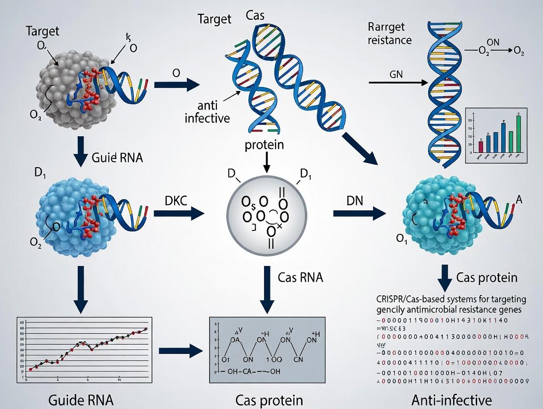
Disarming Superbugs: How CRISPR/Cas Systems Are Revolutionizing the Fight Against Antimicrobial Resistance
This article provides a comprehensive overview for researchers, scientists, and drug development professionals on the use of CRISPR/Cas-based systems to target antimicrobial resistance (AMR) genes.
Disarming Superbugs: How CRISPR/Cas Systems Are Revolutionizing the Fight Against Antimicrobial Resistance
Abstract
This article provides a comprehensive overview for researchers, scientists, and drug development professionals on the use of CRISPR/Cas-based systems to target antimicrobial resistance (AMR) genes. We first explore the foundational principles of CRISPR technology and its logical fit for combating AMR. We then detail current methodologies, including Cas9, Cas12, and Cas13 nucleases for gene inactivation and plasmid curing, alongside delivery strategies like phages and nanoparticles. The discussion addresses critical troubleshooting and optimization challenges, such as off-target effects, specificity, and delivery efficiency. Finally, we compare the efficacy of various CRISPR/Cas systems against traditional and emerging AMR countermeasures, validating their potential. This synthesis aims to guide the development of next-generation, sequence-specific antimicrobials.
The CRISPR Arsenal Against AMR: Understanding the Core Principles and Strategic Rationale
The escalating antimicrobial resistance (AMR) crisis represents a fundamental failure of broad-spectrum antibiotic paradigms. The traditional "one-drug-fits-all" approach exerts immense selective pressure, driving the rapid horizontal gene transfer (HGT) and propagation of resistance determinants across bacterial populations. CRISPR/Cas-based systems emerge as a paradigm-shifting therapeutic and diagnostic framework, offering the precision needed to disarm resistance genes and resensitize pathogens without indiscriminate microbial killing.
Application Notes: CRISPR/Cas Systems for AMR Gene Targeting
Table 1: Current CRISPR/Cas Platforms for AMR Gene Intervention
| System Type | Target Mechanism | Key Advantage | Primary Challenge | Recent In Vitro Efficacy* |
|---|---|---|---|---|
| Cas9 Nuclease | Cleavage of chromosomal AMR genes. | Permanent gene elimination. | Off-target effects; HDR inefficiency in bacteria. | >4-log reduction in mecA-carrying S. aureus (2023). |
| Cas9 Nickase (nCas9) | Single-strand breaks for precise base editing. | Reduced off-target toxicity. | Requires specific PAM sites. | 99.7% blaCTX-M-15 inactivation in E. coli (2024). |
| Catalytically Dead Cas (dCas9) | Silencing via repression (CRISPRi). | Reversible, tunable suppression. | Requires sustained expression. | 1000-fold reduction in ndm-1 expression (2024). |
| Cas13a (C2c2) | Cleavage of AMR gene mRNA transcripts. | Cytoplasmic activity; collateral RNAse effect for diagnostics. | Transcriptional repression only. | 95% reduction in mcr-1 mRNA levels (2023). |
| Cas3 "Shredder" | Processive degradation of large DNA regions. | Efficient against gene clusters or islands. | Excessive DNA damage can trigger SOS response. | Clearance of 50 kb resistance island in K. pneumoniae (2023). |
*Data compiled from recent literature (2023-2024).
Key Insight: The choice of system depends on the resistance mechanism (chromosomal vs. plasmid-borne, enzyme vs. pump), desired outcome (elimination vs. transient suppression), and delivery constraints.
Protocols for Key Experiments
Protocol 1: Design and In Vitro Validation of sgRNAs for Plasmid-Borne β-Lactamase Genes
Objective: To select and validate sgRNAs for targeting prevalent ESBL genes (e.g., blaCTX-M-15) using a Cas9 nuclease system.
Materials:
- Target Sequence: Plasmid DNA harboring blaCTX-M-15.
- Design Tools: CHOPCHOP, Benchling CRISPR tools.
- In Vitro Cleavage Kit: e.g., EnGen Cas9 NLS, NEBuffer r3.1.
- Reagents: Synthesized sgRNA (or Alt-R CRISPR-Cas9 crRNA & tracrRNA), Nuclease-Free Water.
- Analysis: Agarose gel electrophoresis system, TAE buffer, DNA stain.
Procedure:
- sgRNA Design: Identify 5 candidate 20-nt spacer sequences adjacent to 5'-NGG PAM sites within the blaCTX-M-15 open reading frame, prioritizing regions with minimal off-target homology using design tools.
- Reconstitution: Reconstitute lyophilized sgRNA or crRNA:tracrRNA duplex in nuclease-free water to 100 µM.
- RNP Complex Formation: For each reaction, mix 200 ng of target plasmid, 30 nM purified Cas9 protein, and 30 nM of individual sgRNA in 1X NEBuffer r3.1. Final volume: 20 µL. Include a plasmid-only control.
- Incubation: Incubate at 37°C for 1 hour.
- Termination & Analysis: Stop reaction with Proteinase K (0.5 mg/mL, 10 min, 56°C). Run entire product on a 1% agarose gel at 100V for 45 min. Successful cleavage converts supercoiled plasmid to linearized form, visible as a band shift.
Protocol 2: Assessing Bacterial Resensitization via MIC Determination Post-CRISPR Delivery
Objective: To measure the restoration of antibiotic susceptibility following CRISPR-mediated knockout of an AMR gene.
Materials:
- Bacterial Strain: E. coli TOP10 carrying pUC19-blaNDM-1.
- CRISPR Delivery Vector: All-in-one plasmid expressing Cas9 and anti-ndm-1 sgRNA (e.g., pCRISPR-Cas9).
- Controls: Empty vector, non-targeting sgRNA vector.
- Antibiotic Stocks: Meropenem, Ampicillin.
- Culture Media: Cation-adjusted Mueller-Hinton Broth (CAMHB).
- Equipment: 96-well sterile microtiter plates, plate reader.
Procedure:
- Transformation: Transform the target strain with the CRISPR plasmid or controls via electroporation. Select on agar plates with appropriate antibiotic for plasmid maintenance.
- Inoculum Preparation: Grow overnight cultures from single colonies. Dilute to ~5 x 10⁵ CFU/mL in CAMHB.
- MIC Plate Setup: In a 96-well plate, perform two-fold serial dilutions of meropenem (e.g., 128 µg/mL to 0.125 µg/mL) in CAMHB. Add 100 µL of bacterial inoculum to each well. Include growth and sterility controls.
- Incubation & Reading: Incubate plate at 35°C for 16-20 hours. Read optical density at 600 nm.
- Analysis: The MIC is the lowest concentration of antibiotic that inhibits visible growth. Compare MICs between strains carrying the active CRISPR system and controls. A ≥4-fold reduction in MIC indicates successful resensitization.
Visualizations
Diagram 1: CRISPR Systems as a Targeted Solution to the AMR Cycle.
Diagram 2: Cas9 Nuclease Mechanism for AMR Gene Disruption.
The Scientist's Toolkit: Research Reagent Solutions
Table 2: Essential Reagents for CRISPR-Cas AMR Research
| Reagent/Material | Supplier Examples | Function in AMR Research |
|---|---|---|
| Alt-R S.p. HiFi Cas9 Nuclease V3 | Integrated DNA Technologies (IDT) | High-fidelity nuclease for precise, low off-target cleavage of AMR gene sequences. |
| Custom crRNA & tracrRNA | IDT, Sigma-Aldrich | Enables rapid, modular design of guide RNAs against emerging resistance gene variants. |
| EnGen Spy Cas9 NLS | New England Biolabs (NEB) | Nuclear localization signal (NLS)-tagged Cas9 for in vitro cleavage assays and validation. |
| pCas9-CR4 Plasmid | Addgene (plasmid #42876) | All-in-one expression vector for Cas9 and sgRNA in Gram-negative bacteria. |
| pC013-ts Origin Plasmid | Addgene (plasmid #122274) | Temperature-sensitive delivery vector for CRISPR counterselection in bacterial genetics. |
| LentiCRISPR v2 Vector | Addgene (plasmid #52961) | Lentiviral backbone for delivery of CRISPR components into difficult-to-transfect bacterial hosts. |
| Detectr Cas12a (cpf1) Kit | Mammoth Biosciences | For rapid, paper-based diagnostic detection of specific AMR gene sequences. |
| HiScribe T7 Quick High Yield RNA Synthesis Kit | NEB | For in-house synthesis of sgRNA or crRNA transcripts for high-throughput screening. |
| Nucleofector System & Kits | Lonza | Electroporation technology for efficient CRISPR plasmid or RNP delivery into diverse bacterial strains. |
This primer establishes the foundational knowledge of CRISPR/Cas biology and its application as a programmable nuclease system. Within our broader thesis on combating antimicrobial resistance (AMR), CRISPR/Cas systems offer a revolutionary, sequence-specific tool not only for understanding resistance mechanisms but also for directly targeting and eliminating antimicrobial resistance genes (ARGs) from bacterial populations, potentially reversing resistance and restoring antibiotic efficacy.
Part 1: Fundamentals of Adaptive Immunity and Cas Nuclease Function
The Natural CRISPR/Cas Adaptive Immune System
In prokaryotes, the CRISPR/Cas system provides adaptive immunity against invading genetic elements (e.g., plasmids, phages). The system records past infections in the host genome as spacers within the CRISPR array and uses these records to direct sequence-specific cleavage upon re-infection.
Key Stages:
- Adaptation: Cas1-Cas2 complexes capture protospacer sequences from invading DNA and integrate them as new spacers into the CRISPR array.
- Expression: The CRISPR array is transcribed and processed into short CRISPR RNAs (crRNAs).
- Interference: The crRNA guides a Cas nuclease complex to complementary target DNA (protospacer), leading to its cleavage and inactivation.
Repurposing as Programmable Nucleases
The Type II CRISPR/Cas9 system from Streptococcus pyogenes has been simplified for biotechnological use. The system requires two key components:
- Cas9 Nuclease: An endonuclease that creates double-strand breaks (DSBs) in DNA.
- Guide RNA (gRNA): A synthetic fusion of crRNA and trans-activating crRNA (tracrRNA). The 5' ~20-nucleotide spacer sequence confers programmability by base-pairing with the target DNA.
Target recognition requires a short Protospacer Adjacent Motif (PAM) downstream of the target sequence (e.g., 5'-NGG-3' for SpCas9). The Cas9-gRNA complex induces a blunt DSB 3 bp upstream of the PAM.
Table 1: Common CRISPR/Cas Systems and Their Properties
| System & Origin | Canonical Nuclease | PAM Sequence (5'→3') | Cleavage Type (Target Strand) | Primary Application in AMR Research |
|---|---|---|---|---|
| Type II-A (S. pyogenes) | SpCas9 | NGG (or NAG) | Blunt DSB | Gene knockout in resistant pathogens, plasmid curing. |
| Type V-A (Francisella novicida) | FnCas12a (Cpfl) | TTTV (V = A/C/G) | Staggered DSB (5' overhang) | Multiplexed targeting of multiple ARGs. |
| Type II-C (Campylobacter jejuni) | CjCas9 | NNNNRYAC (R = A/G, Y = C/T) | Blunt DSB | Smaller size for delivery via narrow tropism phages. |
| Type VI-A (Leptotrichia shahii) | LshCas13a | Non-coding RNA target | ssRNA cleavage (collateral activity) | Detection and transcriptional silencing of ARG mRNA. |
Diagram 1: CRISPR/Cas9 DNA Targeting Mechanism
Part 2: Application Notes & Protocols for AMR Gene Targeting
Application Note 1: Plasmid Curing to Reverse Resistance
Objective: Eliminate conjugative plasmids carrying ARGs (e.g., bláNDM-1) from a clinical bacterial isolate using a CRISPR/Cas9 plasmid with targeted gRNAs. Rationale: Removing the resistance plasmid restores susceptibility to last-resort antibiotics like carbapenems.
Protocol: Plasmid Curing via Conjugative CRISPR Delivery
gRNA Design & Cloning:
- Design two gRNAs targeting essential regions of the conjugative plasmid (e.g., replication origin oriT and the ARG itself). Avoid off-targets in the host chromosome.
- Clone gRNA sequences into a temperature-sensitive, mobilizable CRISPR/Cas9 plasmid (e.g., pCASP) containing SpCas9.
Conjugative Transfer:
- Prepare donor E. coli strain carrying the pCASP-gRNA plasmid and a helper plasmid for conjugation.
- Mix donor and recipient (target resistant strain) cultures on a filter membrane on non-selective agar.
- Incubate at permissive temperature (30°C) for 4-6 hours to allow conjugation.
Selection & Plasmid Elimination:
- Resuspend cells and plate on agar containing antibiotic selecting for the CRISPR plasmid and an antibiotic to which resistance is conferred by the target plasmid.
- Incubate at restrictive temperature (37°C) to induce CRISPR expression. Successful transconjugants will receive the CRISPR plasmid.
Curing Verification:
- Screen individual colonies by replica plating or PCR for loss of the target ARG.
- Perform antibiotic susceptibility testing (AST) on cured clones to confirm restored sensitivity.
The Scientist's Toolkit: Reagents for Plasmid Curing
| Reagent/Material | Function in the Protocol |
|---|---|
| Temperature-sensitive pCASP Vector | Allows plasmid maintenance at 30°C and Cas9 induction at 37°C. |
| Mobilization Helper Plasmid | Provides in trans conjugation machinery for plasmid transfer. |
| Clinical Bacterial Isolate | The target AMR strain harboring the resistance plasmid. |
| gRNA Oligonucleotides | Designed to target essential sequences on the conjugative plasmid. |
| Antibiotics for Selection | Select for the CRISPR plasmid and counter-select the target resistance plasmid. |
| PCR Primers for ARG | Verify the physical loss of the resistance gene from cured clones. |
| AST Strips/Discs | Confirm phenotypic reversal to antibiotic susceptibility (e.g., lower MIC). |
Diagram 2: Workflow for CRISPR-Based Plasmid Curing
Application Note 2: Sensitizing Biofilm-Associated Infections
Objective: Disrupt biofilms of multidrug-resistant Pseudomonas aeruginosa by targeting chromosomal ARGs and biofilm-related genes. Rationale: Biofilms confer extreme tolerance. Combining CRISPR targeting with sub-inhibitory antibiotics can enhance eradication.
Protocol: Biofilm Disruption Using Cas9 RNP Complexes
RNP Complex Preparation:
- Purify Cas9 Protein: Express His-tagged SpCas9 in E. coli and purify via Ni-NTA chromatography.
- Synthesize gRNA: Use in vitro transcription or purchase synthetic, chemically modified gRNAs targeting a key ARG (e.g., gyrA mutation for fluoroquinolone resistance) and a biofilm regulator (e.g., lasR).
- Form RNP: Mix purified Cas9 and gRNA at a 1:2 molar ratio in nuclease-free buffer. Incubate at 25°C for 10 min.
RNP Delivery into Biofilms:
- Grow 24-hour P. aeruginosa biofilms in a 96-well plate or on a catheter fragment.
- Wash biofilm gently with PBS.
- Treat with RNP complexes (e.g., 500 nM) combined with a peptide-based delivery vehicle (e.g., cell-penetrating peptide or dendrimer) in a sub-inhibitory concentration of ciprofloxacin. Incubate for 4-6 hours.
Assessment of Biofilm Integrity and Viability:
- Biomass: Quantify using crystal violet staining.
- Viability: Use resazurin reduction assay or plate counting of dispersed biofilm cells.
- ARG Disruption: Extract genomic DNA from treated biofilms and perform T7 Endonuclease I (T7EI) assay or deep sequencing to measure editing efficiency at the target locus.
Table 2: Quantitative Outcomes of Biofilm Targeting with Cas9 RNP + Antibiotic
| Treatment Condition (vs. Untreated Biofilm) | Biofilm Biomass Reduction (%) | Viable Cell Count Reduction (Log10 CFU) | Editing Efficiency at gyrA Target (%) |
|---|---|---|---|
| Sub-inhibitory Ciprofloxacin Only | 15 ± 5 | 0.5 ± 0.2 | 0 |
| Cas9 RNP (Anti-gyrA) Only | 20 ± 8 | 1.2 ± 0.3 | 45 ± 10 |
| Cas9 RNP (Anti-lasR) Only | 40 ± 7 | 1.0 ± 0.4 | N/A |
| RNP (Anti-gyrA) + Ciprofloxacin | 65 ± 10 | 3.8 ± 0.5 | 48 ± 12 |
| Scrambled gRNA RNP + Ciprofloxacin | 18 ± 6 | 0.7 ± 0.3 | 0 |
Diagram 3: Biofilm Sensitization Strategy
This primer underscores the dual utility of CRISPR/Cas systems: as a fundamental component of prokaryotic biology and as a precision tool for biomedical research. The provided protocols for plasmid curing and biofilm sensitization exemplify its direct application in the strategic fight against antimicrobial resistance. By enabling the specific targeting and inactivation of ARGs, CRISPR-based technologies present a promising avenue for developing "anti-resistance" therapies that could restore the efficacy of existing antibiotics.
Application Notes
CRISPR/Cas systems, evolved as adaptive immune mechanisms in prokaryotes, are now being repurposed to directly combat antimicrobial resistance (AMR). The conceptual leap involves using these systems to precisely target and inactivate antimicrobial resistance genes (ARGs) within bacterial populations or to sensitize resistant pathogens to conventional antibiotics. This approach moves beyond traditional antibiotic discovery, offering a sequence-specific, programmable weapon against the genetic basis of resistance. Current research focuses on two primary strategies: (i) the use of CRISPR/Cas-based "armed" bacteriophages (phage therapy) to deliver ARG-targeting systems into bacterial populations, and (ii) the development of CRISPR-Cas13a-based diagnostic tools for rapid detection of AMR genotypes to guide treatment. Recent studies demonstrate efficacy both in vitro and in preclinical infection models, showing significant reductions in bacterial load and resistance gene carriage.
Table 1: Recent Quantitative Data on CRISPR/Cas-Based AMR Gene Targeting In Vivo
| Target ARG/Pathogen | CRISPR System | Delivery Vehicle | Animal Model | Key Outcome (vs Control) | Study Year |
|---|---|---|---|---|---|
| mecA (MRSA) | Cas9 | Engineered Phage | Mouse Skin Infection | >99% reduction in MRSA load; restored β-lactam susceptibility | 2023 |
| ndm-1 (Carbapenem-resistant E. coli) | Cas3 | Conjugative Plasmid | Mouse Gut Colonization | 4-log reduction in NDM-1-positive bacterial abundance | 2024 |
| blaKPC (K. pneumoniae) | Cas9 | Lipid Nanoparticles | Mouse Pneumonia Model | 3.5-log CFU reduction in lungs; 80% survival increase | 2023 |
| Multiple ESBL Genes | Cas13a (diagnostic) | N/A (RPA/CRISPR assay) | Clinical Sputum Samples | 100% specificity, 97% sensitivity in 1 hour | 2024 |
Table 2: Comparison of CRISPR-Cas Systems for AMR Intervention
| System | Target | Action Mechanism | Primary Advantage for AMR | Key Challenge |
|---|---|---|---|---|
| Cas9 | DNA | Double-strand break, gene knockout | Permanent elimination of ARG | Off-target effects; requires PAM |
| Cas12a | DNA | Double-strand break, gene knockout | Creates staggered cuts; simpler crRNA | Slower kinetics |
| Cas13a | RNA | Collateral ssRNA cleavage | Can degrade mRNA without genomic alteration; ideal for diagnostics | Transient effect; collateral activity must be controlled |
| Cas3 | DNA | Processive DNA degradation | Large deletions, prevents repair | Difficult to control exact deletion size |
Experimental Protocols
Protocol 1: Design and Assembly of a CRISPR-Cas9 Phage formecATargeting in MRSA
Objective: To construct an engineered bacteriophage capable of delivering a mecA-targeting CRISPR-Cas9 system into Methicillin-Resistant Staphylococcus aureus (MRSA). Materials: Lysogenic Staphylococcus phage (e.g., ΦNM1), mecA-specific spacer sequence oligos, Cas9 gene codon-optimized for S. aureus, E. coli cloning strain, phage propagation strain, Q5 High-Fidelity DNA Polymerase, T4 DNA Ligase, BsaI-HF restriction enzyme, LB broth, SM buffer, PEG 8000, DNase I/RNase A. Procedure:
- Spacer and crRNA Array Cloning: Design a 20-nt spacer sequence complementary to the mecA gene. Synthesize oligos, anneal, and clone into the BsaI site of a shuttle plasmid containing a codon-optimized cas9, a tracrRNA, and a phage-specific integration site.
- Phage Engineering: Propagate the wild-type ΦNM1 phage on a permissive S. aureus strain. Islate phage genomic DNA. Using phage recombinase-mediated recombineering, integrate the CRISPR-Cas9 cassette from the shuttle plasmid into a non-essential region of the phage genome.
- Phage Purification: Infect a liquid culture of the propagating strain with the engineered phage lysate. After lysis, filter sterilize (0.45 µm). Precipitate phage with PEG/NaCl, resuspend in SM buffer, and purify via CsCl density gradient centrifugation.
- Validation: Confirm spacer sequence via Sanger sequencing of the engineered phage DNA. Test targeting efficiency by infecting a broth culture of MRSA (MOI=10) with the engineered phage. Plate serial dilutions on oxacillin-containing and non-containing media after 6h. A significant reduction in CFU on non-antibiotic plates and restored sensitivity (CFU on oxacillin) indicates successful mecA disruption.
Protocol 2: Rapid Detection of ESBL Genes using RPA-CRISPR-Cas13a
Objective: To detect Extended-Spectrum Beta-Lactamase (ESBL) genes (blaCTX-M, blaTEM, blaSHV) from bacterial isolates within 60 minutes. Materials: Cas13a protein, crRNA designed for conserved ESBL gene regions, Recombinase Polymerase Amplification (RPA) kit (TwistAmp Basic), fluorescent reporter RNA (e.g., FAM-UU-BHQ1), nitrocellulose lateral flow strips (if using biotin-labeled reporters), heat block/water bath. Procedure:
- Sample Prep: Boil 1-3 bacterial colonies in 50 µL nuclease-free water for 10 min, centrifuge, and use supernatant as DNA template.
- RPA Amplification: Prepare a 50 µL RPA reaction per manufacturer's instructions using primers specific to the target ESBL gene(s). Incubate at 37-42°C for 15-20 minutes.
- CRISPR-Cas13a Detection: Pre-mix 5 µL of the RPA product with 15 µL of detection mix containing 50 nM Cas13a, 62.5 nM specific crRNA, and 125 nM fluorescent reporter. Incubate at 37°C for 10-30 minutes.
- Output: Measure fluorescence in a plate reader (ex/em ~485/535 nm) at 5-minute intervals. A time-dependent increase in fluorescence indicates positive detection. For lateral flow readout, use a biotin-labeled reporter and FAM-labeled crRNA; a positive test shows both control and test lines.
The Scientist's Toolkit
Table 3: Key Research Reagent Solutions for CRISPR-based AMR Research
| Reagent/Material | Function/Application | Example/Notes |
|---|---|---|
| Cas9 Nuclease (S. aureus optimized) | Executes DNA cleavage of target ARG. | Requires codon-optimization for functional expression in the target bacterial species. |
| CRISPR-Cas13a Detection Kit | For rapid, sensitive detection of ARG transcripts or amplicons. | Commercial kits (e.g., SHERLOCK, DETECTR) combine RPA/LAMP with Cas13a/Cas12. |
| Phage Engineering Kit | Facilitates cloning and integration of CRISPR cassettes into phage genomes. | Often includes recombinase proteins and phage-specific integration plasmids. |
| Synthetic crRNA & tracrRNA | Provides target specificity for Cas9; can be ordered as custom synthetic RNA. | Chemically modified crRNAs can enhance stability in vivo. |
| RPA (TwistAmp) Kit | Isothermal amplification of target ARG sequences for downstream Cas detection. | Enables rapid, equipment-free amplification critical for point-of-care diagnostics. |
| Fluorescent RNA Reporter (FAM-UU-BHQ1) | Signal generation in Cas13a-based assays; cleavage relieves quenching. | The backbone and modifications affect cleavage kinetics and background signal. |
| Conjugative Delivery Plasmid | Enables transfer of CRISPR machinery between bacterial cells via conjugation. | Useful for targeting ARGs in mixed populations or biofilms. |
Visualizations
Strategy for Turning Bacterial Defense into AMR Offense
Workflow for Developing CRISPR-Based AMR Solutions
Mechanisms of Cas9 vs Cas13a in AMR Targeting
Within the broader thesis on developing CRISPR/Cas-based systems to combat antimicrobial resistance (AMR), a critical strategic decision lies in target selection. The genetic localization of a resistance gene—whether on the bacterial chromosome or on mobile plasmids—profoundly influences the dynamics of resistance spread, the efficacy of a CRISPR/Cas intervention, and its evolutionary consequences. This application note provides a comparative analysis and experimental protocols to guide researchers in prioritizing and validating these distinct genetic targets.
Table 1: Key Characteristics of Plasmid-Borne vs. Chromosomal Resistance Genes
| Characteristic | Plasmid-Borne Resistance Genes | Chromosomal Resistance Genes |
|---|---|---|
| Primary Threat | Horizontal Gene Transfer (HGT), rapid dissemination across strains/species. | Vertical inheritance, clonal expansion within a lineage. |
| Genetic Context | Often within mobile genetic elements (MGEs) like transposons, integrons. | Often point mutations in housekeeping genes or acquired gene islands. |
| Copy Number | Variable; can be multiple copies per cell (medium/high copy plasmids). | Typically one or two copies per chromosome. |
| Stability | Can be lost without selection pressure (curing). | Stable, not easily lost. |
| CRISPR/Cas Challenge | Requires delivery to high proportion of population to halt spread. Potential for plasmid escape variants. | Requires high cleavage efficiency within each cell. Risk of selecting CRISPR escape mutants. |
| Therapeutic Goal | "Anti-dissemination": Blocking HGT, reversing resistance in populations. | "Anti-escalation": Suppressing resistant clones, re-sensitizing infections. |
Table 2: 2023-2024 Surveillance Data on Prevalent Resistance Mechanisms by Location
| Resistance Mechanism (Example) | Common Gene(s) | Predominant Location (Estimated %) | Key Pathogens |
|---|---|---|---|
| Extended-Spectrum β-Lactamase (ESBL) | blaCTX-M, blaTEM, blaSHV | Plasmid (>85%) | E. coli, K. pneumoniae |
| Carbapenemase | blaKPC, blaNDM | Plasmid (>95%) | Enterobacterales |
| Metallo-β-lactamase | blaNDM-1 | Plasmid (~100%) | Acinetobacter spp., Pseudomonas |
| Fluoroquinolone Resistance | qnr series | Plasmid (>70%) | Enterobacteriaceae |
| Colistin Resistance | mcr-1 to mcr-10 | Plasmid (~100%) | E. coli, Salmonella |
| Vancomycin Resistance | vanA operon | Plasmid/Transposon (Tn1546) | Enterococcus faecium |
| Methicillin Resistance | mecA (SCCmec element) | Chromosomal (Mobile Island) | Staphylococcus aureus |
| Fluoroquinolone Resistance | Mutations in gyrA/parC | Chromosomal (Mutation) | Neisseria gonorrhoeae |
| Rifampin Resistance | Mutations in rpoB | Chromosomal (Mutation) | Mycobacterium tuberculosis |
Experimental Protocols
Protocol 1: Determining the Genomic Localization of a Resistance Gene
Objective: To experimentally confirm whether a resistance gene of interest is located on the chromosome or on a plasmid.
Materials: See "The Scientist's Toolkit" (Section 5).
Methodology:
Bacterial Culture & Plasmid Curing (Optional):
- Inoculate the resistant strain in LB broth without antibiotic and incubate overnight at 37°C.
- Perform serial passages (e.g., 1:1000 dilution) daily for 5-7 days in antibiotic-free medium.
- Plate dilutions on non-selective agar. Replica-plate or patch 100 colonies onto antibiotic-containing agar and plain agar.
- Isolates that lose resistance suggest plasmid-borne genes.
Plasmid DNA Extraction:
- Using a commercial kit, isolate plasmid DNA from an overnight culture of the resistant strain.
- Perform a separate genomic DNA extraction from the same strain.
PCR Amplification:
- Design primers specific to the target resistance gene.
- Set up two parallel PCR reactions:
- Reaction A: Template = Plasmid DNA.
- Reaction B: Template = Genomic DNA.
- Include positive control (known plasmid-borne gene) and negative controls (no template, susceptible strain DNA).
Analysis & Interpretation:
- Run PCR products on an agarose gel.
- Plasmid-borne gene: Strong amplification from plasmid DNA. Weak or no amplification from genomic DNA (if pure).
- Chromosomal gene: Amplification from genomic DNA only. No amplification from purified plasmid DNA.
- Ambiguous/Integrated gene: Amplification from both. Proceed to Southern Blot or WGS.
Confirmation by Southern Blot or Whole Genome Sequencing (WGS):
- For Southern blot, digest genomic DNA with a rare-cutter restriction enzyme, run on a pulse-field gel, blot, and probe with the resistance gene. Plasmid bands will differ in size from chromosomal fragments.
- WGS followed by de novo assembly and BLAST analysis provides definitive localization.
Protocol 2: Assessing CRISPR/Cas9 Efficacy Against Chromosomal vs. Plasmid Targets
Objective: To compare the killing efficiency and escape mutant frequency when using a CRISPR/Cas9 system targeting a resistance gene on the chromosome versus on a plasmid.
Materials: Two isogenic strains: (1) Chromosomal resistance mutant, (2) Plasmid-bearing strain (cured version as susceptible control). Conjugative plasmid or phage for delivery of CRISPR/Cas9 system. Appropriate selective media.
Methodology:
CRISPR/Cas9 Construct Design:
- Design identical spacer sequences targeting a core region of the resistance gene present in both strains.
- Clone spacers into a delivery vector expressing Cas9 and the sgRNA. Include a non-targeting sgRNA control.
Delivery:
- For the plasmid-borne target, deliver the CRISPR/Cas9 construct via conjugation or electroporation.
- For the chromosomal target, use the same delivery method.
- Include an empty vector control for both strains.
Efficacy Assay:
- Plate transformed/conjugated cells on selective media with and without the cognate antibiotic.
- Incubate and count colonies after 24-48 hours.
- Calculate Killing Efficiency: (1 - (CFU on antibiotic plate / CFU on non-antibiotic plate)) x 100%.
Escape Mutant Analysis:
- From the selective antibiotic plate, pick 20-30 colonies.
- Isolate plasmid and genomic DNA. Perform PCR and Sanger sequence the target region in both.
- For plasmid target: Look for plasmid loss (curing) or sequence mutations/deletions.
- For chromosomal target: Look for in-frame mutations, large deletions, or proof-of-killing (no growth in PCR).
Table 3: Expected Experimental Outcomes
| Metric | Plasmid-Borne Target | Chromosomal Target |
|---|---|---|
| Primary Mechanism of Re-sensitization | Plasmid curing or cleavage without repair. | Chromosomal cleavage leading to cell death (bactericidal) or large deletions. |
| Killing/Efficacy Rate | High (if delivery efficient), but depends on copy number. | High, but requires double-strand break lethality. |
| Escape Mutant Frequency | High: Surviving cells often harbor mutated or recombined plasmids. | Lower but significant: Surviving cells may have inactivating mutations or CRISPR system failure. |
| Escape Mutant Type | Plasmid evaders (rearranged, spacer escape). | Chromosomal mutants (small indels, gene disruption). |
Strategic Decision Pathway for Target Selection
The Scientist's Toolkit
Table 4: Key Research Reagent Solutions for Localization & Targeting Studies
| Item / Reagent | Function in Protocol | Example (Supplier) |
|---|---|---|
| Plasmid Miniprep Kit | Selective isolation of plasmid DNA from bacterial lysates, crucial for differentiating plasmid vs. chromosomal location. | Qiagen QIAprep Spin Miniprep Kit |
| Genomic DNA Extraction Kit | Purification of high-molecular-weight chromosomal DNA, free of plasmid contamination. | Thermo Fisher GeneJET Genomic DNA Purification Kit |
| PCR Master Mix | Amplification of target resistance genes from different DNA templates. | NEB Q5 High-Fidelity 2X Master Mix |
| Conjugative Delivery Vector | Enables transfer of CRISPR/Cas9 machinery via bacterial mating, essential for in situ plasmid targeting studies. | pSW-2 (or similar E. coli mobilizable vector) |
| Electrocompetent Cells | High-efficiency transformation for delivering CRISPR constructs, especially for non-conjugative strains. | Lucigen ElectroTen-Blue |
| CRISPR/Cas9 Cloning Kit | Modular system for sgRNA insertion and Cas9 expression vector assembly. | Addgene Kit #1000000057 (pCas9) |
| Pulsed-Field Gel Electrophoresis System | Separates large DNA fragments (whole plasmids, chromosomal digests) for Southern blot confirmation. | Bio-Rad CHEF-DR II System |
| Selective Agar Media | Contains specific antibiotics to phenotype resistance loss or CRISPR-mediated killing. | Mueller-Hinton Agar w/ antibiotics |
| Whole Genome Sequencing Service | Definitive analysis of genetic context, plasmid maps, and escape mutant mutations. | Illumina Nextera Flex / PacBio HiFi |
The escalating crisis of antimicrobial resistance (AMR) necessitates a paradigm shift from broad-spectrum conventional antibiotics to precision antimicrobials. Within the broader thesis of CRISPR/Cas-based systems for AMR gene targeting, this application note details the core advantages of specificity and evolvability. These systems, particularly CRISPR/Cas13a (targeting RNA) and CRISPR/Cas9 (targeting DNA), offer programmable, sequence-specific elimination of resistance genes or pathogens while sparing the commensal microbiota—a key limitation of conventional drugs. Furthermore, their design is inherently evolvable; guide RNAs can be rapidly redesigned in silico to counter newly emergent resistance genotypes, a process far slower for small-molecule antibiotic development.
Quantitative Comparison: Specificity & Evolvability Metrics
Table 1: Comparative Analysis of Conventional Antibiotics vs. CRISPR/Cas Antimicrobials
| Feature | Conventional Broad-Spectrum Antibiotics | CRISPR/Cas-Based Antimicrobials |
|---|---|---|
| Spectrum of Activity | Broad (often against multiple bacterial genera) | Ultra-narrow (programmable to a specific DNA/RNA sequence, ~20-30 nt) |
| Impact on Commensal Microbiota | High collateral damage (dysbiosis) | High potential for species- or strain-specific targeting |
| Development Timeline for New Variants | 10-15 years (new chemical entity) | Potentially <1 year (new guide RNA design & synthesis) |
| Primary Resistance Mechanism | Target modification, efflux pumps, enzyme inactivation | Target sequence mutation in PAM/protospacer; countered by re-designing gRNA |
| "Evolvability" (Adaptation Speed) | Low (fixed chemical structure) | Very High (sequence reprogrammable via synthetic gRNA) |
| Typical Specificity Validation (in vitro) | MIC/MBC against pure cultures | Next-generation sequencing (NGS) of off-target effects; fluorescence assays with mismatched targets |
Table 2: Representative Experimental Data from Recent Studies (2023-2024)
| Study Target (CRISPR System) | Specificity Metric Reported | Evolvability/Adaptation Demonstrated | Key Quantitative Result |
|---|---|---|---|
| Carbapenemase (blaKPC) gene (Cas9) | No effect on E. coli lacking blaKPC; NGS showed no significant off-targets in genome. | Single gRNA restored carbapenem sensitivity. | >4-log reduction in target bacterial load in murine infection model. |
| Methicillin Resistance (mecA) gene in MRSA (Cas9) | Discrimination of single-nucleotide polymorphism (SNP) in mecA variant. | Two alternative gRNAs designed for common SNP variants. | 99.7% killing of MRSA in planktonic culture; no effect on isogenic MSSA. |
| Pan-aminoglycoside resistance (16S rRNA methyltransferases) (Cas13a) | Cas13a collateral activity contained via engineered phage delivery. | A single crRNA array designed to target 5 different armA gene family alleles. | 90-99% reduction in viable counts across 3 Enterobacteriaceae species. |
| Multidrug-Resistant P. aeruginosa (Cas3) | Phage-delivered system targeted a unique bacterial strain identifier. | Guide re-targeting demonstrated against 3 different clinical strain genotypes. | Specific biofilm eradication (>3-log reduction) without affecting other biofilm members. |
Experimental Protocols
Protocol 3.1: Assessing Specificity: Off-Target Analysis for a CRISPR/Cas9 Antimicrobial
Aim: To validate that a designed CRISPR/Cas9 system targeting an AMR gene (e.g., blaNDM-1) does not cleave genomic off-target sites. Materials: See "Research Reagent Solutions" (Section 5). Method:
- In Silico Prediction: Use tools like Cas-OFFinder or CHOPCHOP to predict potential off-target sites in the host genome (allowing up to 3-5 mismatches).
- Cell-free Cleavage Assay: Synthesize PCR amplicons (300-500 bp) encompassing the top 10 predicted off-target loci and the intended on-target site.
- Reaction Setup: Combine per 20 µL reaction: 100 ng DNA amplicon, 50 nM purified Cas9 nuclease, 50 nM target-specific sgRNA (or non-targeting control), 1X Cas9 reaction buffer. Incubate at 37°C for 1 hour.
- Analysis: Run products on a 2% agarose gel. Cleavage is indicated by the disappearance of the full-length amplicon and appearance of smaller fragments. Quantify band intensity to calculate cleavage efficiency (%).
- Whole-Genome Sequencing (WGS): Treat the bacterial strain (harboring the AMR gene) with the CRISPR/Cas9 system delivered via electroporation or conjugative plasmid. Isolate genomic DNA from treated and control populations. Perform Illumina WGS (30x coverage). Align reads to reference genome and use tools like CRISPResso2 or Bowtie2 to identify insertions/deletions (indels) at the on-target and predicted off-target sites. Significant indel frequency above background (typically >0.1%) at an off-target site indicates a lack of specificity.
Protocol 3.2: Demonstrating Evolvability: Rapid Reprogramming to Counter a Point Mutation
Aim: To design and validate a new sgRNA to restore activity against an AMR gene that has acquired a point mutation escaping the original sgRNA. Materials: See "Research Reagent Solutions" (Section 5). Method:
- Escape Mutant Generation: Grow the target bacterium (e.g., E. coli with blaCTX-M-15) under sub-lethal pressure of the original CRISPR/Cas9 system (e.g., delivered via a temperate phage). Plate survivors and isolate colonies.
- Sequencing: Sanger sequence the target region of the AMR gene from escape mutants to identify the causative point mutation(s).
- Guide RNA Re-design: Using the new mutant target sequence, design a new sgRNA. Adhere to design rules: ensure a PAM (e.g., 5'-NGG-3' for SpCas9) is adjacent to the target region. Prioritize guides with minimal predicted off-targets.
- Synthesis & Cloning: Synthesize the new sgRNA oligonucleotide and clone it into your delivery vector (e.g., plasmid or phage genome) replacing the original guide.
- Efficacy Validation: Deliver the new CRISPR/Cas9 construct against both the original strain and the escape mutant. Perform time-kill curves or minimum inhibitory concentration (MIC) assays in the presence of the relevant antibiotic (e.g., cefotaxime). Successful evolvability is shown by the restoration of killing/efficacy against the escape mutant.
Visualizations
CRISPR Evolvability Workflow
Specificity & Evolvability Comparison
The Scientist's Toolkit: Research Reagent Solutions
Table 3: Essential Materials for CRISPR/Cas AMR Targeting Experiments
| Reagent / Solution | Function & Rationale | Example Product/Provider |
|---|---|---|
| High-Fidelity Cas9 Nuclease (Purified) | Ensures precise DNA cleavage with minimal off-target activity for specificity assays. | Alt-R S.p. HiFi Cas9 Nuclease V3 (Integrated DNA Technologies) |
| In Vitro Transcription Kit (for gRNA/crRNA) | Generates high-yield, pure guide RNAs for cell-free cleavage assays and rapid prototyping. | MEGAshortscript T7 Transcription Kit (Thermo Fisher) |
| CRISPR-Cas9 Delivery Vector (Phage or Plasmid) | Enables efficient delivery of the system into target bacterial cells for in vivo validation. | pCRISPR or engineered λ phage-based vectors (Addgene, commercial phage kits) |
| Off-Target Prediction Software | Identifies potential genomic off-target sites to guide specificity analysis and gRNA design. | Cas-OFFinder (open source), IDT's off-target predictor (web tool) |
| Next-Generation Sequencing Kit | Allows whole-genome sequencing to empirically validate on- and off-target effects. | Illumina DNA Prep Kit (Illumina) |
| CRISPR Analysis Software | Quantifies indel frequencies from sequencing data to measure editing efficiency and specificity. | CRISPResso2 (open source) |
| Synthetic Oligonucleotides for gRNA Cloning | Rapid, cost-effective source for cloning new guide sequences to demonstrate evolvability. | Custom DNA Oligos (Twist Bioscience, Sigma-Aldrich) |
| Electrocompetent Target Bacteria | Essential for transforming CRISPR plasmids into hard-to-transfect clinical bacterial isolates. | In-house prepared or commercial high-efficiency electrocompetent cells. |
From Lab to Pathogen: CRISPR/Cas Delivery Systems and Practical Applications for AMR Gene Disruption
Antimicrobial resistance (AMR) poses a catastrophic threat to global health. Within a broader thesis on CRISPR/Cas-based countermeasures, this application note details the strategic selection and deployment of three distinct Cas nucleases—Cas9, Cas12, and Cas13—for targeting AMR genes in bacterial pathogens and mobile genetic elements. Each system offers unique mechanisms of action suitable for different AMR gene classes and experimental objectives.
Comparative Analysis of Key Cas Systems
Table 1: Core Characteristics of Cas9, Cas12, and Cas13 Systems
| Feature | Cas9 (SpCas9) | Cas12a (Cpf1) | Cas13a (LshCas13a) |
|---|---|---|---|
| Target Molecule | dsDNA | dsDNA | ssRNA |
| PAM/PFS Requirement | 5'-NGG-3' (SpCas9) | 5'-TTTV-3' (LbCas12a) | 3' of protospacer, non-G |
| Cleavage Mechanism | Blunt dsDNA breaks | Staggered dsDNA breaks | Collateral ssRNA cleavage |
| Key Applications in AMR | Gene knockout, CRISPRi, plasmid curing | Multiplex gene editing, plasmid degradation | Transcript degradation, nucleic acid detection |
| Delivery Methods (Bacteria) | Plasmid, ribonucleoprotein (RNP) | Plasmid, RNP | Plasmid, RNP |
| Noted Efficiency in AMR Models | 60-95% (gene knockout) | 70-90% (multiplex editing) | >99% (transcript knockdown) |
Table 2: Quantitative Performance in Model AMR Gene Studies
| Cas System | Target AMR Gene | Model Organism | Reported Efficacy | Key Metric |
|---|---|---|---|---|
| Cas9 | mecA (MRSA) | S. aureus | ~90% kill rate | Bacterial killing in vitro |
| Cas9 (CRISPRi) | blaNDM-1 | E. coli | 100-fold reduction in MIC | Minimum Inhibitory Concentration |
| Cas12a | tet(M), erm(B) | Enterococcus faecalis | 85% co-cleavage | Plasmid curing frequency |
| Cas13a | blaCTX-M mRNA | K. pneumoniae | 99% transcript reduction | qRT-PCR (ΔCq) |
Application Notes & Detailed Protocols
Protocol A: Cas9-Mediated Knockout of Chromosomal β-Lactamase Genes
Objective: To permanently disrupt the blaKPC gene in a carbapenem-resistant K. pneumoniae isolate.
Research Reagent Solutions:
| Reagent/Material | Function |
|---|---|
| pCas9-KPC-sgRNA Plasmid | Expresses SpCas9 and target-specific sgRNA. |
| Electrocompetent K. pneumoniae Cells | For efficient plasmid DNA transformation. |
| SOC Recovery Medium | Enhances cell viability post-electroporation. |
| Kanamycin (50 µg/mL) | Selects for plasmid-containing transformants. |
| Carbapenem (Meropenem) Discs | Verifies loss of resistance phenotype. |
| T7 Endonuclease I Assay Kit | Detects indels at target locus. |
| PfuUltra II Fusion HS DNA Polymerase | Amplifies target locus for sequencing validation. |
Methodology:
- sgRNA Design: Design a 20-nt spacer sequence upstream of a 5'-NGG-3' PAM within the blaKPC open reading frame. Clone into pCas9 vector using BsaI restriction sites.
- Transformation: Introduce 100 ng of purified plasmid into 50 µL of electrocompetent K. pneumoniae via electroporation (2.5 kV, 200Ω, 25µF). Recover in 1 mL SOC for 2 hours at 37°C.
- Selection & Screening: Plate on LB agar + Kanamycin. Incubate 16h at 37°C. Screen 10 colonies by colony PCR (using primers flanking target site) and Sanger sequence to confirm indel mutations.
- Phenotypic Validation: Perform disc diffusion assay with meropenem (10 µg) according to CLSI guidelines. Compare zone of inhibition to wild-type isolate.
Protocol B: Cas12a Multiplexed Targeting of Conjugative Plasmid-Borne AMR Genes
Objective: To cure an IncF plasmid harboring blaCTX-M-15 and aac(6')-Ib-cr from an E. coli clinical isolate.
Diagram 1: Cas12a RNP plasmid curing workflow.
Methodology:
- crRNA Design & RNP Complex Formation:
- Design two 22-nt crRNAs targeting the plasmid replication (rep) and resolution (res) genes, adjacent to 5'-TTTV-3' PAMs.
- Synthesize crRNAs and complex with purified LbCas12a protein (Integrated DNA Technologies) at a 3:1 molar ratio in Nuclease-Free Duplex Buffer. Incubate 10 min at 25°C.
- RNP Electroporation: Wash target E. coli culture in ice-cold 10% glycerol. Mix 10 µL of cells with 5 µL of RNP complex (5 µM final). Electroporate using conditions optimized for Gram-negatives.
- Recovery and Plasmid Loss Enrichment: Recover cells in LB for 4 hours. Serial dilute and plate on non-selective LB agar. Incubate overnight.
- Screening: Patch 100 colonies onto LB and LB + Ceftazidime (2 µg/mL). Isolates growing only on non-selective media are potential cured strains. Confirm via plasmid extraction and PCR for blaCTX-M-15.
Protocol C: Cas13a-Based Transcriptional Suppression (CRISPRa) of Efflux Pump Genes
Objective: To knock down expression of the mexB gene of the MexAB-OprM efflux pump in P. aeruginosa, restoring antibiotic susceptibility.
Diagram 2: Cas13a collateral RNA cleavage mechanism.
Methodology:
- Inducible Cas13a System: Clone LwaCas13a under an anhydrotetracycline (aTc)-inducible promoter in a broad-host-range vector. Clone the mexB-targeting crRNA downstream of a constitutive promoter.
- Conditional Expression: Transform the construct into P. aeruginosa via conjugation. Grow cultures to mid-log phase and induce with 100 ng/mL aTc for 6 hours.
- Transcript Quantification: Harvest cells. Extract total RNA, treat with DNase. Perform qRT-PCR for mexB using rpoD as a housekeeping control. Calculate fold-change (2^-ΔΔCq) relative to uninduced control.
- Phenotypic Assay: Perform broth microdilution for levofloxacin (a MexAB-OprM substrate) according to CLSI M07. Compare MICs between induced and uninduced cultures.
Strategic Selection Guide & Decision Tree
Table 3: Selection Guide for AMR Strategy
| Primary Goal | Recommended System | Rationale | Key Consideration |
|---|---|---|---|
| Permanent elimination of chromosomal resistance gene | Cas9 (Nuclease) | Creates irreversible double-strand breaks, leading to frameshift mutations. | Off-target effects in genome; requires functional repair system. |
| Transcriptional repression (CRISPRi) of multiple genes | dCas9 (Catalytically Dead) | Efficient, programmable block of transcription elongation. | Reversible effect; requires tight repression control. |
| Elimination of multiple plasmids or ICEs | Cas12 (Multiplexable) | Processes its own crRNAs, enabling multiplexing with a single array; cleaves dsDNA. | Requires T-rich PAM; staggered cuts may aid repair. |
| Sensitive detection of AMR genes (diagnostics) | Cas12/Cas13 (Collateral Activity) | Exhibits trans-cleavage upon target recognition, enabling amplification-free detection. | Used for surveillance, not therapeutic. |
| Knockdown of mRNA without genomic change | Cas13 | Targets RNA directly, ideal for transient sensitization or studying essential AMR genes. | Effect is transient; high expression needed. |
Concluding Remarks
The strategic deployment of CRISPR/Cas systems requires alignment of the molecular target (DNA vs. RNA), desired outcome (permanent edit vs. transient modulation), and delivery constraints. Cas9 remains the gold standard for precise chromosomal editing, Cas12 excels at multiplexed plasmid targeting, and Cas13 offers unique RNA-level intervention. Integrating these tools provides a versatile arsenal for direct AMR gene disruption, functional genomics, and novel therapeutic development, forming a critical component of the thesis on next-generation AMR countermeasures.
Designing sgRNAs for Maximum Efficiency Against Key Resistance Determinants
Within the broader thesis on CRISPR/Cas-based systems for targeting antimicrobial resistance (AMR) genes, this application note provides a focused protocol for designing single guide RNAs (sgRNAs) with maximum on-target efficiency against key bacterial resistance determinants. The precision of sgRNA design is the critical first step in developing effective CRISPR-Cas antimicrobials, as it directly dictates the specificity and cleavage activity of the Cas nuclease against genes encoding extended-spectrum beta-lactamases (ESBLs), carbapenemases, and other priority resistance mechanisms.
Core Principles for High-Efficiency sgRNA Design
Optimal sgRNA design integrates multiple sequence and structural parameters to predict and maximize Cas9 (or Cas12a) cleavage activity. The following factors must be evaluated concurrently.
Sequence-Based Parameters
- GC Content: Aim for 40-60% GC content within the 20-nt spacer sequence. This range promotes stable DNA-RNA heteroduplex formation without excessive stability that can reduce specificity.
- Protospacer Adjacent Motif (PAM): The PAM sequence is Cas-protein-specific (e.g., 5'-NGG-3' for Streptococcus pyogenes Cas9). The sgRNA must be designed immediately 5' to the PAM on the target strand.
- Seed Region: The 8-12 nucleotides proximal to the PAM are the "seed region" and require perfect complementarity for efficient cleavage. Mismatches here drastically reduce activity.
- Avoidance of Self-Complementarity: The sgRNA sequence should be analyzed for internal hairpins or dimerization potential that could interfere with its loading into the Cas protein.
Genomic Context & Specificity
- Off-Target Prediction: Use established algorithms (e.g., from Benchling, CHOPCHOP, or CRISPOR) to scan the bacterial genome for sequences with up to 3-5 mismatches, particularly outside the seed region. Prioritize sgRNAs with zero or minimal predicted off-targets.
- Target Site Selection: Favor targets within the first 75% of the coding sequence of the resistance gene, prioritizing conserved functional domains to increase the likelihood of a disruptive indel.
Quantitative Design Rules
Recent empirical studies on bacterial targets have quantified the impact of specific nucleotides at defined positions relative to the PAM. The table below summarizes key positional weightings for SpCas9 sgRNA efficiency.
Table 1: Position-Specific Nucleotide Preferences for High-Efficiency SpCas9 sgRNAs
| Position (from PAM, 5'→3') | Most Favorable Nucleotide(s) | Relative Weight (Impact on Efficiency) | Notes |
|---|---|---|---|
| -1 (adjacent to PAM) | G, A | High | A strong determinant; G is optimal. |
| -2 | G | High | Positively correlated with activity. |
| -3 | G, C | Moderate | |
| -4 to -7 | A, T | Low to Moderate | Avoid poly-G/C stretches. |
| -8 to -12 (Core Seed) | No mismatches | Critical | Absolute requirement for perfect match to target. |
| -13 to -20 | C | Low | Minimal impact individually. |
| Overall GC Content | 40-60% | High | Integrates across all positions. |
Detailed Protocol: A Workflow for sgRNA Design &In SilicoValidation
Objective: To design and rank candidate sgRNAs targeting the blaKPC carbapenemase gene.
Materials & Reagents
Table 2: Research Reagent Solutions & Essential Materials
| Item/Category | Specific Product/Resource (Example) | Function in Protocol |
|---|---|---|
| Target Sequence Source | NCBI Nucleotide Database (Gene ID: ... for blaKPC), Bacterial Isolate Genome File (FASTA) |
Provides the precise DNA target sequence for sgRNA design. |
| sgRNA Design Platform | Benchling (SaaS), CRISPOR web tool, CHOPCHOP | Integrates algorithms for on-target efficiency scoring and genome-wide off-target prediction. |
| In Vitro Validation Kit | Alt-R S.p. Cas9 Nuclease 3NLS, Alt-R CRISPR-Cas9 sgRNA Synthesis Kit (IDT) | For synthesizing and testing top candidate sgRNAs in cleavage assays. |
| Cloning Kit (if needed) | pCRISPR-COLE1 or similar E. coli expression vector, Gibson Assembly Master Mix | For cloning validated sgRNAs into delivery vectors. |
| Analysis Software | Geneious Prime, SnapGene, Python with Biopython | For sequence alignment, manipulation, and analysis of results. |
Step-by-Step Procedure
Step 1: Acquire Target Gene Sequence.
- Retrieve the full coding sequence (CDS) of the target resistance gene (e.g., blaKPC-2, blaNDM-1, mecA) from a trusted database like NCBI RefSeq. For clinical applications, align against your specific bacterial isolate's genome sequence.
Step 2: Identify All Possible PAM Sites.
- Using sequence analysis software, scan both strands of the target CDS for all instances of the relevant PAM (e.g.,
NGGfor SpCas9). - Record the 20-23 nucleotides immediately 5' to each PAM on the target strand. This is the potential sgRNA spacer sequence.
Step 3: Filter and Rank Candidates Using Design Rules.
- For each candidate spacer, calculate its GC percentage. Filter out candidates with GC < 40% or > 60%.
- Score each candidate using the positional weights in Table 1. Assign positive scores for preferred nucleotides at positions -1, -2, -3.
- Manually inspect and discard candidates with:
- Poly-T tracts (≥4 T's), which can terminate Pol III transcription.
- Significant secondary structure within the spacer.
- BLAST the spacer sequence against the host bacterial genome to flag obvious, large-scale homologies outside the target.
Step 4: Perform Comprehensive Off-Target Analysis.
- Input the top 5-10 candidate spacer sequences into the CRISPOR or Benchling off-target prediction tool.
- Select the appropriate reference genome (e.g., Escherichia coli str. K-12 MG1655).
- Set parameters to allow up to 3 mismatches. Analyze the results table.
- Prioritize candidates with:
- Zero predicted off-target sites with ≤2 mismatches.
- No off-targets within other functional resistance genes or essential genes.
- A high "specificity score" (e.g., CFD score in CRISPOR).
Step 5: Select Final Candidates and Design Oligos.
- Select 3-4 top-ranked sgRNAs for empirical testing.
- For in vitro transcription (IVT) or chemical synthesis, design oligos by adding the appropriate constant scaffold sequence to the 5' end of your chosen 20-nt spacer. For example, for SpCas9 using the U6 promoter, the forward oligo is:
5'-[T7 promoter]-G[20-nt spacer]-GTTTTAGAGCTAGAA-3'.
Experimental Validation Workflow
The selected sgRNAs must be validated through a hierarchical experimental cascade.
Protocol 1:In VitroCleavage Assay
Purpose: To confirm the intrinsic biochemical activity of the Cas protein programmed with each sgRNA.
Procedure:
- Synthesize the target DNA fragment (~500 bp) containing the blaKPC target site via PCR. Purify the amplicon.
- Assemble cleavage reactions:
- 100 ng target DNA
- 50 nM purified SpCas9 protein
- 100 nM chemically synthesized sgRNA (or IVT sgRNA)
- 1X Cas9 Nuclease Reaction Buffer
- Incubate at 37°C for 1 hour.
- Stop the reaction with Proteinase K and incubate at 56°C for 10 min.
- Analyze products on a 2% agarose gel. A successful cleavage will convert the supercoiled or linear full-length band into two smaller fragments.
- Quantification: Use gel quantification software to calculate the percentage of cleaved DNA. sgRNAs achieving >80% cleavage in vitro advance to the next stage.
Protocol 2: Plasmid Interference Assay
Purpose: To test sgRNA efficiency in living bacteria against a plasmid-borne resistance gene.
Procedure:
- Clone the blaKPC gene into a selectable (e.g., AmpR) plasmid.
- Co-transform this target plasmid alongside a second compatible plasmid expressing the Cas9 protein and one of the candidate sgRNAs into a model E. coli strain.
- Plate transformations on media containing antibiotics to select for both plasmids and the target plasmid.
- Incubate and count colonies. Also plate on media selecting only for the Cas9/sgRNA plasmid.
- Calculate efficiency: The "plasmid interference efficiency" is the percentage reduction in colony count on double-selection plates versus single-selection plates. High-efficiency sgRNAs will drastically reduce the survival of cells maintaining the target plasmid due to Cas9-mediated cleavage.
This systematic approach to sgRNA design, integrating quantitative sequence rules, comprehensive off-target screening, and a staged validation workflow, is essential for advancing CRISPR-Cas systems from research tools into precise therapeutics against antimicrobial resistance. By prioritizing sgRNAs with maximal on-target efficiency and minimal off-target effects, researchers can build a solid foundation for the subsequent stages of delivery vehicle optimization and in vivo efficacy testing outlined in the broader thesis.
Within the broader thesis on CRISPR/Cas-based systems for targeting antimicrobial resistance (AMR) genes, the development of effective delivery vectors is a critical bottleneck. Bacteriophages (phages), natural bacterial viruses, present a promising solution. Their inherent bactericidal activity and specificity for bacterial hosts make them ideal "Trojan Horse" vectors for delivering CRISPR-Cas payloads designed to selectively disrupt AMR genes or eliminate resistant bacterial populations. This Application Note details the protocols and research reagents for leveraging phages in this context.
Table 1: Recent Preclinical Applications of Phage-Delivered CRISPR-Cas Systems Against AMR (2022-2024)
| Target Bacteria | AMR Gene(s) Targeted | CRISPR System | Delivery Method (Phage) | Efficacy (In Vitro/In Vivo) | Key Outcome |
|---|---|---|---|---|---|
| Escherichia coli | blaNDM-1, blaCTX-M | Cas9 | Engineered T7 phage | >4-log reduction in vitro; 90% survival in murine peritonitis model | Re-sensitization to β-lactams observed. |
| Klebsiella pneumoniae | blaKPC | Cas9 | Engineered λ phage | ~99.9% bacterial killing in biofilm assay | Significant reduction in biofilm biomass. |
| Staphylococcus aureus (MRSA) | mecA | Cas9 | Engineered ΦNM1 phage | >3-log reduction in bacterial load in mouse skin infection model | Synergy observed with conventional antibiotics. |
| Acinetobacter baumannii | blaOXA-23 | Cas12a (Cpf1) | Engineered APK phage | 99.7% killing in vitro; reduced mortality in Galleria mellonella model | Broader host range phage utilized effectively. |
| Pseudomonas aeruginosa | Multiple (via targeting of essential gene) | Cas3 (CRISPR-Cas3 system) | Engineered JBD30 phage | ~99.99% killing in vitro; prolonged survival in murine lung infection | Exploited "self-replicating" Cas3 system for enhanced killing. |
Detailed Experimental Protocols
Protocol 1: Engineering a CRISPR-Cas Phage Vector
Objective: To integrate a CRISPR-Cas expression cassette into a temperate phage genome for targeted AMR gene disruption.
Materials: See Scientist's Toolkit below. Method:
- CRISPR Array Design & Cloning:
- Design a 20-nt spacer sequence complementary to the target AMR gene (e.g., blaNDM-1). Synthesize oligonucleotides, anneal, and clone into the CRISPR plasmid (e.g., pCas9) downstream of the promoter.
- Verify sequence by Sanger sequencing.
- Phage Genome Preparation:
- Propagate the temperate phage (e.g., λ phage for E. coli) on a permissive host. Isolate phage genomic DNA using a phenol-chloroform extraction kit.
- Homologous Recombination in Plaque (HR-P):
- Electroporate the CRISPR-Cas plasmid into an E. coli host expressing recombination proteins (e.g., RecET or Redαβγ system).
- Infect these cells with the wild-type phage at low MOI (~0.1) to allow homologous recombination between flanking homology arms on the plasmid and the phage genome.
- Plate for plaques. Screen plaques by PCR using primers flanking the integration site.
- Phage Purification & Validation:
- Amplify a positive recombinant phage clone. Purify using cesium chloride gradient ultracentrifugation.
- Validate CRISPR-Cas functionality by spot-testing the engineered phage on lawns of bacteria harboring the AMR gene versus a control strain. Measure zone of inhibition/bacterial clearing.
Protocol 2: Assessing Efficacy Against Biofilms
Objective: To evaluate the activity of CRISPR-Cas phage against AMR bacteria in a biofilm model.
Materials: 96-well polystyrene plates, crystal violet, fluorescent viability stains (SYTO9/PI), confocal microscopy. Method:
- Biofilm Formation: Grow the target AMR bacterial strain (e.g., K. pneumoniae blaKPC+) in a 96-well plate for 24-48h at 37°C to form a mature biofilm.
- Phage Treatment: Gently wash formed biofilms with fresh medium. Treat with engineered CRISPR-Cas phage at a defined MOI (e.g., 10) in a small volume. Include wild-type phage and phage-free buffer as controls.
- Quantification:
- Biomass: After 24h incubation, stain biofilms with 0.1% crystal violet, solubilize in acetic acid, and measure OD590nm.
- Viability: Use a live/dead bacterial viability kit. Image using confocal microscopy to visualize biofilm architecture and the proportion of dead cells.
- Data Analysis: Express treated biofilm biomass and viability as a percentage of the untreated control.
The Scientist's Toolkit: Research Reagent Solutions
Table 2: Essential Reagents for Phage-Delivered CRISPR-Cas Research
| Item | Function/Description | Example Product/Catalog |
|---|---|---|
| CRISPR-Cas Plasmid Kit | Modular vector for spacer insertion, containing Cas9/nuclease and selectable marker. | pCRISPR-Cas9 (Addgene #113319) |
| Phage DNA Isolation Kit | For high-purity, high-molecular-weight phage genomic DNA prep. | Norgen Phage DNA Isolation Kit |
| Recombineering System | Enzymes for efficient homologous recombination in bacterial hosts (critical for phage engineering). | GeneBridge Red/ET Kit |
| Plaque Assay Materials | Top agar, host bacterial strain, and culture media for phage titering and isolation. | LB Broth, LB Agar, Soft Agar (0.5%) |
| CsCl Gradients | For ultracentrifugation-based purification of engineered phage particles. | Cesium Chloride, Ultra Pure |
| Bacterial Viability Stain | Dual-fluorescence stain for live/dead cell differentiation in biofilms. | LIVE/DEAD BacLight Bacterial Viability Kit |
| qPCR Master Mix with Probes | For quantifying phage genomic copy number and bacterial load in treated samples. | TaqMan Fast Advanced Master Mix |
| Host Bacterial Strains | Isogenic pairs with/without the target AMR gene for specificity testing. | ATCC/BEI Resources |
Visualizations
Diagram 1: Workflow for Engineering CRISPR-Cas Phage Vectors
Diagram 2: Mechanism of Phage-Delivered CRISPR-Cas Action
Application Notes
The deployment of CRISPR/Cas systems to combat antimicrobial resistance (AMR) requires efficient, targeted delivery to pathogenic bacterial populations. Conjugative plasmids and engineered nanoparticles represent two distinct, complementary delivery strategies, each with unique advantages and limitations within an AMR-targeting thesis framework.
Conjugative Plasmids exploit natural bacterial mating mechanisms to transfer CRISPR machinery horizontally. This self-propagation is ideal for targeting AMR genes within complex bacterial communities, such as biofilms or the gut microbiome. Recent studies demonstrate the use of mobilizable CRISPR/Cas "cargo plasmids," which are transferred by a helper conjugative plasmid, to deliver anti-resistance cassettes into multidrug-resistant pathogens.
Nanoparticles (NPs), particularly lipid- and polymer-based, offer a non-replicative, controlled delivery alternative. They protect CRISPR payloads (e.g., Cas9/sgRNA ribonucleoprotein complexes or encoding DNA) from degradation and can be functionalized for specific targeting. This method is crucial for in vivo applications where precise dosing and minimal off-target effects on commensals are paramount.
Quantitative Comparison of Delivery Vehicles
Table 1: Comparative Analysis of Delivery Vehicles for CRISPR/anti-AMR Applications
| Parameter | Conjugative Plasmids | Engineered Nanoparticles (e.g., Lipid NPs) |
|---|---|---|
| Primary Mechanism | Bacterial conjugation (Type IV secretion system) | Encapsulation & fusion/endocytosis |
| Payload Capacity | High (>10 kb) | Moderate (~2-10 kb for DNA; RNP limited by size) |
| Host Range | Determined by plasmid origin of transfer (oriT) & pili | Broad; can be tuned with surface ligands |
| Transfer Efficiency | Variable; 10⁻³ to 10⁻¹ per donor in vitro | High (>80% encapsulation efficiency) |
| Persistence | Self-replicating; can be sustained or made suicidal | Transient; payload is diluted upon cell division |
| Immunogenicity Risk | Low (biological system) | Moderate to High (depending on material) |
| Key Advantage for AMR | Autonomous spread in populations, biofilm penetration | Controlled, tunable delivery; suitable for systemic use |
| Major Limitation | Potential for unintended horizontal gene transfer | Large-scale production complexity, potential cytotoxicity |
Protocols
Protocol 1: Engineering a Mobilizable CRISPR/Cas9 Plasmid for Conjugative Delivery
Objective: To construct a non-conjugative, mobilizable plasmid carrying a CRISPR/Cas9 system targeting a specific β-lactamase gene (e.g., blaNDM-1) and a counter-selectable marker.
Materials:
- Source Bacterial Strain: E. coli donor strain harboring a helper conjugative plasmid (e.g., RP4).
- Recipient Strain: AMR pathogen (e.g., E. coli NDM-1).
- Mobilizable Vector Backbone: Plasmid containing an oriT sequence compatible with the helper plasmid's conjugation machinery.
- CRISPR Components: Cas9 gene, sgRNA targeting blaNDM-1, and a repair template for homology-directed repair (if for gene correction).
- Selective Agents: Antibiotics for donor (helper plasmid), cargo plasmid, and recipient selection.
Procedure:
- Clone the anti-AMR CRISPR cassette into the mobilizable vector backbone. The cassette should include:
- A constitutively expressed Cas9.
- A sgRNA under a polymerase III promoter targeting the AMR gene.
- An origin of transfer (oriT) from a plasmid like RP4.
- A conditional toxin-antitoxin system (e.g., ccdB/ccdA) for post-conjugation counterselection.
- Transform the constructed mobilizable plasmid into the donor E. coli strain containing the helper conjugative plasmid.
- Conjugate donor and recipient strains via a filter mating protocol: a. Grow donor and recipient cultures to mid-log phase. b. Mix donor and recipient at a 1:2 ratio on a sterile membrane filter placed on non-selective agar. c. Incubate for 4-6 hours at 37°C to allow conjugation. d. Resuspend cells and plate on agar containing antibiotics that select for the recipient and the mobilizable plasmid (but not the donor or helper plasmid).
- Screen transconjugants for loss of the AMR phenotype via antibiotic susceptibility testing (e.g., disc diffusion) and confirm genomic cleavage by PCR and sequencing.
Protocol 2: Formulating Lipid Nanoparticles (LNPs) for Cas9 RNP Delivery to Bacterial Cells
Objective: To prepare and apply LNPs encapsulating Cas9 ribonucleoprotein (RNP) complexes targeting an AMR gene for in vitro delivery.
Materials:
- Cas9 Protein: Purified recombinant S. pyogenes Cas9.
- sgRNA: In vitro transcribed or chemically synthesized, targeting the AMR gene.
- LNP Components: Ionizable cationic lipid (e.g., DLin-MC3-DMA), phospholipid (DSPC), cholesterol, and PEG-lipid (DMG-PEG 2000).
- Formulation Equipment: Microfluidic mixer (e.g., NanoAssemblr).
Procedure:
- Prepare Cas9 RNP: Mix Cas9 protein with sgRNA at a 1:1.2 molar ratio in nuclease-free buffer. Incubate at 25°C for 10 min to form the RNP complex.
- Prepare Lipid Solution: Dissolve ionizable lipid, DSPC, cholesterol, and PEG-lipid in ethanol at a defined molar ratio (e.g., 50:10:38.5:1.5).
- Prepare Aqueous Phase: Dilute the Cas9 RNP complex in citrate buffer (pH 4.0).
- Formulate LNPs using microfluidic mixing: a. Set the flow rate ratio (aqueous:organic) to 3:1. b. Rapidly mix the two phases in the mixing chamber. c. Collect the resulting LNP suspension in a collection vial.
- Dialyze the LNP suspension against PBS (pH 7.4) for 4 hours at 4°C to remove ethanol and establish a neutral pH.
- Characterize LNPs: Measure particle size (target ~80-100 nm) and polydispersity index via dynamic light scattering, and determine encapsulation efficiency using a Ribogreen assay.
- Delivery: Incubate bacterial cultures (e.g., stationary-phase Acinetobacter baumannii) with LNP-RNPs at varying concentrations for 4-6 hours. Assess AMR gene editing efficiency via colony PCR, T7E1 assay, and subsequent phenotypic susceptibility testing.
Diagrams
Title: Conjugative Delivery of CRISPR to Target AMR Genes
Title: LNP Formulation Workflow for Cas9 RNP Delivery
The Scientist's Toolkit
Table 2: Essential Research Reagents and Materials
| Item | Function/Application |
|---|---|
| Helper Conjugative Plasmid (RP4) | Provides in trans conjugation machinery (T4SS, pilus) for mobilizing cargo plasmids. |
| oriT-containing Vector | Backbone for cargo plasmid; contains origin of transfer recognized by helper plasmid machinery. |
| Conditional Toxin-Antitoxin System | Enables counterselection post-conjugation to eliminate donor cells and ensure transconjugant purity. |
| Ionizable Cationic Lipid | Key LNP component; promotes encapsulation of nucleic acids/RNPs and endosomal escape. |
| Microfluidic Mixer | Enables reproducible, scalable production of monodisperse LNPs with high encapsulation efficiency. |
| Ribogreen Assay Kit | Quantifies encapsulated nucleic acid payload within LNPs. |
| T7 Endonuclease I (T7E1) | Detects Cas9-induced indel mutations at the target AMR gene locus post-delivery. |
| Cas9 Nuclease, recombinant | Active enzyme for in vitro RNP complex formation. |
Thesis Context: This document provides application notes and protocols for CRISPR/Cas-based systems within a broader research thesis aimed at targeting and mitigating antimicrobial resistance (AMR) genes. These strategies are pivotal for developing novel anti-resistance interventions.
Application Note: Plasmid Curing for Re-Sensitization
Plasmid curing involves the selective elimination of resistance-conferring plasmids from bacterial populations, restoring susceptibility to antibiotics.
Key Quantitative Data: Table 1: Efficacy of CRISPR/Cas Plasmid Curing Strategies
| Target Plasmid (Resistance) | CRISPR System | Delivery Method | Curing Efficiency (%) | Key Antibiotic Re-Sensitized | Reference (Year) |
|---|---|---|---|---|---|
| pKpQIL (blaCTX-M-15) | Cas9 | Conjugation | 99.8 | Cefotaxime | Gholizadeh et al. (2023) |
| pUC19 (ampR) | Cas12a | Electroporation | 95.2 | Ampicillin | Wan et al. (2024) |
| IncX3 (mcr-1) | Cas9 | Phage | >99.9 | Colistin | Rodrigues et al. (2023) |
| pSA-1 (tetM) | Cas9 | Nanoparticle | 87.5 | Tetracycline | Zhang et al. (2024) |
Protocol: Conjugative Delivery of CRISPR/Cas9 for Plasmid Curing
- Design and Cloning: Design gRNAs targeting essential replication or maintenance genes (e.g., repA) on the target resistance plasmid. Clone spacer sequences into a conjugative plasmid carrying Cas9 and the gRNA expression cassette.
- Donor Strain Preparation: Transform the constructed plasmid into an appropriate E. coli donor strain (e.g., HB101 containing the conjugation machinery).
- Conjugation: Mix donor and recipient (target bacterium, e.g., Klebsiella pneumoniae) strains at a 1:2 ratio on a sterile filter placed on non-selective LB agar. Incubate at 37°C for 4-6 hours.
- Selection and Screening: Resuspend cells and plate on agar containing antibiotics selective for the delivered CRISPR plasmid and the target bacterial species, but not for the resistance marker on the target plasmid. Incubate for 24-48 hours.
- Efficiency Assessment: Patch individual colonies onto plates with and without the antibiotic to which resistance was lost (e.g., cefotaxime). Calculate curing efficiency as (colonies sensitive to antibiotic / total colonies screened) x 100%.
- Validation: Confirm plasmid loss via plasmid extraction gel electrophoresis and PCR amplification of the target region.
Application Note: Gene Silencing via CRISPRi
CRISPR interference (CRISPRi) uses a catalytically "dead" Cas9 (dCas9) to block transcription, allowing for tunable, reversible silencing of chromosomal AMR genes without cleaving DNA.
Key Quantitative Data: Table 2: Silencing Efficiency of CRISPRi on Chromosomal AMR Genes
| Target Gene (Resistance) | dCas9 Variant | Promoter for gRNA | Silencing Efficiency (Fold Reduction) | Growth Impact | Reference |
|---|---|---|---|---|---|
| blaNDM-1 | dCas9 | J23119 | 450x | None | Li et al. (2023) |
| mecA (MRSA) | dCas9-SoxS | PltetO-1 | 120x | Bacteriostatic | Cui et al. (2024) |
| ampC | dCas9 | Ptac | 85x | None | Wang et al. (2023) |
Protocol: Inducible CRISPRi for Silencing mecA in MRSA
- Vector Construction: Clone an anhydrotetracycline (aTc)-inducible dCas9 (e.g., dCas9-SoxS for enhanced repression) and a constitutive gRNA targeting the promoter or coding region of the mecA gene into a single staphylococcal shuttle plasmid.
- Transformation: Introduce the constructed plasmid into methicillin-resistant Staphylococcus aureus (MRSA) via electroporation or phage transduction.
- Induction and Culture: Grow transformed MRSA to mid-log phase. Split culture and induce the experimental arm with 100 ng/mL aTc. Maintain an uninduced control.
- RNA Extraction and qRT-PCR: Harvest cells 2-3 hours post-induction. Extract total RNA, synthesize cDNA, and perform qRT-PCR using primers for mecA and a housekeeping gene (e.g., gyrB). Calculate fold-change via the 2^(-ΔΔCt) method.
- Phenotypic Assessment: Perform MIC assays against oxacillin for induced and uninduced cultures using broth microdilution (CLSI guidelines). A significant increase in sensitivity indicates successful silencing.
Application Note: Bacteriostatic vs. Bactericidal Outcomes
The outcome of CRISPR/Cas targeting—bacteriostatic (inhibits growth) or bactericidal (kills)—depends on the target's essentiality and Cas9's activity.
Key Quantitative Data: Table 3: Outcomes of Targeting Different Genetic Elements
| CRISPR Target Type | Example Target | Cas System | Primary Outcome | Measurable Reduction in Viability (CFU/mL) | Key Determinant |
|---|---|---|---|---|---|
| Essential Chromosomal Gene | gyrA | Cas9 | Bactericidal | >3-log10 reduction | Essentiality, Double-strand break (DSB) lethality |
| Non-Essential AMR Gene | blaSHV-18 | Cas9 | Bacteriostatic* | <1-log10 reduction | Successful repair by NHEJ, gene disruption |
| Plasmid (Multicopy) | tetA on ColE1-like plasmid | Cas12a | Bacteriostatic (Curing) | Varies with curing rate | Plasmid elimination, not host death |
| Note: *Can become bactericidal if targeting disrupts a critical fitness gene or with multiple, simultaneous DSBs. |
Protocol: Differentiating Static vs. Cidal Effects
- Strain and Target Selection: Engineer two gRNAs: one targeting an essential gene (e.g., gyrA) and one targeting a non-essential resistance gene (e.g., aac(6')-Ib).
- Cas9 Delivery: Deliver each CRISPR/Cas9 construct separately into the same bacterial strain via a highly efficient method (e.g., electroporation). Include a non-targeting gRNA control.
- Time-Kill Assay: After delivery, dilute cultures to ~10^5 CFU/mL in fresh medium. Plate for viable counts (CFU/mL) immediately (T=0) and at 2, 4, 6, and 24 hours post-treatment.
- Data Analysis: Plot log10 CFU/mL versus time. A ≥3-log10 decrease in CFU/mL from the initial inoculum at 24 hours defines a bactericidal effect (expected for essential gene targeting). A <3-log10 decrease defines a bacteriostatic effect (expected for non-essential gene targeting or plasmid curing).
Visualizations
Diagram 1: Workflow for CRISPR-based plasmid curing
Diagram 2: CRISPRi mechanism for silencing AMR gene transcription
Diagram 3: Determinants of bacteriostatic vs. bactericidal CRISPR outcomes
The Scientist's Toolkit
Table 4: Key Research Reagent Solutions for CRISPR-AMR Experiments
| Reagent/Material | Function & Application | Key Consideration |
|---|---|---|
| dCas9 (D10A, H840A) Expression Vector | Expresses catalytically inactive Cas9 for CRISPRi silencing experiments. | Ensure compatible origin of replication and promoter for host bacterium. |
| Conjugative Plasmid Backbone (e.g., pRK2013, pCU1) | Enables mobilization of CRISPR machinery from donor to recipient strain via conjugation. | Requires tra genes and appropriate selection markers. |
| Chemically Competent E. coli Donor Strains (e.g., HB101, S17-1 λpir) | Specialized strains with conjugation machinery for plasmid transfer. | Choose based on plasmid compatibility and chromosomal integration profile. |
| aTc (Anhydrotetracycline) | Small molecule inducer for tightly regulated, inducible promoters (e.g., PltetO-1). | Use at optimized concentrations to minimize off-target effects on bacterial growth. |
| gRNA Cloning Kit (e.g., Golden Gate, BsaI-based) | Modular system for rapid and efficient insertion of spacer sequences into expression vectors. | High efficiency is critical for library-scale experiments. |
| RNP Complexes (Cas9 protein + sgRNA) | Direct delivery of pre-assembled ribonucleoproteins for rapid, DNA-free editing. | Reduces off-target effects and avoids DNA integration concerns. |
| Phage-derived Delivery Particles | Highly efficient, species-specific delivery of CRISPR payloads to difficult-to-transform bacteria. | Requires knowledge of host receptor and phage biology. |
| MIC Strip Test or Broth Microdilution Panels | Gold-standard for determining minimum inhibitory concentration pre- and post-intervention. | Follow CLSI/EUCAST guidelines for reproducible results. |
Overcoming Hurdles: Optimizing Specificity, Efficiency, and Safety in CRISPR-Based AMR Interventions
Application Notes
The persistence and spread of antimicrobial resistance (AMR) genes pose a critical threat to global health. CRISPR/Cas-based systems offer a promising, precise strategy for the targeted silencing or elimination of AMR gene reservoirs within bacterial populations. However, the efficacy and safety of this approach are contingent upon minimizing off-target DNA cleavage, which could lead to unintended genetic consequences and reduce the system's specificity. This protocol focuses on integrating two synergistic strategies to achieve high-precision targeting of AMR genes: the use of engineered high-fidelity Cas9 variants and computationally optimized single-guide RNA (sgRNA) design.
High-Fidelity Cas Variants: Wild-type SpCas9 can tolerate multiple mismatches between the sgRNA and genomic DNA, leading to off-target effects. Engineered variants like SpCas9-HF1, eSpCas9(1.1), and HypaCas9 incorporate mutations that reduce non-specific electrostatic interactions with the DNA phosphate backbone, thereby increasing fidelity while largely retaining on-target activity. For applications targeting chromosomal AMR genes (e.g., blaNDM-1, mcr-1), these variants are essential to avoid unintended cleavage of essential genes or regulatory elements in the host bacterium or co-resident microbiota.
Computational sgRNA Design: In silico sgRNA selection is the first and most critical step for specificity. Tools like ChopChop, CRISPOR, and CCTop cross-reference potential sgRNA sequences against the appropriate genome database to predict on-target efficiency and score potential off-target sites. For AMR gene targeting, specific considerations include: 1) avoiding regions of high homology with core genomes, 2) prioritizing sgRNAs that target conserved domains of the AMR gene to prevent escape mutants, and 3) evaluating the genomic context (e.g., GC content, chromatin accessibility in the host strain).
Synergistic Application: The combination of a high-fidelity Cas variant with a rigorously selected, computationally validated sgRNA creates a multiplicative effect in reducing off-target risk. This integrated approach is vital for preclinical research aimed at developing CRISPR-based antimicrobials or sensitizing resistant bacteria to existing antibiotics, ensuring that the therapeutic effect is mediated through the intended genetic target.
Table 1: Comparison of High-Fidelity SpCas9 Variants
| Variant Name | Key Mutations | Reported On-Target Efficiency (Relative to WT) | Reported Off-Target Reduction (Fold vs WT) | Primary Mechanism of Fidelity Enhancement |
|---|---|---|---|---|
| SpCas9-HF1 | N497A, R661A, Q695A, Q926A | 60-100% (target-dependent) | >85% at known sites | Weakened non-specific DNA backbone interactions |
| eSpCas9(1.1) | K848A, K1003A, R1060A | 70-100% (target-dependent) | >90% at known sites | Reduced positive charge for lower non-specific binding |
| HypaCas9 | N692A, M694A, Q695A, H698A | >70% on most targets | ~80% reduction | Stabilized proofreading conformation (REC3 domain) |
| Sniper-Cas9 | F539S, M763I, K890N | Often higher than WT | ~78% reduction | Improved discrimination via altered conformational dynamics |
| evoCas9 | Derived from directed evolution | Broadly similar to WT | >90% at known sites | Multiple mutations enhancing specificity |
Table 2: Key Metrics for Computational sgRNA Design Tools
| Tool Name | Primary Function | Key Output Metrics | Best For |
|---|---|---|---|
| CRISPOR | Off-target prediction & on-target scoring | Doench '16 efficiency score, CFD off-target score, MIT specificity score | Comprehensive analysis with multiple scoring algorithms |
| ChopChop | sgRNA design & off-target finding | Efficiency score, Off-target count (with mismatches), Genomic visualization | Rapid, user-friendly design for various organisms |
| CCTop | Off-target identification & visualization | Mismatch profile, Potential off-target sites ranked by probability | In-depth off-target profiling and assessment |
| GuideScan | Design for coding/non-coding regions | Specificity score, Genomic context analysis (TSS, exons) | Designing sgRNAs for specific genomic features |
| ATUM | Design with secondary structure prediction | gRNA Score, Off-target index, Predicted RNA folding stability | Considering sgRNA nucleic acid structure |
Experimental Protocols
Protocol 1:In SilicoDesign and Selection of sgRNAs for AMR Gene Targeting
Objective: To identify high-specificity sgRNAs targeting a chosen AMR gene (e.g., vanA) using computational tools.
- Input Sequence: Retrieve the nucleotide sequence of the target AMR gene (e.g., from NCBI Nucleotide database). Include ~500 bp of flanking genomic sequence if known.
- Design Candidates: Use ChopChop (https://chopchop.cbu.uib.no/). Input the sequence, select the relevant bacterial reference genome (e.g., Enterococcus faecium), and run the analysis.
- Filter for Efficiency: From the results, extract all sgRNA sequences with an efficiency score > 50.
- Assess Specificity: Input the filtered sgRNA sequences into CRISPOR (http://crispor.tefor.net/). Specify the precise reference genome. Record the MIT specificity score and the total number of predicted off-target sites with ≤ 3 mismatches.
- Final Selection: Prioritize sgRNAs that meet all criteria: a) Efficiency score > 60, b) MIT specificity score > 70, c) Zero predicted off-target sites with 0-1 mismatches, and d) Minimal off-target sites with 2-3 mismatches, preferably in non-coding regions. Select the top 3-5 candidates for experimental validation.
Protocol 2: Validation of sgRNA Specificity Using Targeted Deep Sequencing (GUIDE-seq)
Objective: To empirically identify genome-wide off-target sites for a chosen sgRNA/Cas9 nuclease pair in a bacterial strain harboring the AMR gene.
- Prepare Components: Clone the selected sgRNA expression cassette into a plasmid encoding a high-fidelity Cas9 variant (e.g., eSpCas9(1.1)). Synthesize the double-stranded oligonucleotide (dsODN) GUIDE-seq tag as per original publication.
- Delivery & Integration: Co-transform the target bacterial strain (e.g., E. coli with blaCTX-M-15) with the Cas9/sgRNA plasmid and the dsODN GUIDE-seq tag via electroporation. Include a negative control (Cas9 plasmid without sgRNA).
- Culture & Harvest: Allow recovery for 4-6 hours, then culture overnight. Harvest genomic DNA using a bacterial DNA extraction kit.
- Library Preparation & Sequencing: Perform tag-specific PCR amplification as described in Tsai et al., Nat Biotechnol, 2015. Fragment the genomic DNA, prepare a sequencing library compatible with Illumina platforms, and sequence to high depth (≥ 5 million reads per sample).
- Data Analysis: Process sequencing reads using the standard GUIDE-seq analysis pipeline (available on GitHub) to identify genomic sites enriched with the dsODN tag. These sites represent double-strand breaks (DSBs) catalyzed by the Cas9/sgRNA complex. Compare sites between the experimental and control samples to generate a list of verified on-target and off-target cleavage sites.
Protocol 3: Functional Assessment of On-Target Cleavage Against an AMR Gene
Objective: To measure the loss of antimicrobial resistance following CRISPR/Cas targeting.
- Strain & Transformation: Use a clinical isolate or engineered lab strain carrying the plasmid-borne or chromosomal AMR gene. Transform with a plasmid expressing a high-fidelity Cas9 variant and the selected, validated sgRNA. Include controls: empty vector and non-targeting sgRNA.
- Cleavage Assay (T7E1): 48 hours post-transformation, harvest cells and extract genomic DNA. PCR-amplify the target region surrounding the sgRNA cut site. Purify the amplicon and subject it to a re-annealing process to allow heteroduplex formation. Digest with the T7 Endonuclease I (T7E1) enzyme, which cleaves mismatched DNA. Analyze fragments by agarose gel electrophoresis. Cleavage bands indicate indel mutations at the target site.
- Phenotypic Resistance Test: Perform a standard broth microdilution assay (CLSI guidelines) for the relevant antibiotic. Compare the Minimum Inhibitory Concentration (MIC) of the strain expressing the CRISPR/Cas system against the control strains. A significant decrease (e.g., ≥ 4-fold) in MIC confirms functional silencing of the AMR gene.
- Next-Generation Sequencing (NGS) Verification: For the experimental strain, perform targeted NGS of the on-target region. This will quantify the exact percentage of indels and confirm the precision of the cut site.
Visualization
Diagram 1: Workflow for High-Fidelity AMR Gene Targeting
Diagram 2: Mechanism of Enhanced Fidelity in Cas9 Variants
The Scientist's Toolkit
Table 3: Essential Research Reagent Solutions for High-Fidelity CRISPR/Cas AMR Research
| Item | Function & Application | Example/Notes |
|---|---|---|
| High-Fidelity Cas9 Expression Plasmid | Deliver optimized Cas9 variant (e.g., eSpCas9(1.1)) into bacterial cells. Critical for reducing off-target cleavage. | Addgene plasmids #71814 (eSpCas9), #72247 (SpCas9-HF1). |
| sgRNA Cloning Kit | Streamline the insertion of computationally designed sgRNA sequences into the expression backbone. | Commercial kits (e.g., from ToolGen, Synthego) or Golden Gate assembly systems. |
| GUIDE-seq dsODN Tag | A short, double-stranded oligonucleotide tag that integrates into Cas9-induced DSBs, enabling genome-wide off-target detection. | Synthesized per Tsai et al. protocol; critical for empirical specificity validation. |
| T7 Endonuclease I (T7E1) | Enzyme used to detect indel mutations at the target site by cleaving heteroduplex DNA in re-annealed PCR products. | Common assay for initial on-target activity validation before NGS. |
| Next-Generation Sequencing (NGS) Library Prep Kit | For preparing targeted amplicon libraries to quantify indel frequencies and confirm GUIDE-seq identified sites. | Kits from Illumina (Nextera), NEB, or Swift Biosciences. |
| Broth Microdilution Panels | Standardized 96-well plates for determining the Minimum Inhibitory Concentration (MIC) of antibiotics. Essential for phenotypic validation of AMR gene knockout. | Available from suppliers like Thermo Fisher (Sensititre) or prepared in-house per CLSI guidelines. |
| Electrocompetent Cells | High-efficiency bacterial strains (e.g., E. coli, specific pathogens) prepared for transformation via electroporation, the primary delivery method for CRISPR plasmids. | Commercial strains or prepared in-house from clinical isolates. |
Within the research framework of developing CRISPR/Cas-based systems for targeting antimicrobial resistance (AMR) genes, the in vivo delivery of these therapeutic cargos remains the primary bottleneck. This application note details the critical challenges of vector tropism, host immune recognition, and cargo loading capacity, and provides contemporary protocols to address them in preclinical models for AMR gene disruption.
Table 1: Current Viral Vector Platforms for CRISPR/Cas AMR Gene Targeting
| Vector | Packaging Capacity (kb) | Tropism (Primary) | Immunogenicity Profile | Efficiency for In Vivo AMR Target |
|---|---|---|---|---|
| AAV | ~4.7 | Broad (serotype-dependent) | Low (pre-existing immunity varies) | Moderate to High (depends on serotype) |
| Lentivirus | ~8-10 | Dividing cells (pseudotyping expands) | Moderate (integrating) | High (for hematopoietic targets) |
| Adenovirus | ~8-36 | Broad (high hepatic) | High (innate & adaptive) | Moderate (limited by immunity) |
| Lipid Nanoparticles (LNP) | >10 | Hepatotropic (systemic), tunable | Low to Moderate (dose-dependent) | High for liver (e.g., targeting plasmid-borne AMR) |
| Bacteriophage | Variable | Bacteria-specific | Low (in human host) | High for bacterial reservoir decolonization |
Table 2: Strategies for Immune Evasion and Their Efficacy
| Strategy | Mechanism | Reported Reduction in Neutralization | Key Consideration for AMR Research |
|---|---|---|---|
| Polymer Shielding (e.g., PEGylation) | Creates hydrophilic steric barrier | Up to 80% (vs. pre-existing anti-AAV antibodies) | Can reduce cellular uptake; requires optimization. |
| Capsid Engineering (Directed Evolution) | Selects for capsids evading neutralization | >100-fold increase in transduction in pre-immune models | Crucial for re-dosing in chronic AMR colonization. |
| Empty Capsid Decoy Co-administration | Saturates neutralizing antibodies | ~60% rescue of transduction | Simple to implement; requires high decoy dose. |
| Transient Immunomodulation | Suppress adaptive response (e.g., with mTOR inhibitors) | Enables stable re-administration in murine models | Risk of general immunosuppression in infection context. |
Experimental Protocols
Protocol 1:In VivoTropism Profiling of AAV Serotypes for Lung AMR Target Delivery
Objective: To compare the transduction efficiency of different AAV serotypes in lung epithelium, a key site for AMR gene reservoirs (e.g., in P. aeruginosa), following systemic and intranasal administration. Materials: AAV2, AAV5, AAV6, AAV9 serotypes packaging a CRISPR/Cas9 construct (e.g., SaCas9) and gRNA against a model AMR gene (e.g., blaTEM-1), luciferase reporter; BALB/c mice; IVIS Imaging System; tissue homogenizer. Procedure:
- Administration: Divide mice (n=5/group) into systemic (retro-orbital, 1e11 vg) and intranasal (5e10 vg in 25 µL) administration groups for each serotype.
- Longitudinal Imaging: At days 3, 7, 14, and 28 post-administration, inject mice with D-luciferin (150 mg/kg, i.p.) and image using IVIS under anesthesia.
- Tissue Harvest & Quantification: Euthanize mice at day 28. Harvest lungs, liver, spleen, heart. Homogenize tissues and perform qPCR for vector genome copies/µg of host DNA.
- Analysis: Compare luminescence signal and vector biodistribution across serotypes and routes. Target: identify serotype with highest lung:liver ratio for targeted AMR editing.
Protocol 2: Evaluating CRISPR/LNP Formulation Stability and Loading Capacity
Objective: To formulate and characterize lipid nanoparticles (LNPs) co-encapsulating mRNA encoding Cas9 and a gRNA targeting an AMR gene, assessing encapsulation efficiency and in vitro potency. Materials: Ionizable lipid (e.g., DLin-MC3-DMA), DSPC, Cholesterol, PEG-lipid; Cas9 mRNA (modified nucleotides); gRNA (chemically modified); microfluidic mixer; nanoparticle tracking analyzer (NTA); Ribogreen assay; HEK293 cells harboring a GFP reporter interrupted by an AMR gene (e.g., mecA). Procedure:
- LNP Formulation: Using a staggered herringbone microfluidic mixer, combine ethanol phase (ionizable lipid, DSPC, cholesterol, PEG-lipid) with aqueous phase (Cas9 mRNA and gRNA in citrate buffer, pH 4.0) at 3:1 flow rate ratio.
- Characterization: Dialyze formed LNPs against PBS. Use NTA to determine particle size and PDI. Use Ribogreen assay (with/without Triton X-100) to determine RNA encapsulation efficiency (%).
- In Vitro Potency Assay: Treat reporter HEK293 cells with LNPs at various RNA concentrations (e.g., 0.1, 0.5, 1.0 µg/mL). Analyze GFP restoration via flow cytometry at 72h post-transfection as a measure of AMR gene disruption.
- Calculation: Determine the effective loading (functional Cas9:gRNA complexes per particle) based on encapsulation efficiency and editing outcome.
Diagrams
Title: Vector Selection and Engineering for AMR Target Tropism
Title: Immune Evasion Pathways and Countermeasure Strategies
The Scientist's Toolkit: Research Reagent Solutions
| Item | Function in AMR-CRISPR Delivery Research | Example/Note |
|---|---|---|
| Ionizable Cationic Lipids | Core component of LNPs; enables mRNA encapsulation and endosomal escape. | DLin-MC3-DMA, SM-102. Critical for liver-targeted AMR plasmid disruption. |
| AAV Serotype Library | Enables empirical testing of tissue tropism for different AMR reservoirs (lung, gut, skin). | AAV-DJ (broad), AAV6.2 (lung), AAVrh10 (CNS). |
| Capsid-Specific Neutralizing Antibody Assay | Quantifies pre-existing immunity to AAV serotypes in animal models or human sera. | Essential for designing in vivo studies and predicting clinical translatability. |
| Chemically Modified gRNA | Increases stability, reduces immunogenicity, and improves editing efficiency of RNP complexes. | 2'-O-methyl, phosphorothioate bonds. Key for LNP or direct delivery. |
| Barcoded gRNA Libraries | For pooled in vivo screens to identify host factors affecting delivery/editing in AMR models. | Allows tracing of individual gRNA fate post-administration. |
| Endosomal Escape Reporter | Quantifies the efficiency of cargo release into the cytoplasm, a major barrier for non-viral vectors. | e.g., Gal8-mCherry assay. Used to optimize LNP formulations. |
| Next-Generation Sequencing (NGS) Kits for INDEL Analysis | Gold-standard for quantifying on-target editing and off-target effects at AMR gene loci. | Illumina MiSeq amplicon sequencing. Required for preclinical safety assessment. |
Anti-CRISPR (Acr) proteins are small, highly diverse proteins produced by bacteriophages and other mobile genetic elements to inhibit the CRISPR-Cas adaptive immune systems of bacteria. Within the thesis context of developing CRISPR/Cas-based systems to target antimicrobial resistance (AMR) genes, understanding Acrs is critical for two primary reasons:
- Overcoming Bacterial Defenses: If engineered Cas systems are delivered via phages or other vectors to target and cleave AMR genes, target bacteria may employ their native CRISPR-Cas systems to destroy the therapeutic vector. Acrs can be co-delivered to temporarily inhibit the host CRISPR system, enhancing delivery efficiency.
- Spatiotemporal Control: In sensitive applications like microbiome editing, uncontrolled Cas activity could lead to off-target effects. Acrs provide a precise "off-switch" or modulator for CRISPR-Cas tools, enhancing safety and specificity.
Key Application Areas:
- Phage Therapy Enhancement: Engineering therapeutic phages with Acr genes to evade bacterial CRISPR immunity when targeting antibiotic-resistant pathogens.
- Precision Microbiome Engineering: Using Acrs to regulate CRISPR-based edits within complex bacterial communities, allowing selective manipulation without widespread collateral damage.
- Improved Gene Drive Systems: Implementing Acr-based fail-safes in CRISPR-based gene drives designed to spread AMR gene-disrupting constructs in bacterial populations.
- Functional Genomics: Using Acr proteins to transiently inhibit endogenous CRISPR-Cas systems in bacterial strains, facilitating genetic manipulation and transformation.
Table 1: Characterized Anti-CRISPR Protein Families and Their Targets
| Anti-CRISPR Family | Primary Target CRISPR-Cas System(s) | Known Mechanism of Inhibition | Typical Size (aa) | Potential Application in AMR Research |
|---|---|---|---|---|
| AcrIIA1-AcrIIA28 | Type II-A (e.g., SpyCas9) | DNA mimicry, blocking PAM interaction, inhibiting R-loop formation, promoter binding | 80-150 | Control of SpyCas9-based AMR gene editing tools; enhance phage delivery |
| AcrIIC1-AcrIIC5 | Type II-C (e.g., NmeCas9) | Dimerization to block DNA binding, direct nuclease inhibition | 100-120 | Modulate compact Cas9 variants used in delivery-constrained scenarios |
| AcrVA1-AcrVA5 | Type V-A (e.g., Cas12a) | Inducing Cas12a dimerization, inhibiting target DNA binding, allosteric inhibition | 140-200 | Control of multi-gene targeting via Cas12a's collateral activity in AMR cassettes |
| AcrIE1-AcrIE11 | Type I-E (e.g., Cascade) | Preventing Cas3 recruitment, stabilizing Cas8, blocking DNA binding | 90-180 | Inhibit native bacterial Type I systems during therapeutic intervention |
| AcrIIIB1 | Type III-B | Inhibits RNA cleavage and cyclic oligoadenylate signaling | ~140 | Research tool for studying complex phage-bacteria interactions in AMR contexts |
Table 2: Efficacy Metrics of Selected Anti-CRISPR Proteins in Experimental Models
| Acr Protein | Target Cas Protein | Experimental Model | Reported Inhibition Efficiency (%)* | Key Assay | Reference (Example) |
|---|---|---|---|---|---|
| AcrIIA4 | SpyCas9 | E. coli transformation assay | >99.9 | Plasmid interference assay | Bondy-Denomy et al., 2013 |
| AcrIIA2 | SpyCas9 | Human HEK293T cells | ~95 | GFP reporter knockout assay | Shin et al., 2017 |
| AcrVA1 | LbCas12a | E. coli cell-free TXTL | >99 | DNA cleavage visualization | Marino et al., 2020 |
| AcrIIC3 | NmeCas9 | N. meningitidis | ~90 | Native transformation assay | Lee et al., 2022 |
| AcrIE1 | Cascade (I-E) | in vitro DNA binding | ~80 | EMSA | Borges et al., 2018 |
*Efficiencies are context-dependent and represent values from cited key studies.
Detailed Experimental Protocols
Protocol 1: In Vitro Cas9 Nuclease Inhibition Assay Using AcrIIA4
Purpose: To quantitatively assess the inhibition of SpyCas9 RNPs by purified AcrIIA4 protein. Materials: Purified SpyCas9 protein, tracrRNA, target-specific crRNA, target DNA plasmid, purified AcrIIA4 protein, reaction buffer (20 mM HEPES pH 7.5, 150 mM KCl, 10 mM MgCl₂, 1 mM DTT), agarose gel electrophoresis supplies.
- RNP Complex Formation: Pre-complex 100 nM SpyCas9 with 120 nM each of crRNA and tracrRNA in reaction buffer. Incubate 10 min at 25°C.
- Acr Pre-incubation: To the formed RNP, add AcrIIA4 at a molar ratio ranging from 0:1 to 10:1 (Acr:Cas9). Incubate for 15 min at 37°C.
- Cleavage Reaction: Initiate by adding 10 nM target plasmid DNA. Incubate at 37°C for 30 min.
- Reaction Termination: Add Proteinase K and SDS to final concentrations of 0.5 mg/mL and 0.1%, respectively. Incubate at 55°C for 15 min.
- Analysis: Run samples on a 1% agarose gel. Stain with ethidium bromide or SYBR Safe. Quantify DNA bands (uncut vs. cut) using gel analysis software. Calculate % inhibition relative to a no-Acr control.
Protocol 2: Bacterial Plasmid Interference Assay for Acr Discovery/Validation
Purpose: To test the ability of a putative Acr gene to protect a target plasmid from CRISPR-Cas-mediated destruction in E. coli. Materials: E. coli strain expressing a functional CRISPR-Cas system with a known spacer, "target plasmid" containing the matching protospacer and PAM, "test plasmid" carrying the putative acr gene, control empty vector, selective agar plates (e.g., Amp, Cm, Spec).
- Co-transformation: Competent cells of the CRISPR-Cas expressing strain are co-transformed with a mixture of:
- Target Plasmid (e.g., AmpR, 50 ng)
- Test Plasmid (e.g., CmR, carrying acr gene, 50 ng) OR Control Plasmid (empty vector, CmR).
- Selection: Transformants are plated on double-selection agar (Amp + Cm). The target plasmid alone cannot transform the cells due to CRISPR targeting. Its survival indicates Acr activity.
- Quantification: Count colony-forming units (CFUs) after 16-20h at 37°C. Calculate the Interference Efficiency as: (CFUs with test plasmid / CFUs with control plasmid) x 100. High values (>10-100 fold increase) indicate potent Acr activity.
- Validation: Isolate plasmids from surviving colonies and sequence to confirm the presence of intact target plasmid.
Diagrams
Diagram Title: Acr-Mediated Phage Defense Counteraction
Diagram Title: Acr Discovery & Validation Workflow
The Scientist's Toolkit: Research Reagent Solutions
Table 3: Essential Materials for Anti-CRISPR Research
| Item | Function/Description | Example Supplier/Product |
|---|---|---|
| Purified CRISPR-Cas Proteins | Target enzymes for in vitro inhibition assays. | SpyCas9 (NEB), AsCas12a (IDT), E. coli Cascade (purified in-house). |
| Acr Expression Vectors | Plasmids for heterologous expression and purification of Acr proteins. | pET-based vectors (Novagen) for bacterial expression. |
| CRISPR-Expressing Bacterial Strains | Strains with functional endogenous Type I or II systems for interference assays. | E. coli MLG (Type I-E), N. meningitidis 8013 (Type II-C). |
| Fluorescent Reporter Assay Kits | For quantitative Acr activity measurement in mammalian or bacterial cells. | GFP disruption assay in HEK293T; flow cytometry readout. |
| Cell-Free Transcription-Translation (TXTL) System | Rapid, high-throughput screening of Acr activity against multiple Cas variants. | PURExpress (NEB) or homemade E. coli extract. |
| EMSA/Gel Shift Kits | To study Acr mechanisms via DNA/RNA binding or protein-protein interaction analysis. | LightShift Chemiluminescent EMSA Kit (Thermo). |
| Phage Genomic DNA Libraries | Source material for bioinformatic and functional discovery of novel acr genes. | Environmental phage DNA isolations; commercial genomic libraries. |
Preventing Horizontal Gene Transfer of CRISPR Components
The deployment of CRISPR/Cas systems to selectively target and eliminate antimicrobial resistance (AMR) genes in complex microbial populations represents a promising therapeutic strategy. A critical biosafety consideration for such in-situ applications is preventing the horizontal gene transfer (HGT) of the engineered CRISPR components themselves to non-target bacteria, including pathogens. Uncontrolled transfer could lead to unintended genetic alterations, drive the evolution of evasion mechanisms, or potentially spread engineered systems to environmental microbes. This document outlines application notes and protocols designed to biocontain CRISPR/Cas systems by mitigating risks associated with HGT via conjugation, transformation, and transduction.
Table 1: Efficacy of Different HGT Prevention Strategies
| Strategy | Mechanism | Reported Reduction in HGT* (%) | Key Limitations |
|---|---|---|---|
| Auxotrophy-Based Containment | Deletion of essential metabolic gene (e.g., *dapB); supplement provided in vitro. | 99.9999 (conjugation) | Requires controlled environment; possible environmental suppressors. |
| Toxin-Antitoxin (TA) Systems | Plasmid-encoded toxin and antitoxin. Toxin degrades faster; loss of plasmid kills host. | 99.99 (plasmid transfer) | Potential for toxin-resistant mutants. |
| CRISPRi Self-Targeting | CRISPRi targets essential gene on the delivery vector itself. | 99.9 (transformation) | Requires continuous repression; possible escape. |
| Kill-Switch (Inducible) | Chemically inducible expression of lethal gene (e.g., *hok, ccdB). | 99.999 (field conditions) | Leaky expression can reduce host fitness. |
| Non-Canonical Genetic Codes | Recoding essential plasmid gene to require synthetic amino acid. | 100 in model studies | Requires engineered host with orthogonal translation system. |
*HGT = Horizontal Gene Transfer. Percent reduction values are compiled from recent literature and represent best-case laboratory results under specified conditions.
Detailed Experimental Protocols
Protocol 3.1: Assessing Conjugative Transfer of CRISPR Plasmids
Objective: To quantify the frequency of plasmid-borne CRISPR system transfer from a donor to a recipient strain via conjugation. Materials:
- Donor strain: E. coli harboring mobilizable CRISPR/Cas plasmid with selective marker (e.g., Amp^R).
- Recipient strain: Chromosomally marked, plasmid-free strain with different selective marker (e.g., Rif^R).
- LB broth and LB agar plates.
- Selective agar plates: LB + Ampicillin (100 µg/mL) + Rifampicin (50 µg/mL). Procedure:
- Grow donor and recipient cultures overnight in LB with appropriate antibiotics.
- Subculture 1:100 in fresh LB (no antibiotic) and grow to mid-exponential phase (OD600 ~0.6).
- Mix donor and recipient cells at a 1:1 ratio (by volume, typically 100 µL each) in a sterile microcentrifuge tube.
- For the mating assay, either:
- Spot method: Place 100 µL of mix on a pre-warmed, non-selective LB agar plate. Incubate upright for 1-2 hours at 37°C.
- Liquid method: Incubate mix in a shaking incubator for 1-2 hours at 37°C.
- Resuspend cells in 1 mL of sterile saline or LB. Perform serial dilutions (10^-1 to 10^-6).
- Plate appropriate dilutions on:
- Donor count plates: LB + antibiotic selecting for donor (Amp). Counts CFUs/mL of donors.
- Recipient count plates: LB + antibiotic selecting for recipient (Rif). Counts CFUs/mL of recipients.
- Transconjugant selection plates: LB + Amp + Rif. Counts CFUs/mL of transconjugants.
- Calculation: Conjugation Frequency = (Number of Transconjugants) / (Number of Recipients)
Protocol 3.2: Implementing a Dual-Layer Auxotrophy + CRISPRi Biocontainment System
Objective: To engineer a CRISPR/Cas delivery strain with two redundant, independent mechanisms preventing its survival upon HGT. Part A: Creating an Auxotrophic Donor
- Target Gene: Delete dapB (involved in diaminopimelate synthesis, essential for cell wall) in the donor chromosome using lambda Red recombineering or CRISPR-mediated editing.
- Complementation: Provide the essential gene product in trans on a non-mobilizable, conditionally replicating plasmid (e.g., temperature-sensitive origin) or supplement the growth media with 0.1 mM diaminopimelate (DAP).
- Verification: Confirm the strain cannot grow on media lacking DAP.
Part B: Engineering CRISPRi Self-Targeting on the Mobilizable Vector
- Design: Clone a constitutively expressed gRNA targeting an essential gene (e.g., *gyrA) onto the mobilizable CRISPR/Cas plasmid itself. The gRNA spacer must be a perfect match to the gene on the plasmid's host chromosome.
- Assembly: Clone a dCas9 gene (nuclease-deficient Cas9) under an inducible promoter (e.g., arabinose) onto the same mobilizable plasmid, upstream of the gRNA.
- Logic: In the original, auxotrophic donor, dCas9 expression is repressed. If the plasmid transfers to a new, prototrophic host, induction of dCas9 (by environmental arabinose or leaky expression) will silence the new host's gyrA via the plasmid-encoded gRNA, leading to cell death or growth arrest.
- Testing: Perform conjugation assay (Protocol 3.1) from the dual-contained donor into a prototrophic recipient. Plate transconjugants on media with and without DAP and with/without inducer (arabinose). Effective containment should yield no transconjugants under all non-permissive conditions.
Diagrams
Diagram 1 Title: HGT Risks and Biocontainment Strategies for CRISPR AMR Systems
Diagram 2 Title: Protocol Workflow: Testing HGT Prevention Efficacy
Research Reagent Solutions Toolkit
Table 2: Essential Reagents for HGT Prevention Research
| Reagent / Material | Supplier Examples | Function in Experiments |
|---|---|---|
| Diaminopimelate (DAP) | Sigma-Aldrich, Chem-Impex | Chemical supplement for dapB auxotrophic strains; allows controlled growth. |
| Anhydrotetracycline (aTc) | Takara Bio, Clontech | Inducer for Tet-On/Tet-Off systems used in kill-switch or CRISPRi regulation. |
| Arabinose | Thermo Fisher, MilliporeSigma | Inducer for pBAD promoter, commonly used to control dCas9 expression in containment systems. |
| Mobilizable CRISPR/Cas Vector (e.g., pKJK5-based) | Addgene, custom synthesis | Backbone for constructing HGT-prone delivery plasmids to test containment strategies. |
| Conditionally Replicating Plasmid (oriTS, R6K pir) | Addgene, chromosomal insertion | For auxotrophic complementation; prevents plasmid persistence upon transfer to a new host. |
| Toxin-Antitoxin System Clones (hok/sok, ccdB/ccdA) | Addgene, DSMZ | Ready-to-use genetic modules for post-segregational killing in kill-switch designs. |
| Synthetic Amino Acid (e.g., BOC-L-Lysine) | ChemGood, Cambridge Isotope | Required for strains using non-canonical genetic codes; enables biological isolation. |
| Conjugation Inhibition Controls (e.g., Sodium Azide) | Sigma-Aldrich | Chemical control to confirm conjugation-dependent transfer in assays. |
| Selective Antibiotics (Amp, Rif, Kan, etc.) | Thermo Fisher, Research Products Intl. | For selection of donor, recipient, and transconjugant populations in HGT assays. |
| Phage P1 Lysate | ATCC, prepared in-lab | For conducting transduction-based HGT experiments to assess containment. |
Within the broader thesis on CRISPR/Cas-based systems for targeting antimicrobial resistance (AMR) genes, optimizing the in vivo pharmacokinetics (PK) and dosage regimens of these therapeutic nucleic acids is paramount. Unlike traditional small-molecule antibiotics, CRISPR/Cas systems—comprising Cas nuclease protein and guide RNA (gRNA)—present unique delivery, distribution, metabolism, and excretion challenges. This application note details the protocols and considerations essential for characterizing the PK of CRISPR/Cas antimicrobials and designing efficacious dosage regimens to achieve durable resistance reversal in animal infection models.
Key Pharmacokinetic Parameters for CRISPR/Cas Systems
The following table summarizes the critical PK parameters that must be quantified to model in vivo behavior and establish a PK/PD (Pharmacodynamics) relationship for anti-AMR CRISPR/Cas therapies.
Table 1: Essential Pharmacokinetic Parameters for CRISPR/Cas Therapeutics
| Parameter | Definition | Relevance to CRISPR/Cas Systems | Typical Target/Challenge |
|---|---|---|---|
| Bioavailability (F) | Fraction of administered dose reaching systemic circulation. | Critical for non-IV routes (e.g., inhalation, intraperitoneal). Low for naked nucleic acids; requires engineered delivery vehicles (LNPs, viral vectors). | Aim for >20% for locally administered therapies targeting lung/ GI tract infections. |
| Volume of Distribution (Vd) | Apparent volume into which a drug disperses. | Large Vd indicates extensive tissue distribution. Cas9/gRNA must reach target pathogens in specific organs (e.g., gut, lungs). | High Vd desired for systemic infections; low Vd may suffice for localized delivery. |
| Clearance (CL) | Volume of plasma cleared of drug per unit time. | Rapid renal clearance of free nucleic acids/proteins. Encapsulation prolongs circulation half-life. | Must be low enough to maintain therapeutic concentrations at the infection site. |
| Half-life (t1/2) | Time for plasma concentration to reduce by 50%. | Determines dosing frequency. LNPs can extend t1/2 of CRISPR components to several hours. | Target t1/2 >6h for once- or twice-daily dosing regimens in murine models. |
| Maximum Concentration (Cmax) | Peak plasma concentration after dosing. | High Cmax may drive off-target effects; must be balanced with efficacy. | Optimize to exceed minimum efficacious concentration (MEC) at target site. |
| Area Under Curve (AUC) | Total drug exposure over time. | Correlates with therapeutic effect (e.g., % bacterial load reduction or resistance gene elimination). | Primary PK driver for efficacy; used to calculate PD indices (AUC/MIC). |
Detailed Protocol: PK Study of LNP-Encapsulated Anti-AMR CRISPR/Cas in a Murine Infection Model
Protocol 3.1: Serial Blood and Tissue Sampling for PK Analysis
Objective: To determine the plasma concentration-time profile and tissue distribution of LNP-formulated Cas9 mRNA and gRNA targeting a plasmid-borne beta-lactamase gene in a K. pneumoniae septicemia mouse model.
Materials & Reagents:
- Animals: 80 BALB/c mice (20g average weight), infected via tail vein injection with 10^7 CFU of K. pneumoniae (NDM-1 strain).
- Test Article: LNP formulation containing Cas9 mRNA and gRNA targeting blaNDM-1 gene.
- Dosing: Single intravenous bolus via tail vein at 1 mg/kg total RNA dose.
- Key Reagent Solutions: See "The Scientist's Toolkit" below.
Method:
- Group Allocation: Randomly assign mice to 16 time-point groups (n=5 per group): Pre-dose, 0.08h, 0.25h, 0.5h, 1h, 2h, 4h, 6h, 8h, 12h, 24h, 48h, 72h, 96h, 120h, 168h post-dose.
- Sample Collection:
- At each time point, anesthetize mice and collect ~150 µL of blood via retro-orbital bleed into EDTA-coated tubes.
- Immediately centrifuge at 4°C, 5000xg for 5 min. Harvest plasma into pre-chilled tubes.
- Euthanize animals and perfuse with sterile PBS. Harvest target organs (liver, spleen, kidneys, lungs). Weigh and snap-freeze in liquid N2.
- Sample Processing:
- Plasma: Add 1 mL TRIzol LS reagent to 250 µL plasma. Extract total RNA per manufacturer's protocol. Include a synthetic, non-targeting RNA spike-in for normalization.
- Tissues: Homogenize 50 mg tissue in 1 mL TRIzol. Extract total RNA.
- Quantitative Analysis:
- Perform reverse transcription using Cas9 mRNA-specific primers and gRNA-specific stem-loop RT primers.
- Quantify levels via qRT-PCR (TaqMan probes for Cas9) and digital PCR (for absolute quantification of gRNA copies) against a standard curve of known quantities.
- Express data as concentration (ng/mL or copies/µL in plasma; copies/mg in tissue).
Data Analysis:
- Use non-compartmental analysis (NCA) software (e.g., Phoenix WinNonlin) to calculate PK parameters: AUC0-t, AUC0-∞, Cmax, Tmax, t1/2, Vd, CL.
- Plot mean plasma concentration vs. time profiles.
Protocol 3.2: PK/PD Bridging Study for Dosage Regimen Optimization
Objective: To correlate CRISPR/Cas exposure (AUC) with pharmacodynamic outcomes (bacterial load and resistance gene copy number) to inform dosing regimen.
Method:
- Dose-Ranging Study: Administer five different single IV doses (0.1, 0.3, 1, 3, 5 mg/kg) of the LNP-CRISPR formulation to infected mice (n=8 per group).
- PD Endpoint Measurement: At 24h and 72h post-dose (n=4 per time point):
- Quantify bacterial burden (CFU/g) in spleen and liver by plating homogenates on selective agar.
- Isolve total DNA from tissue homogenates. Quantify the target blaNDM-1 gene copy number relative to a bacterial housekeeping gene via qPCR.
- PK/PD Modeling: Link the measured AUC from each dose group (extrapolated from the PK study) to the log reduction in CFU/g or gene copy number using an Emax model: E = (Emax × AUC) / (AUC50 + AUC), where E is effect, Emax is maximum effect, and AUC50 is the exposure producing 50% of Emax.
- Regimen Simulation: Using the derived PK parameters and the PK/PD relationship, simulate various multi-dose regimens (e.g., q24h, q12h, loading/maintenance) to predict the time above a target efficacious concentration or the cumulative AUC required to achieve a 2-log CFU reduction over 7 days.
Visualizations
Title: PK/PD Integration for Dosage Optimization
Title: In Vivo PK/PD Study Workflow
The Scientist's Toolkit: Key Research Reagent Solutions
Table 2: Essential Materials for CRISPR/Cas PK/PD Studies
| Item | Function & Relevance | Example/Supplier Note |
|---|---|---|
| LNP Delivery Systems | Protect CRISPR payload from degradation, enhance cellular uptake, and modify biodistribution. Critical for achieving therapeutic in vivo concentrations. | Customizable ionizable lipids (e.g., DLin-MC3-DMA). Suppliers: Avanti Polar Lipids, Precision NanoSystems. |
| Synthetic RNA Standards | Absolute quantification of Cas9 mRNA and gRNA in biological matrices via dPCR/qRT-PCR. Requires modified (e.g., chemically stabilized) versions matching the therapeutic sequence. | Custom synthesis from TriLink BioTechnologies, Thermo Fisher. |
| Stem-loop RT Primers for gRNA | Specifically reverse transcribe the short, structured gRNA for highly sensitive cDNA synthesis prior to qPCR. Essential for accurate gRNA quantification. | Designed per Chen et al. (2005) method. Synthesized by IDT. |
| Digital PCR (dPCR) Master Mix | Enables absolute, non-relative quantification of target nucleic acids without a standard curve. Ideal for low-abundance gRNA detection in tissue samples. | QIAcuity dPCR System (QIAGEN) or QuantStudio Absolute Q (Thermo Fisher). |
| Pathogen-Selective Agar | Allows specific enumeration of the target bacterial pathogen from heterogeneous tissue homogenates for accurate PD endpoint (CFU) measurement. | Chromogenic agar plates selective for target species (e.g., CHROMagar). |
| Pharmacokinetic Modeling Software | Performs non-compartmental analysis (NCA) and PK/PD modeling to calculate key parameters and simulate dosing regimens from raw concentration-time data. | Phoenix WinNonlin (Certara), PKSolver (free add-in for Excel). |
Benchmarking Success: Validating CRISPR/Cas Efficacy and Comparing it to Alternative AMR Strategies
Application Notes
Within a thesis investigating CRISPR/Cas-based systems for targeting antimicrobial resistance (AMR) genes, in vitro validation is a critical step to demonstrate therapeutic potential. This involves moving from genomic analysis and in silico design to functional proof-of-concept in bacterial cultures. The core assays include:
- Minimum Inhibitory Concentration (MIC) Reduction: Measures the decrease in the concentration of an antibiotic required to inhibit bacterial growth after treatment with a CRISPR/Cas system targeting a specific resistance gene. A successful reduction indicates restoration of antibiotic susceptibility.
- Plasmid Elimination (Curing): Assesses the ability of a CRISPR/Cas system to selectively cleave and eliminate plasmids harboring AMR genes, thereby eradicating the resistance trait from a bacterial population without affecting chromosomal DNA.
- Resensitization Assays: A broader assessment combining MIC reduction with time-kill curves and population analysis profiles (PAPs) to confirm that the loss of resistance is stable and translates to effective bacterial killing by the rescued antibiotic.
These assays collectively provide quantitative evidence that CRISPR-mediated targeting can reverse a clinically relevant resistance phenotype, laying the groundwork for subsequent in vivo studies.
Key Research Reagent Solutions
| Item | Function in Assays |
|---|---|
| CRISPR/Cas Delivery Vector (e.g., plasmid, phage, conjugative plasmid) | Carries the engineered CRISPR system (Cas nuclease and guide RNA) into target bacterial cells. Essential for all assays. |
| Selective Growth Media (with/without antibiotics) | Used to culture specific bacterial strains, maintain plasmid selection pressure, and perform MIC and curing assays. |
| Cation-Adjusted Mueller-Hinton Broth (CAMHB) | Standardized medium for performing broth microdilution MIC assays, ensuring reproducible results. |
| Resazurin Cell Viability Dye | An indicator dye used in microtiter plate assays; a color change from blue to pink/purple indicates bacterial growth, aiding in MIC endpoint determination. |
| Plasmid Isolation & Purification Kits | For extracting plasmids from treated and control cultures to confirm physical elimination via gel electrophoresis or PCR. |
| qPCR/Droplet Digital PCR (ddPCR) Reagents | For absolute quantification of plasmid copy number per cell before and after treatment, providing sensitive curing data. |
Experimental Protocols
Protocol 1: MIC Reduction Assay via Broth Microdilution
Objective: To determine the change in MIC of a target antibiotic after delivering an anti-AMR gene CRISPR/Cas system. Materials: Target bacterial strain (e.g., E. coli harboring pAMR), CRISPR/Cas delivery vehicle, CAMHB, 96-well sterile microtiter plates, target antibiotic stock solution, multichannel pipette, plate reader (OD~600~). Procedure:
- Bacterial Preparation: Grow the target strain ± CRISPR/Cas treatment overnight. Subculture to mid-log phase (OD~600~ ≈ 0.5) and adjust to ~5 x 10⁵ CFU/mL in CAMHB.
- Antibiotic Serial Dilution: Prepare a 2X concentration series of the target antibiotic in CAMHB across a 96-well plate (e.g., 11 wells, 100 µL/well). Use the highest clinically relevant concentration as the start. Include a growth control (no antibiotic) and a sterility control (medium only).
- Inoculation: Add 100 µL of the adjusted bacterial suspension to each antibiotic-containing well and the growth control. Add 100 µL of sterile CAMHB to the sterility control.
- Incubation & Reading: Cover plate, incubate statically at 35±2°C for 16-20 hours. Measure OD~600~ with a plate reader.
- MIC Determination: The MIC is the lowest concentration of antibiotic that inhibits visible growth (OD~600~ ≤ 0.1 above sterility control). Compare the MIC of CRISPR-treated cultures to non-treated and vector-only controls.
Table 1: Representative MIC Reduction Data for CRISPR-Targeted bla~NDM-1~ in E. coli
| Treatment Condition | MIC for Meropenem (µg/mL) | Fold Reduction |
|---|---|---|
| No Plasmid (Susceptible) | ≤0.25 | (Baseline) |
| + pAMR (Control) | 32 | 1x |
| + pAMR + Empty Vector | 32 | 1x |
| + pAMR + CRISPR Anti-bla~NDM-1~ | 0.5 | 64x |
Protocol 2: Plasmid Elimination (Curing) Assay
Objective: To quantify the loss of a target AMR plasmid post-CRISPR/Cas treatment. Materials: Treated bacterial cultures, plasmid miniprep kits, agarose gel electrophoresis system, PCR/qPCR reagents, selective agar plates (with antibiotic matching plasmid marker). Procedure:
- Treatment & Outgrowth: Deliver the anti-plasmid CRISPR/Cas system. Allow for outgrowth (4-6 hours), then dilute and plate on non-selective agar to obtain single colonies. Incubate overnight.
- Replica Plating/Colony PCR: Pick ~100 individual colonies. Replica plate them onto agar with and without the antibiotic selecting for the plasmid. Alternatively, perform colony PCR using primers specific for the plasmid backbone.
- Curing Efficiency Calculation:
Curing Efficiency (%) = [(Colonies on non-selective agar - Colonies on selective agar) / Colonies on non-selective agar] x 100.
- Direct Plasmid Quantification (Optional): Perform plasmid isolation from bulk cultures pre- and post-treatment. Analyze by gel electrophoresis (band intensity) or, more precisely, by ddPCR using primers for the plasmid and a single-copy chromosomal gene to determine plasmid copy number loss.
Table 2: Plasmid Curing Efficiency Analysis
| Treatment Group | Colonies on Non-Selective Agar | Colonies on Selective Agar | Curing Efficiency (%) |
|---|---|---|---|
| No Treatment | 100 | 98 | 2% |
| Empty Vector | 100 | 97 | 3% |
| CRISPR Anti-Plasmid | 105 | 15 | 85.7% |
Protocol 3: Resensitization Time-Kill Curve Assay
Objective: To evaluate the bactericidal activity of a rescued antibiotic following CRISPR-mediated resensitization. Materials: Treated/control cultures, target antibiotic, sterile flasks/tubes, CAMHB, serial dilution materials, colony counting agar plates. Procedure:
- Sample Preparation: Prepare cultures (CRISPR-treated, untreated control, susceptible strain) in CAMHB at ~5 x 10⁵ CFU/mL in separate flasks.
- Antibiotic Addition: Add the target antibiotic at the breakpoint concentration or 2x the new MIC to each flask. Maintain a growth control (no antibiotic) for each.
- Time-Point Sampling: Immediately take a 100 µL sample (T=0) from each flask. Serially dilute (10-fold) and plate for viable counts (CFU/mL). Repeat at T=2h, 4h, 6h, and 24h.
- Analysis: Plot Log~10~ CFU/mL versus time. Successful resensitization is demonstrated by a ≥3 log~10~ CFU/mL reduction in the CRISPR-treated group by 24h compared to the T=0 count, mirroring the killing curve of the susceptible strain.
Table 3: Time-Kill Curve Data for Resensitization to Ciprofloxacin
| Time Point | Untreated Resistant (Log~10~ CFU/mL) | CRISPR-Treated (Log~10~ CFU/mL) | Susceptible Control (Log~10~ CFU/mL) |
|---|---|---|---|
| 0 hours | 5.8 | 5.7 | 5.7 |
| 2 hours | 5.9 | 4.9 | 4.5 |
| 6 hours | 6.2 | 3.1 | 2.8 |
| 24 hours | 8.1 | 1.5 (99.97% kill) | 1.2 |
Visualizations
Experimental Workflow for In Vitro Validation
Broth Microdilution MIC Protocol Steps
Molecular Pathway of CRISPR-Mediated Plasmid Curing
Application Notes
Within the broader thesis on developing CRISPR/Cas-based antimicrobials, in vivo validation is the critical transition from in vitro genetic targeting to demonstrating therapeutic efficacy in complex biological systems. These models assess the system's ability to penetrate biofilms, colonize infection sites, engage bacterial targets, and ultimately reduce pathogen load and improve host survival, while evaluating safety and off-target effects.
Key Applications:
- Biofilm Models: Demonstrate the ability of CRISPR/Cas delivery systems (e.g., phage, nanoparticles) to penetrate extracellular polymeric substance (EPS) and kill or disarm antibiotic-resistant bacteria within structured communities on abiotic (catheter, implant) or biotic (epithelial) surfaces.
- Acute Infection Models: Evaluate the pharmacokinetics, biodistribution, and rescue efficacy of anti-resistance CRISPR therapeutics in systemic (e.g., septicemia) or localized (e.g., pneumonia, abscess) infections.
- Reservoir & Colonization Models: Test the efficacy of CRISPR-based strategies in decolonizing resistant pathogens from host niches like the gastrointestinal tract or skin, preventing subsequent infection.
Experimental Protocols
Protocol 2.1: Static Microtiter Plate Biofilm Model for CRISPR/Cas Phage Efficacy
Aim: To quantify the eradication of antibiotic-resistant biofilms by engineered bacteriophages delivering anti-resistance CRISPR/Cas systems.
Materials:
- Bacterial strain (e.g., methicillin-resistant Staphylococcus aureus [MRSA])
- Engineered CRISPR/Cas-phage construct targeting a resistance gene (e.g., mecA)
- Control phage (wild-type or scramble CRISPR)
- Tryptic Soy Broth (TSB) + 1% glucose (for biofilm induction)
- 96-well flat-bottom polystyrene plates
- Phosphate Buffered Saline (PBS)
- 0.1% Crystal Violet (CV) solution
- 30% Acetic acid
- Microplate reader
Procedure:
- Biofilm Formation: Grow bacteria to mid-log phase, dilute to ~1x10^6 CFU/mL in TSB+1% glucose. Aliquot 200 µL per well into a 96-well plate. Incubate statically for 24-48h at 37°C.
- Treatment: Carefully aspirate planktonic cells and medium. Gently wash biofilms twice with 200 µL PBS. Add 200 µL of fresh medium containing the engineered CRISPR/Cas phage or control phage at a defined multiplicity of infection (MOI, e.g., 0.1, 1, 10). Include phage-free medium control.
- Incubation: Incubate for an additional 24h at 37°C.
- Biofilm Biomass Quantification (CV Staining):
- Aspirate treatment, wash twice with PBS.
- Fix biofilms with 200 µL of 99% methanol for 15 min. Discard methanol, air-dry plate.
- Stain with 200 µL of 0.1% CV for 15 min.
- Wash extensively under running tap water until no dye runs off. Air-dry.
- Destain with 200 µL of 30% acetic acid for 15 min on a shaker.
- Transfer 125 µL of destained solution to a new plate.
- Measure optical density at 595 nm (OD595) using a microplate reader.
- Viable Cell Count (CFU Enumeration):
- In parallel plates, after treatment, disrupt biofilms by scraping and vigorous pipetting in PBS.
- Serially dilute the homogenate and plate on agar.
- Count CFU after 24h incubation.
Data Analysis: Compare OD595 (total biomass) and CFU/mL (viable cells) between treatment and control groups. Statistical analysis (e.g., t-test, ANOVA) is required.
Protocol 2.2: Murine Thigh Infection Model for CRISPR Therapeutic Efficacy
Aim: To evaluate the in vivo antibacterial activity of a CRISPR/Cas system targeting an antimicrobial resistance gene in a localized infection.
Materials:
- Animals: Immunocompetent mice (e.g., 6-8 week old, female, CD-1).
- Bacteria: Luciferase-expressing, antibiotic-resistant strain (e.g., E. coli harboring blaNDM-1 plasmid).
- Therapeutic: CRISPR/Cas delivery vector (e.g., conjugative plasmid, engineered phage, lipid nanoparticle-formulated CRISPR RNA).
- Controls: Saline, scramble CRISPR control, conventional antibiotic comparator.
- Equipment: In vivo imaging system (IVIS) for bioluminescence, calipers.
Procedure:
- Preparation: Render mice neutropenic via cyclophosphamide injection (150 mg/kg, i.p., 4 days and 1 day pre-infection).
- Infection: Grow bacteria to mid-log phase, wash, and resuspend in PBS. Inject 100 µL containing ~1x10^7 CFU into the posterior thigh muscle of each mouse.
- Treatment: At a defined time post-infection (e.g., 2h), administer therapeutic or control agent via appropriate route (i.p., i.v., or local). Multiple doses may be given over 24-48h.
- Monitoring:
- Bioluminescence: Image mice at 0, 2, 6, 24, and 48h post-treatment using IVIS. Quantify total flux (photons/sec) from the region of interest.
- Bacterial Burden: At study endpoint (e.g., 24h), euthanize mice, excise and homogenize thighs. Serially dilute homogenate and plate for CFU counts.
- Clinical Scoring: Monitor weight, activity, and local swelling.
- Pharmacokinetics/Pharmacodynamics (Optional): Collect blood/tissue at intervals to measure vector/CRISPR component levels via qPCR.
Data Analysis: Compare mean log10 CFU/thigh and bioluminescence signals between groups. A reduction of ≥1 log10 CFU compared to control is considered significant. Survival studies use Kaplan-Meier analysis.
Data Presentation
Table 1: Efficacy of Anti-mecA CRISPR/Phage in MRSA Biofilm Eradication
| Treatment (MOI=10) | Mean OD595 (CV Stain) ± SD | Reduction in Biomass | Mean Log10 CFU/mL ± SD | Log Reduction vs Control |
|---|---|---|---|---|
| Growth Control (No Phage) | 2.35 ± 0.21 | - | 8.74 ± 0.32 | - |
| Wild-Type Phage | 1.89 ± 0.18 | 19.6% | 7.21 ± 0.41 | 1.53 |
| CRISPR/Phage (mecA) | 0.67 ± 0.11 | 71.5% | 5.02 ± 0.28 | 3.72 |
| Scramble CRISPR Phage | 2.01 ± 0.19 | 14.5% | 8.15 ± 0.37 | 0.59 |
Table 2: In Vivo Efficacy of NDM-1 Targeting CRISPR Nanoparticle in Murine Thigh Infection
| Treatment Group (Single Dose) | Mean Log10 CFU/Thigh at 24h ± SEM | Δ vs Vehicle Control | Survival at 7 Days (%) | Bioluminescence Reduction at 24h |
|---|---|---|---|---|
| Vehicle (Saline) | 7.88 ± 0.24 | - | 0 | - |
| Meropenem (50 mg/kg) | 5.12 ± 0.31 | 2.76 | 40 | 65% |
| CRISPR-NP (Scramble) | 7.45 ± 0.28 | 0.43 | 10 | 8% |
| CRISPR-NP (anti-blaNDM-1) | 4.95 ± 0.27 | 2.93 | 80 | 92% |
Visualizations
Title: Murine Thigh Model Workflow
Title: CRISPR Targets AMR Gene to Restore Sensitivity
The Scientist's Toolkit: Essential Research Reagents & Materials
| Item | Function in Biofilm/Animal Models | Example/Note |
|---|---|---|
| Engineered Bacteriophage | Delivery vector for CRISPR cassettes; specifically infects target bacteria within biofilms or in vivo. | Must be purified, titered, and free of endotoxin for in vivo use. |
| CRISPR Lipid Nanoparticles (LNPs) | Encapsulate and protect CRISPR ribonucleoproteins (RNPs) or plasmids; enable delivery to infection sites. | Formulation critical for stability, biodistribution, and cellular uptake. |
| Bioluminescent Pathogen Strain | Enables real-time, non-invasive monitoring of bacterial burden and spread in living animals. | e.g., S. aureus Xen36; requires an in vivo imaging system (IVIS). |
| Neutropenia-Inducing Agent | Renders immunocompromised host for establishing consistent, progressive bacterial infections. | Cyclophosphamide is common; follow approved animal protocols. |
| Crystal Violet & Acetic Acid | Stain and destain biofilms for semi-quantitative measurement of total adhered biomass. | Standard for static biofilm assays; can be automated. |
| Tissue Homogenizer | Mechanically disrupts excised tissue (e.g., thigh, lung) to release bacteria for CFU enumeration. | Pre-set programs ensure consistent homogenization across samples. |
| qPCR Reagents & Probes | Quantify copies of CRISPR vectors or depletion of target bacterial DNA in tissue homogenates. | Essential for pharmacokinetic and pharmacodynamic analysis. |
Within the broader thesis on CRISPR/Cas-based systems for targeting antimicrobial resistance (AMR) genes, this analysis compares three leading biotechnological antimicrobial strategies. While the core thesis focuses on CRISPR’s precision in gene editing and silencing, this application note contextualizes its therapeutic potential against established and emerging biological agents: bacteriophages and their derived lysins. Each approach offers distinct mechanisms, advantages, and developmental challenges in combating drug-resistant pathogens.
Table 1: Core Characteristics of Antimicrobial Modalities
| Feature | CRISPR/Cas Systems | Phage Therapy | Engineered Lysins |
|---|---|---|---|
| Primary Target | Specific DNA/RNA sequences (e.g., AMR genes, virulence factors) | Specific bacterial surface receptors (host range) | Peptidoglycan bonds in bacterial cell wall |
| Mode of Action | Gene disruption (cleavage) or transcriptional silencing | Infection, replication, lysis, and progeny release | Enzymatic hydrolysis of cell wall, causing osmotic lysis |
| Spectrum | Highly sequence-specific; narrow or programmable | Narrow (strain-specific) to moderate (polyvalent phages) | Broad (often genus-specific; e.g., anti-staphylococcal) |
| Resistance Risk | Low to Moderate (if targeting essential genes) | Moderate (bacterial mutation of receptors) | Low (targets conserved, essential structures) |
| Delivery Challenge | High (requires vector for Cas and gRNA) | Moderate (phage tropism & pharmacokinetics) | Low (purified recombinant protein) |
| Key Advantage | Programmable precision; can reverse resistance | Self-replicating, can evolve with bacteria | Rapid killing, acts on biofilms, low resistance |
| Clinical Stage | Preclinical (in vivo models) | Phase I/II trials (e.g., for P. aeruginosa, E. coli) | Phase III (e.g., exebacase for S. aureus bacteremia) |
| Major Hurdle | Off-target effects, delivery efficiency, immunogenicity | Phage purification, regulatory pathways, host immunity | Immunogenicity, short half-life, optimal dosing |
Table 2: Representative Quantitative Efficacy Data from Recent Studies (2023-2024)
| Modality | Pathogen Model | Key Metric | Result | Citation (Source) |
|---|---|---|---|---|
| CRISPR/Cas9 (delivered by phage) | MRSA in murine skin infection | Log reduction in CFU (vs. control) | ~3.5-log reduction at 24h | Nature Comm., 2023 |
| Phage Cocktail | Carbapenem-resistant A. baumannii in murine pneumonia | Survival rate at 96h | 87.5% vs. 12.5% (untreated) | mBio, 2024 |
| Engineered Lysin (CF-370) | P. aeruginosa in murine neutropenic thigh | Bacterial load reduction | 4.2-log reduction vs. placebo | Antimicrob. Agents Ch., 2023 |
| CRISPR/Cas13a (antimicrobial) | ESBL E. coli in vitro | Specific killing of target strain | >99.9% killing in 5h | Cell Rep., 2023 |
Application Notes & Protocols
Application Note 1: CRISPR/dCas9 for Transcriptional Silencing of blaNDM-1 in E. coli
- Objective: Utilize catalytically dead Cas9 (dCas9) fused to a repressor domain to silence expression of the NDM-1 carbapenemase gene.
- Principle: dCas9-gRNA complex binds to the promoter or coding sequence of blaNDM-1, blocking transcription without cleaving DNA, thereby resensitizing bacteria to carbapenems.
- Protocol Summary:
- gRNA Design: Design two gRNAs targeting the -10 box region of the blaNDM-1 promoter. Cloned into a plasmid expressing dCas9-KRAB repressor.
- Transformation: Electroporate the construct into NDM-1-positive E. coli.
- Validation: After 16h induction, assess via:
- qRT-PCR: Measure blaNDM-1 mRNA levels relative to housekeeping gene.
- MIC Assay: Perform broth microdilution with meropenem. Expect ≥8-fold reduction in MIC.
- Off-target Analysis: Perform RNA-seq on treated vs. untreated cells to assess global transcriptional changes.
Application Note 2: Phage-Antibiotic Synergy (PAS) Protocol Against Biofilms
- Objective: Eradicate established P. aeruginosa biofilms using a combination of a lytic phage and sub-inhibitory concentrations of ciprofloxacin.
- Principle: Sub-MIC antibiotics can stress bacteria, increasing phage replication rates and biofilm penetration, leading to synergistic killing.
- Protocol Summary:
- Biofilm Formation: Grow P. aeruginosa PAO1 in a 96-well peg plate for 48h in tryptic soy broth.
- Treatment: Transfer pegs to fresh plates containing:
- Well A: PBS control.
- Well B: Phage only (10^8 PFU/mL).
- Well C: Ciprofloxacin only (0.25x MIC).
- Well D: Combination (Phage + Ciprofloxacin).
- Incubation: Treat for 24h at 37°C.
- Quantification: Sonicate pegs to disaggregate biofilm, serially dilute, and plate for CFU counts. Report log CFU/peg reduction.
Application Note 3: In Vitro Lytic Activity Assay for a Novel Lysin
- Objective: Determine the minimum effective concentration (MEC) and killing kinetics of a purified recombinant lysin against Staphylococcus aureus.
- Principle: Lysin is added to a logarithmic-phase bacterial culture, and viability is measured over time via optical density and plating.
- Protocol Summary:
- Bacterial Preparation: Grow S. aureus to mid-log phase (OD600 ~0.5) in MH broth, wash, and resuspend in buffer (pH 7.4, 1mM DTT).
- Lysin Seriation: Prepare lysin in buffer at 2x final concentration (e.g., 0.1 to 50 µg/mL).
- Reaction: Mix equal volumes of bacteria (10^6 CFU/mL final) and lysin in a 96-well plate. Include buffer-only control.
- Kinetic Reading: Monitor OD600 every 2 minutes for 30 min in a plate reader.
- Viability Count: At T=0, 5, 15, 30 min, remove aliquots, serially dilute, and plate for CFU. The MEC is the lowest concentration causing >3-log reduction in 15 min.
Visualization: Experimental Workflows and Logical Relationships
Title: CRISPR-Cas Workflow for AMR Gene Targeting
Title: Phage vs Lysin Antimicrobial Mechanisms
The Scientist's Toolkit: Research Reagent Solutions
Table 3: Essential Materials for Featured Experiments
| Item | Function in Experiment | Example Product/Catalog |
|---|---|---|
| dCas9-KRAB Expression Plasmid | Provides the programmable transcriptional repressor scaffold. | Addgene #110821 (pAC-dCas9-KRAB) |
| T7 Endonuclease I / Surveyor Nuclease | Detects CRISPR/Cas9-induced indel mutations at target genomic loci. | NEB #M0302 / IDT #706025 |
| High-Efficiency Electrocompetent Cells | Essential for transforming large CRISPR plasmids into target bacterial strains. | Lucigen #60210-2 (E. coli 10G) |
| PEG-it Phage Precipitation Solution | Concentrates and purifies bacteriophage lysates from culture supernatants. | System Biosciences #LV810A-1 |
| Recombinant Lysin (Positive Control) | Purified lysin for standardizing in vitro lytic activity assays. | ATCC PRA-3008 (ClyS) |
| EnzChek Lysozyme Assay Kit | Fluorometric assay to measure peptidoglycan hydrolysis activity of lysins. | Thermo Fisher Scientific #E22013 |
| Calcium-Dependent Dithiothreitol (DTT) | Reducing agent used in lysin activity buffers to maintain enzyme function. | Sigma-Aldrich #DTT-RO |
| Crystal Violet / Resazurin | For staining and quantifying bacterial biofilms in microtiter plates. | Sigma-Aldrich #C6158 / #R7017 |
| Microbial cfDNA Isolation Kit | Isolates cell-free DNA from serum to track phage/CRISPR kinetics in vivo. | QIAGEN #55114 |
Application Notes
The rise of antimicrobial resistance (AMR) necessitates novel therapeutic strategies. Within the context of CRISPR/Cas-based systems for targeting AMR genes, this analysis compares three distinct approaches: CRISPR-based antimicrobials, Antimicrobial Peptides (AMPs), and conventional Small Molecule Antibiotics. Each modality operates via a fundamentally different mechanism, offering unique advantages and challenges in eradicating resistant pathogens and modulating the resistome.
Table 1: Quantitative Comparison of Anti-AMR Modalities
| Feature | CRISPR-Cas Antimicrobials (e.g., Cas9, Cas13) | Antimicrobial Peptides (AMPs) | Small Molecule Antibiotics |
|---|---|---|---|
| Primary Target | Specific DNA or RNA sequences (e.g., AMR genes, virulence genes) | Bacterial membrane (majority); some intracellular targets | Specific bacterial proteins/enzymes (e.g., ribosomes, topoisomerases) |
| Spectrum of Activity | Highly programmable; can be narrow or broad based on guide design | Often broad-spectrum, can target Gram-positive & Gram-negative | Varies (narrow to broad) |
| Typical MIC Range | Not standardly defined; efficacy measured by log reduction (>3-4 log CFU decrease in models) | 1-10 µg/mL (varies widely by peptide and organism) | 0.01 - >100 µg/mL (highly compound-dependent) |
| Rate of Resistance Development | Theoretically low due to sequence-specific targeting | Low to moderate; requires major membrane or transport alterations | High for many current classes (e.g., β-lactams, fluoroquinolones) |
| Key Advantage | Programmable precision, can selectively deplete resistance genes from a population | Rapid killing, biofilm disruption, immunomodulatory effects | Well-established pharmacokinetics/dynamics, oral bioavailability |
| Key Challenge | Delivery efficiency in vivo, potential for off-target effects, bacterial evasion of CRISPR systems | Proteolytic degradation, potential toxicity (hemolysis), high production cost | Existing widespread resistance, collateral damage to microbiota |
| Therapeutic Index | Potentially high if delivered specifically; host toxicity should be minimal | Can be narrow due to eukaryotic membrane toxicity | Generally wide, but class-dependent |
| Current Clinical Stage | Preclinical research & early biotechnology development | Several in Phase 1-3 trials (e.g., murepavadin, pexiganan) | Marketed, but efficacy diminishing |
Experimental Protocols
Protocol 1: Assessing CRISPR-Cas9 Efficacy Against a Plasmid-Borne AMR Gene In Vitro
- Objective: To measure the depletion of a bacterial population harboring a targeted β-lactamase (blaNDM-1) gene.
- Materials: E. coli strain with pUC57-blaNDM-1 plasmid, Cas9 expression plasmid (pCas9), sgRNA plasmid targeting blaNDM-1 (pTarget), electroporation apparatus, LB broth/agar, carbenicillin (Carb), kanamycin (Kan), chloramphenicol (Cam).
- Procedure:
- Transformation: Co-electroporate pCas9 and the pTarget (blaNDM-1-specific sgRNA) into the E. coli strain. Include controls: pCas9 + non-targeting sgRNA.
- Selection and Outgrowth: Recover cells in SOC medium for 2h, then plate on LB + Kan + Cam to select for both plasmids. Incubate overnight at 30°C (temperature-sensitive Cas9 system).
- Efficacy Assay: Pick 5 colonies from each group and inoculate into 5mL LB + Kan + Cam. Grow to mid-log phase. Perform serial dilutions and spot-plate on two plate types: a) LB + Kan + Cam (total transformants) and b) LB + Kan + Cam + Carb (transformants retaining functional blaNDM-1).
- Quantification: Count CFUs after 24h. Calculate the percentage of Carb-resistant colonies. CRISPR-targeting should show a significant reduction in Carb-resistant CFUs compared to the non-targeting control, indicating plasmid clearance or gene disruption.
Protocol 2: Evaluating Synergy Between AMPs and CRISPRi for Resensitization
- Objective: To determine if CRISPR interference (CRISPRi) repression of an efflux pump gene sensitizes P. aeruginosa to a sub-inhibitory concentration of an AMP.
- Materials: P. aeruginosa PAO1, inducible CRISPRi system (dCas9 expressed from pIND-dCas9, sgRNA targeting mexB of the MexAB-OprM efflux pump), polymyxin B (PMB), LB broth, arabinose (inducer), 96-well microtiter plates.
- Procedure:
- Strain Preparation: Grow CRISPRi strain and a non-targeting sgRNA control strain in LB + antibiotics ± 0.2% arabinose to induce dCas9 expression for 4h.
- Checkerboard Assay: In a 96-well plate, prepare 2-fold serial dilutions of PMB along the rows. Dilute bacterial cultures to ~5x10^5 CFU/mL and add along the columns. Final arabinose concentration is maintained at 0.1%.
- Incubation & Analysis: Incubate plate at 37°C for 18-20h. Measure OD600. Calculate the Fractional Inhibitory Concentration Index (FICI). FICI ≤0.5 indicates synergy. Expect synergy only in the mexB-targeting strain induced with arabinose, demonstrating resensitization via efflux pump repression.
Visualization
Title: Workflow for CRISPR-Based Targeting of AMR Genes
Title: Comparative Mechanisms of Action Against Bacteria
The Scientist's Toolkit: Key Research Reagent Solutions
| Reagent/Material | Function in Anti-AMR Research |
|---|---|
| dCas9 (Nuclease-deficient Cas9) | Core protein for CRISPR interference (CRISPRi); represses transcription of target AMR genes without cleaving DNA, allowing for resensitization studies. |
| CRISPR-Cas9 Plasmid Systems (e.g., pCas9) | All-in-one vectors for expressing Cas9 and sgRNA in bacteria; essential for conducting gene knockout/plasmid curing experiments. |
| Phage or Conjugative Delivery Particles | Vehicles for delivering CRISPR payloads to specific bacterial strains in vitro and in complex communities, addressing the critical challenge of in vivo delivery. |
| Synthetic Antimicrobial Peptides (≥95% purity) | High-purity peptides for in vitro MIC/MBC assays, synergy testing, and mechanism-of-action studies (e.g., membrane depolarization assays). |
| Fluorescent DNA/RNA Dyes (e.g., SYTOX Green, Propidium Iodide) | Used to assess AMP-induced membrane damage or to visualize nucleic acid cleavage in CRISPR-Cas13 (targets RNA) assays via fluorescence. |
| Inducible Promoter Systems (e.g., arabinose-pBAD, anhydrotetracycline) | Allows precise temporal control over Cas protein or sgRNA expression, crucial for studying essential AMR genes and minimizing fitness costs during culture. |
| Microbial Synergy Checkerboard Plates | Pre-formatted 96-well plates for efficient setup of combination therapy assays (e.g., CRISPRi + antibiotic, AMP + small molecule). |
| qPCR Assays for AMR Gene Copy Number | Quantifies the depletion of plasmid-borne AMR genes post-CRISPR treatment or tracks chromosomal gene expression changes after CRISPRi knockdown. |
Application Notes
The deployment of CRISPR-Cas systems as sequence-specific antimicrobials ("CRISPRi" or "CRISPR-Cas antimicrobials") represents a paradigm shift in combating antimicrobial resistance (AMR). The core thesis is that by directly targeting and eliminating AMR genes or bacterial viability genes, selective pressure for resistance to traditional antibiotics is removed. However, a significant paradox emerges: the bacterial targets themselves can evolve resistance to the CRISPR attack. These Application Notes detail the mechanisms of this resistance and provide protocols for its study within AMR gene-targeting research.
Mechanisms of Bacterial Resistance to CRISPR-Cas Systems
Resistance primarily arises through mutations that prevent CRISPR-Cas components from binding or cleaving their DNA targets.
Table 1: Quantified Frequencies and Mechanisms of CRISPR Resistance
| Resistance Mechanism | Example Sequence Change | Reported Frequency (Range) | Impact on CRISPR Function |
|---|---|---|---|
| Spacer Mutation | SNP/deletion in PAM (e.g., NGG → NGC) | 10-3 to 10-5 per cell | Prevents Cas9 recognition and binding. |
| Protospacer Mutation | SNP within seed region (bps 3-12) | 10-6 to 10-8 per cell | Reduces cleavage efficiency; can abolish it. |
| Anti-CRISPR (Acr) Protein Acquisition | Expression of AcrIIA4, AcrIIA2, etc. | Variable; dependent on horizontal gene transfer. | Directly inhibits Cas9 protein activity. |
| CRISPR Array Loss | Excision of spacer or entire array | ~10-2 under strong selection | Eliminates guide RNA production. |
Experimental Protocols
Protocol 1: Measuring the Frequency of CRISPR-Escape Mutants In Vitro
Objective: Quantify the rate at which bacteria survive CRISPR-Cas targeting and characterize the genetic basis of escape.
Materials:
- Bacterial strain harboring the target AMR gene (e.g., mcr-1 in an E. coli backbone).
- CRISPR-Cas delivery vector (e.g., plasmid expressing Cas9 and sgRNA targeting mcr-1).
- Control vector (non-targeting sgRNA).
- Selective agar plates (with/without antibiotic corresponding to the targeted AMR gene).
- Luria-Bertani (LB) broth and agar.
- PCR and Sanger sequencing reagents.
Procedure:
- Transformation: Introduce the CRISPR-Cas targeting plasmid and the control plasmid into separate aliquots of competent target bacteria via electroporation.
- Recovery: Incubate cells in non-selective LB broth for 1 hour at 37°C.
- Plating for Survivors: Plate serial dilutions of the recovery cultures onto two plate types: a. LB agar + plasmid selection (e.g., chloramphenicol) to determine total transformant count (Ntotal). b. LB agar + plasmid selection + the antibiotic whose resistance gene is targeted (e.g., colistin). This selects for cells that retain the AMR gene despite CRISPR attack.
- Incubation: Incubate plates at 37°C for 16-24 hours.
- Frequency Calculation: CRISPR-escape frequency = (CFU on plate b) / (CFU on plate a). Compare to control plasmid frequency.
- Genetic Validation: Isolate 20-50 colonies from plate b. Perform PCR amplification of the targeted genomic locus (including PAM and protospacer) and sequence to identify inactivating mutations.
Protocol 2: Screening for Anti-CRISPR Protein Activity in Clinical Isolates
Objective: Detect the presence of functional Anti-CRISPR (Acr) proteins in bacterial isolates that survive CRISPR exposure.
Materials:
- Library of clinical isolates.
- Reporter strain: A biosensor strain containing a functional CRISPR-Cas system targeting a fluorescent reporter gene (e.g., GFP). Loss of fluorescence indicates active CRISPR-Cas.
- Conjugation or transformation tools for mobilizing genetic material from isolates into the reporter strain.
- Flow cytometer or fluorescence plate reader.
Procedure:
- Prepare Genomic DNA: Extract genomic DNA from the panel of clinical isolates.
- Clone Potential acr Loci: Use PCR to amplify putative acr gene regions (based on known homology) and clone into an expression vector compatible with the reporter strain.
- Introduce into Reporter Strain: Transform the expression vectors (and an empty vector control) into the CRISPR-active reporter strain.
- Assay for CRISPR Inhibition: Measure fluorescence intensity of cultures after 4-6 hours of induction. a. High fluorescence relative to the empty vector control indicates the cloned genetic material expresses a protein that inhibits the resident CRISPR-Cas system, protecting the GFP gene.
- Validation: Confirm by co-expressing with a known CRISPR target on a separate plasmid and assessing plasmid retention rates.
Visualizations
Diagram 1: Bacterial Resistance Pathways to CRISPR Attack
Diagram 2: Protocol for CRISPR-Escape Mutant Frequency Assay
The Scientist's Toolkit: Research Reagent Solutions
Table 2: Essential Materials for CRISPR Anti-Resistance Research
| Reagent/Material | Function in Research | Example Supplier/ID |
|---|---|---|
| Nuclease-Active Cas9 Expression Plasmid | Provides the DNA-cleavage enzyme. Requires codon-optimization for the bacterial species used. | Addgene #62225 (pCas9). |
| sgRNA Cloning Vector (with tracrRNA) | Backbone for inserting custom 20-nt spacer sequences targeting specific AMR genes. | Addgene #62655 (pTargetF). |
| Chemically Competent E. coli (DH5α, MG1655) | Standard cloning and propagation strain. | Thermo Fisher Scientific (C404010, etc.). |
| Clinical Isolate Competent Cell Preparation Kit | Enables CRISPR plasmid introduction into diverse, often refractory, bacterial isolates. | Lucigen BXCEL100. |
| High-Fidelity PCR Kit (for PAM/spacer amplification) | Accurate amplification of target loci from escape mutants for sequencing. | NEB Q5 Hot Start. |
| Anti-CRISPR Protein Expression Plasmids | Positive controls for CRISPR inhibition assays (e.g., AcrIIA2, AcrIIA4). | Addgene #139452, #139453. |
| Fluorescent Reporter Strain with Integrated Target | Biosensor for quantifying CRISPR activity and inhibition in vivo. | Custom-built (e.g., E. coli with GFP targeted by chromosomal CRISPR). |
| Next-Generation Sequencing Service (Amplicon-Seq) | For deep sequencing of target regions from a population to map all escape mutations. | Illumina MiSeq. |
Conclusion
CRISPR/Cas systems represent a paradigm shift in targeting AMR, offering unprecedented gene-level precision to disarm pathogens and resensitize them to existing antibiotics. This exploration has detailed the foundational rationale, diverse methodological toolkits, critical optimization pathways, and rigorous validation frameworks necessary for advancement. While challenges in delivery, specificity, and potential resistance remain, the strategic comparison with other novel therapies underscores CRISPR's unique advantage of programmability. Future directions must focus on translating in vitro success into safe and effective clinical applications, developing broad-spectrum cocktail formulations, and integrating CRISPR tools with diagnostics for personalized anti-infective regimens. Success in this domain could fundamentally alter our therapeutic approach to the global AMR crisis.