Biochar vs. SCFAs: Comparative Strategies to Curb Antibiotic Resistance Gene Spread in Biomedical Applications
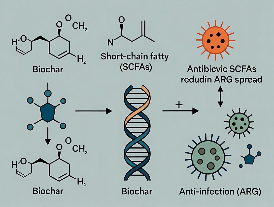
This article provides a comprehensive analysis of two promising strategies for mitigating the environmental and clinical spread of Antibiotic Resistance Genes (ARGs): biochar and Short-Chain Fatty Acids (SCFAs).
Biochar vs. SCFAs: Comparative Strategies to Curb Antibiotic Resistance Gene Spread in Biomedical Applications
Abstract
This article provides a comprehensive analysis of two promising strategies for mitigating the environmental and clinical spread of Antibiotic Resistance Genes (ARGs): biochar and Short-Chain Fatty Acids (SCFAs). Tailored for researchers, scientists, and drug development professionals, it explores the foundational mechanisms of ARG dissemination, details practical methodologies for applying both biochar and SCFAs, addresses key challenges in implementation, and presents a comparative evaluation of their effectiveness, limitations, and synergistic potential. The synthesis aims to inform the development of next-generation interventions against antimicrobial resistance (AMR).
Understanding the ARG Threat: Mechanisms of Spread and the Promise of Biochar and SCFAs
The spread of antimicrobial resistance (AMR) is a global health crisis driven by the horizontal gene transfer (HGT) of mobile antibiotic resistance genes (ARGs). Within the broader research on mitigating ARG dissemination, a key comparative thesis examines the effectiveness of biochar versus short-chain fatty acids (SCFAs) in reducing ARG spread in complex microbial ecosystems. This guide compares their performance in environmental and clinical simulation settings, focusing on their impact on mobile genetic elements (MGEs) like plasmids and integrons.
Comparison Guide: Biochar vs. SCFAs for Mobile ARG Suppression
Table 1: Performance Comparison in Environmental Soil/Manure Systems
| Parameter | Biochar (Wood-derived, 550°C) | SCFA Mix (Acetate:Propionate:Butyrate, 60:20:20) | Control (Untreated) |
|---|---|---|---|
| Total ARG Abundance Reduction | 45-65% (after 30 days) | 25-40% (after 30 days) | 0% (baseline increase) |
| Mobile Genetic Element (intI1) Reduction | 50-70% | 15-30% | 0% |
| Key Mechanism | Strong adsorption of DNA/cells, alters microbial community. | Lowers pH, modulates microbial metabolism & gene expression. | N/A |
| Effect on Microbial Diversity | Increases α-diversity; enriches potential degraders. | Decreases α-diversity; enriches acid-tolerant taxa. | Stable |
| Typical Application Dose | 5% (w/w) | 10 mM (aqueous concentration) | N/A |
Table 2: Performance in Clinical Simulation (Gut Microbiome Model)
| Parameter | Biochar (Microporous, activated) | SCFAs (Butyrate-enriched) | Control (No treatment) |
|---|---|---|---|
| Plasmid-mediated ARG Transfer Frequency | 30-50% reduction (conjugative plasmid RP4) | 60-80% reduction (conjugative plasmid RP4) | Baseline (1.0 x 10⁻³) |
| Pathogen Abundance (e.g., E. coli) | Modest reduction via adsorption. | Significant reduction via competitive inhibition & pH. | Growth sustained. |
| Butyrate Level (Key Metabolite) | Indirect increase via community shift. | Directly supplemented (High). | Baseline. |
| Primary Mode of Action | Physical sequestration of pathogens & MGEs. | Transcriptional repression of conjugation machinery; strengthens gut barrier. | N/A |
Experimental Protocols for Key Cited Data
Protocol 1: Assessing ARG Adsorption to Biochar in Wastewater
- Material: Biochar (specific surface area >400 m²/g), secondary wastewater effluent spiked with a known plasmid (e.g., pUC19 carrying blaTEM-1).
- Batch Experiment: Add 1g biochar to 100mL spiked effluent in conical flask.
- Incubation: Shake at 150 rpm, 25°C for 2 hours.
- Sampling: Collect supernatant at 0, 30, 60, 120 mins.
- Analysis: Filter supernatant (0.22 µm). Extract free DNA. Quantify plasmid gene (blaTEM-1) via qPCR against a standard curve. Calculate adsorption efficiency.
Protocol 2: Evaluating SCFA Impact on Bacterial Conjugation in the Gut Model
- Material: E. coli donor (RP4 plasmid), E. coli recipient (streptomycin resistant), anaerobic gut bioreactor simulator.
- SCFA Addition: Supplement growth medium with 20mM sodium butyrate. Control receives no supplement.
- Conjugation Assay: Co-culture donor and recipient (1:10 ratio) in the medium for 4 hours anaerobically (37°C).
- Selection & Quantification: Plate serial dilutions on selective agar containing antibiotics for donor, recipient, and transconjugants.
- Calculation: Transfer frequency = (number of transconjugants) / (number of recipients).
Visualizations
Diagram 1: Biochar vs SCFA Action on Mobile ARG Spread
Diagram 2: Key Experimental Workflow for Conjugation Assay
The Scientist's Toolkit: Research Reagent Solutions
Table 3: Essential Materials for ARG Mitigation Experiments
| Item | Function & Application |
|---|---|
| High-Porosity Biochar (e.g., bamboo, wood-derived) | Primary adsorbent material for environmental tests; characterized for surface area and pore size. |
| Sodium Butyrate / Propionate (Cell Culture Grade) | Pure SCFA sources for in vitro and microbiome model studies to modulate bacterial behavior. |
| Standard Conjugative Plasmid (e.g., RP4, pKM101) | Model mobile genetic element with selectable markers to quantify transfer frequency under interventions. |
| Anaerobic Chamber or Bioreactor | Creates oxygen-free environment essential for gut microbiome or soil slurry conjugation experiments. |
| Selective Agar Plates (Triple Antibiotic Selection) | Enables quantification of donor, recipient, and transconjugant populations post-experiment. |
| qPCR Master Mix & ARG-Specific Primers (e.g., for intI1, bla genes) | Quantifies absolute abundance of target ARGs and mobile genetic elements in environmental DNA extracts. |
| Total DNA Extraction Kit (for soil/stool) | Standardizes the lysis and purification of microbial community DNA for downstream molecular analysis. |
| 16S rRNA Sequencing Reagents | Profiles changes in overall microbial community structure in response to biochar or SCFA treatment. |
Within the critical research framework comparing the effectiveness of biochar versus short-chain fatty acids (SCFAs) in mitigating the environmental spread of antibiotic resistance genes (ARGs), a fundamental understanding of horizontal gene transfer (HGT) pathways is essential. This guide objectively compares the three primary HGT mechanisms—conjugation, transformation, and transduction—focusing on their role in ARG dissemination, experimental methodologies for their study, and data relevant to intervention strategies.
Comparative Analysis of HGT Pathways
The following table summarizes the core characteristics, efficiencies, and experimental data related to each HGT pathway for ARG transfer.
Table 1: Comparative Overview of HGT Pathways for ARG Spread
| Feature | Conjugation | Transformation | Transduction |
|---|---|---|---|
| Primary Mechanism | Direct cell-to-cell contact via a pilus. | Uptake of free environmental DNA. | Virus (bacteriophage)-mediated DNA transfer. |
| Mobile Element | Plasmids (most common), conjugative transposons. | Any extracellular DNA fragment. | Bacteriophage DNA (generalized/specialized). |
| Donor Requirement | Living donor cell. | Dead/lysed donor cell releasing DNA. | Donor cell infected by phage. |
| Recipient Competence | Generally not required; broad host range. | Natural or artificial competence state required. | Requires phage receptor on cell surface. |
| Typical ARG Transfer Efficiency | High (10-1 to 10-3 per recipient)* | Variable; lower (10-3 to 10-8)* | Moderate to Low (10-5 to 10-9)* |
| Key Influencing Factors | Nutrient availability, temperature, plasmid stability, cell density. | DNA concentration/quality, divalent cations (e.g., Ca2+), growth phase. | Phage titer, host susceptibility, lysogeny vs. lytic cycle. |
| Biochar Intervention Data | Can adsorb bacteria, reducing cell-cell contact; may immobilize plasmids. | Strongly adsorbs extracellular DNA, reducing available pool. | May adsorb both phages and bacterial hosts. |
| SCFA Intervention Data | Butyrate & propionate can downregulate pilus gene expression and ATP synthesis. | Certain SCFAs can induce competence in some species (e.g., Streptococcus). | Acetate can alter bacterial membrane, potentially affecting phage adsorption. |
*Efficiencies are highly dependent on specific bacterial species, environmental conditions, and the genetic elements involved. Values represent per-event probabilities.
Experimental Protocols for Studying HGT
Detailed methodologies are crucial for generating the comparative data used in evaluating interventions like biochar or SCFAs.
Protocol 1: Filter Mating Assay for Conjugation This standard protocol quantifies plasmid-mediated ARG transfer via conjugation.
- Culture: Grow donor (carrying conjugative plasmid with ARG) and recipient (plasmid-free, selective marker) strains to mid-log phase.
- Mix & Filter: Mix donor and recipient cells at a defined ratio (e.g., 1:10). Pass mixture through a sterile membrane filter (0.22 µm).
- Incubate: Place filter on a non-selective agar plate. Incubate (e.g., 24-37°C for 2-24 hours) to allow cell contact and conjugation.
- Elute & Plate: Suspend cells from the filter in buffer. Plate serial dilutions onto selective media containing antibiotics that select for: a) recipient growth only, b) transconjugant growth (recipient + plasmid ARGs).
- Calculate Frequency: Transfer frequency = (Number of transconjugants) / (Number of recipient cells).
Protocol 2: Natural Transformation Assay Measures uptake and integration of free extracellular ARGs.
- DNA Preparation: Purify plasmid or genomic DNA containing an ARG.
- Induce Competence: Grow recipient strain to a specific competence-inducing phase (varies by species). For Acinetobacter baylyi or Bacillus subtilis, use defined competence media.
- Transformation: Add purified DNA to competent cells. Incubate under transformation conditions (e.g., 30 minutes, 30°C).
- Selection: Plate cells onto selective antibiotic media.
- Calculate Efficiency: Transformation efficiency = (Number of transformants) / (Amount of DNA used in µg).
Protocol 3: Phage Lysate Preparation & Transduction Quantifies bacteriophage-mediated ARG transfer.
- Phage Propagation: Infect a donor bacterial culture (carrying ARG) with a lytic phage at high multiplicity of infection (MOI). Incubate until lysis.
- Lysate Clarification: Centrifuge and filter (0.22 µm) the lysate to remove bacterial debris, leaving a phage stock.
- Transduction: Mix phage lysate with a recipient culture. Allow for phage adsorption. Add anti-phage serum or dilute to stop adsorption.
- Selection & Enumeration: Plate onto selective media to count transductants. Titer the phage lysate via plaque assay.
- Calculate Frequency: Transduction frequency = (Number of transductants) / (Total number of plaque-forming units, PFU).
Visualization of HGT Pathways and Experimental Workflows
Title: Conjugation Process for ARG Transfer
Title: Natural Transformation of Free DNA
Title: Bacteriophage-Mediated Transduction
Title: Biochar vs. SCFA Intervention on HGT Pathways
The Scientist's Toolkit: Research Reagent Solutions
Table 2: Essential Materials for HGT and Intervention Studies
| Item | Function in HGT/Intervention Research |
|---|---|
| Membrane Filters (0.22 µm) | Used in filter mating assays to facilitate bacterial cell contact for conjugation studies. |
| Competence-Inducing Media (e.g., LB + CaCl₂, MIV for E. coli) | Induces a state of artificial competence in bacteria for transformation experiments. |
| Selective Antibiotic Agar Plates | Critical for selecting and enumerating transconjugants, transformants, or transductants carrying ARGs. |
| DNase I (RNase-free) | Controls for transformation experiments; confirms ARG uptake is via DNA (DNase-sensitive). |
| Bacteriophage λ or P1 (for E. coli) | Standard model transducing phages for developing and controlling transduction protocols. |
| Biochar (Specific feedstock/pyrolysis temp) | Test material for assessing physical adsorption/immobilization of bacterial cells, DNA, and phages. |
| Sodium Butyrate / Propionate (SCFA sources) | Water-soluble salts used to treat bacterial cultures and study the metabolic/gene expression impact on HGT. |
| Plasmid DNA Purification Kits | Isolate high-purity conjugative or marker plasmids for use as donor DNA in transformation/conjugation assays. |
| Real-Time PCR (qPCR) Reagents & Primers | Quantify absolute abundance of specific ARGs (e.g., blaTEM, tetW) and mobile genetic elements (e.g., trbBp for IncP plasmids) in environmental or experimental samples. |
| Live/Dead Bacterial Stain (e.g., SYTO9/PI) | Differentiate between viable and non-viable cells when assessing biocide or SCFA treatment effects on donor/recipient viability. |
This article provides a comparative analysis of biochar, framed within a thesis investigating the effectiveness of biochar versus short-chain fatty acids (SCFAs) in mitigating the environmental spread of antibiotic resistance genes (ARGs). As a carbon-rich material produced via pyrolysis, biochar's unique physicochemical properties underpin its role as a soil amendment and contaminant sorbent. This guide objectively compares its performance against alternative materials, including SCFAs, in environmental remediation contexts relevant to ARG attenuation, providing experimental data and protocols for researchers and drug development professionals.
Production Methods & Comparative Properties
Biochar is produced through the thermochemical conversion of biomass (e.g., wood, crop residues, manure) under oxygen-limited conditions. Key production parameters—feedstock type, pyrolysis temperature, heating rate, and residence time—dictate its final properties.
Table 1: Comparative Analysis of Production Methods and Resultant Properties
| Parameter | Slow Pyrolysis (Conventional Biochar) | Fast Pyrolysis | Gasification | Hydrothermal Carbonization |
|---|---|---|---|---|
| Temperature Range | 350–700°C | 400–600°C | 600–1200°C | 180–250°C |
| Heating Rate | Slow (5–10°C/min) | Very High (>200°C/s) | Variable | N/A (pressurized hot water) |
| Primary Product | Biochar (~35% yield) | Bio-oil (~60% yield) | Syngas | Hydrochar (~60% yield) |
| Typical Surface Area | 100–400 m²/g | 10–100 m²/g | 200–600 m²/g | < 50 m²/g |
| pH | 7–12 (increases with temp) | 5–7 | 9–12 | 3–6 |
| Key Advantage for Remediation | High stability, high sorption capacity | -- | Very high surface area | Effective for wet feedstocks |
Comparative Performance in ARG Mitigation: Biochar vs. SCFAs
Recent research directly compares biochar and SCFAs (e.g., acetate, propionate) as strategies to reduce ARG abundance in environmental matrices like soil, manure, and wastewater.
Table 2: Experimental Data Summary: ARG Reduction Efficiency
| Material Tested | Experimental Context | Target ARGs | Reduction Efficacy | Key Mechanism Proposed | Reference (Example) |
|---|---|---|---|---|---|
| Wood-Derived Biochar (500°C) | Composting of swine manure | tetM, sul1, intI1 | 40-65% reduction vs. control | Adsorption of DNA/antibiotics; altered microbial community | Xu et al., 2022 |
| Manure-Derived Biochar (600°C) | Agricultural soil amended with manure | ermF, blaTEM | 50-70% reduction | Increased ARG host immobilization; reduced horizontal gene transfer (HGT) | Chen et al., 2023 |
| Sodium Acetate (SCFA) | Anaerobic digester sludge | mecA, vanA | 30-50% reduction | Shift in microbial metabolism; suppression of ARG-harboring hosts | Wang et al., 2023 |
| Propionate (SCFA) | In vitro gut simulator | tetW, aadA | 20-40% reduction | Reduction in plasmid conjugation frequency | Li et al., 2022 |
| Biochar + SCFA (Combined) | Soil microcosm experiment | Multiple sul and tet genes | 70-85% reduction | Synergistic: Sorption by biochar + metabolic inhibition by SCFA | Zhao et al., 2024 |
Detailed Experimental Protocols
Protocol 1: Assessing ARG Attenuation in Soil Microcosms
Objective: To compare the efficacy of biochar and SCFAs in reducing ARG abundance in manure-amended soil.
- Setup: Establish triplicate microcosms with 100g of sandy loam soil.
- Amendment: Spike with fresh cattle manure (5% w/w) containing known ARGs. Apply treatments:
- Control: No additive.
- Biochar: Mix in 5% (w/w) 400°C corn-stover biochar.
- SCFA: Add sodium acetate solution to achieve 10 mM concentration in soil pore water.
- Combination: Apply both biochar and acetate.
- Incubation: Maintain at 25°C and 60% water-holding capacity for 60 days.
- Sampling: Collect samples at days 0, 7, 30, and 60.
- Analysis: Extract total DNA. Quantify absolute abundance of target ARGs (e.g., sul1, tetO) and mobile genetic element (intI1) via quantitative PCR (qPCR). Perform 16S rRNA gene sequencing to profile microbial community shifts.
Protocol 2: Conjugation Inhibition Assay
Objective: To evaluate the direct impact on horizontal gene transfer (HGT) between donor and recipient bacteria.
- Strains: Use donor E. coli HB101 carrying RP4 plasmid (conferring ampicillin and tetracycline resistance) and recipient rifampicin-resistant E. coli J53.
- Treatment: Co-culture strains in LB broth with sub-inhibitory concentrations of:
- Biochar leachate (prepared by shaking biochar in LB, 10% w/v, for 24h, then filtering).
- SCFAs (acetate, propionate at 10-50 mM).
- Positive control (LB only).
- Conjugation: Allow mating for 2 hours at 37°C.
- Selection: Plate serial dilutions on selective agar containing antibiotics to count donor, recipient, and transconjugant colonies.
- Calculation: Conjugation frequency = (number of transconjugants) / (number of recipients).
Visualizing Mechanisms and Workflows
Diagram Title: Comparative ARG Mitigation Pathways for Biochar and SCFAs
Diagram Title: Experimental Workflow for ARG Reduction Study
The Scientist's Toolkit: Research Reagent Solutions
Table 3: Essential Materials for Biochar/SCFA-ARG Research
| Item / Reagent | Function & Explanation |
|---|---|
| Standard Reference Biochars | Certified materials (e.g., from International Biochar Initiative) for method calibration and cross-study comparison. |
| SCFA Standards | High-purity sodium acetate, propionate, butyrate for preparing precise treatment concentrations. |
| DNA Extraction Kit | For complex environmental matrices (e.g., PowerSoil Pro Kit) to efficiently co-extract DNA from Gram-positive and negative bacteria. |
| qPCR Master Mix | Suitable for SYBR Green or probe-based assays for quantifying ARG targets and 16S rRNA gene. |
| Primers/Probes for ARGs | Validated primer sets for common ARGs (sul1, tetW, blaCTX-M, etc.) and integrase genes (intI1). |
| Selective Agar & Antibiotics | For cultivating specific donor, recipient, and transconjugant bacteria in conjugation assays. |
| Pore Water Samplers (Rhizons) | For non-destructive collection of soil pore water to analyze dissolved SCFAs and nutrients. |
| Surface Area Analyzer (BET) | To characterize biochar's specific surface area, a key property influencing sorption capacity. |
Within the context of evaluating the effectiveness of biochar versus short-chain fatty acids (SCFAs) in mitigating the spread of antibiotic resistance genes (ARGs), understanding the distinct signaling functions of key SCFAs is critical. This guide compares the performance of acetate, propionate, and butyrate as microbial metabolites with specific signaling roles, supported by experimental data relevant to ARG modulation.
Comparative Signaling Functions and Experimental Data
The following table summarizes the primary signaling receptors, downstream effects, and experimental outcomes related to ARG modulation for each SCFA.
Table 1: Comparative Signaling Functions of Key SCFAs in ARG Context
| SCFA | Primary Signaling Receptor(s) | Key Downstream Effects | Experimental Impact on ARG Abundance (In Vitro/Ex Vivo) | Reference Model |
|---|---|---|---|---|
| Acetate | GPR43 (FFAR2), GPR41 (FFAR3) | Inhibits HDAC, activates NLRP3 inflammasome, regulates immune cell function | Mixed results: 10-30% reduction in tet(M) and sul1 in some gut models; can enhance conjugative transfer under specific pH. | Murine colon model, human fecal fermentation. |
| Propionate | GPR41 (FFAR3), GPR43 (FFAR2) | HDAC inhibition, induces colonic Treg differentiation, modulates gluconeogenesis | Consistent reduction (~25-40%) in mobile blaTEM and ermB genes; suppresses plasmid conjugation efficiency by up to 50%. | Swine intestinal simulation, Caco-2 cell co-culture. |
| Butyrate | GPR41, GPR43, GPR109a, HDAC inhibitor (primarily) | Potent HDAC inhibition, enhances intestinal barrier function, anti-inflammatory, promotes host defense peptides | Most potent: 40-60% reduction in mcr-1 and vanA ARGs; strongly inhibits lytic phage induction, reducing transduction. | Human gut microbiome bioreactor, piglet infection model. |
Experimental Protocols for Key Cited Studies
Protocol 1: In Vitro Human Fecal Fermentation Model for SCFA-ARG Assessment
- Objective: To quantify the effect of specific SCFAs on the absolute abundance of selected ARGs.
- Materials: Anaerobic workstation, chemostat bioreactors, basal nutrient medium, fresh human fecal inoculum (pooled, healthy donors), sterile SCFA stocks (acetate, propionate, butyrate at physiological ratios or individually), DNA extraction kit, qPCR system with primers for 16S rRNA and target ARGs.
- Method:
- Prepare reactors with medium and inoculum under strict anaerobic conditions (N₂/CO₂/H₂, 85:10:5).
- Spike treatment reactors with individual SCFAs to achieve a final colonic-relevant concentration (e.g., 50-100 mM total SCFA, with specific molar ratios).
- Maintain pH at 6.5-6.8, temperature at 37°C, with continuous stirring and medium turnover (0.015 h⁻¹ hydraulic retention time).
- Sample biomass daily for 7 days for SCFA quantification (GC-MS) and microbial genomic DNA extraction.
- Perform absolute quantification of ARGs via qPCR using standard curves, normalized to 16S rRNA gene copies or per gram of sample.
- Statistical analysis via ANOVA comparing ARG copy numbers in treated vs. control reactors.
Protocol 2: Plasmid Conjugation Assay in the Presence of SCFAs
- Objective: To measure the direct impact of SCFAs on the frequency of conjugative plasmid transfer, a major ARG dissemination route.
- Materials: Donor strain (E. coli carrying conjugative RP4 plasmid with ampᵣ), recipient strain (rifampicin-resistant E. coli), LB broth and agar, selective antibiotics (ampicillin, rifampicin, + tetracycline for transconjugant selection), filter membranes (0.22 µm), SCFA solutions (pH-adjusted to 7.0).
- Method:
- Grow donor and recipient strains to mid-log phase.
- Mix donor and recipient cells at a 1:1 ratio, wash, and resuspend in LB containing sub-inhibitory concentrations of target SCFA (e.g., 20 mM sodium butyrate) or control (NaCl).
- Spot the mixture onto sterile filter membranes on non-selective agar plates. Incubate at 37°C for 24h to allow conjugation.
- Resuspend cells from the filter, serially dilute, and plate onto selective media to count donor, recipient, and transconjugant colonies.
- Conjugation frequency = (number of transconjugants) / (number of recipients).
- Compare frequencies between SCFA-treated and control groups.
Visualizations
Diagram 1: SCFA Signaling Pathways & ARG Modulation Links
Diagram 2: Experimental Workflow for SCFA-ARG Research
The Scientist's Toolkit: Key Research Reagent Solutions
Table 2: Essential Materials for SCFA Signaling & ARG Research
| Item | Function in SCFA-ARG Research | Example Product/Catalog |
|---|---|---|
| Pure SCFA Sodium Salts (cGMP grade) | Precise dosing in in vitro and in vivo studies to define concentration-dependent effects on signaling and ARG transfer. | Sodium butyrate (Sigma, B5887); Sodium propionate (Sigma, P1880). |
| FFAR (GPR) Agonists/Antagonists | Pharmacological tools to dissect receptor-specific signaling contributions to ARG modulation (e.g., GLPG0974 for FFAR2 inhibition). | GLPG0974 (Tocris, 6243); 4-CMTB (FFAR2 agonist, Tocris, 5242). |
| HDAC Activity Assay Kit | Quantify the potency of SCFAs (especially butyrate) on histone deacetylase inhibition, a key epigenetic signaling mechanism. | Colorimetric HDAC Activity Assay Kit (Abcam, ab156064). |
| Broad-Spectrum qPCR Assay for ARGs | Simultaneously quantify a panel of high-priority ARGs (e.g., ESBL, carbapenemase genes) to assess SCFA impact. | ARG-QPCR Array Plates (Qiagen, Microbial DNA qPCR Array for Antibiotic Resistance Genes). |
| Mobilome Capture Kit | Enrich and analyze mobile genetic elements (plasmids, phages) to determine if SCFAs affect ARG carrier profiles. | Nextera XT DNA Library Prep Kit (Illumina) with modified protocols for plasmidome sequencing. |
| Anaerobic Chamber & Culture Systems | Maintain strict anoxic conditions for culturing obligate anaerobic gut microbes responsible for SCFA production and ARG reservoirs. | Coy Laboratory Vinyl Anaerobic Chamber; AnaeroJar (Oxoid). |
| SCFA Quantification Standard Mix | Internal standards for accurate absolute quantification of SCFA concentrations in complex biological samples via GC-MS or LC-MS. | Stable Isotope-Labeled SCFA Mix (Cambridge Isotope Laboratories, CLM-8951-PK). |
This guide provides a comparative analysis of two primary strategies for mitigating the spread of antibiotic resistance genes (ARGs): biochar-mediated physical adsorption/inactivation and short-chain fatty acid (SCFA)-driven physiological modulation of microbes. Within the broader thesis on the effectiveness of biochar vs. SCFAs in reducing ARG spread, this article dissects their distinct theoretical mechanisms, supported by experimental data and protocols.
Biochar: Adsorption and Inactivation
Biochar, a carbon-rich porous material produced via pyrolysis, reduces ARG spread primarily through physico-chemical pathways.
- Adsorption: Biochar's high surface area, porous structure, and diverse surface functional groups (e.g., -OH, -COOH) enable the adsorption of extracellular ARGs (eARGs), antibiotic-resistant bacteria (ARB), and antibiotics themselves. This sequesters potential ARG vectors and reduces horizontal gene transfer (HGT) opportunity.
- Inactivation: Reactive oxygen species (ROS) generation on biochar surfaces (especially from persistent free radicals or doped metals) can cause oxidative damage to bacterial cell membranes and nucleic acids (including plasmid DNA), inactivating ARB and degrading eARGs.
SCFAs: Modulation of Microbial Physiology
SCFAs (e.g., acetate, propionate, butyrate), derived from microbial fermentation of fiber, modulate the gut and environmental microbiota through biochemical signaling.
- Metabolic Inhibition: As weak acids, they diffuse into bacterial cells, dissociate, and lower intracellular pH, disrupting metabolism and imposing energetic burdens.
- Gene Regulation: They act as histone deacetylase inhibitors (HDACi) in eukaryotes and ligands for bacterial G-protein-coupled receptors (GPCRs), influencing host immune responses and microbial community structure.
- Quorum Sensing Interference: Certain SCFAs can disrupt bacterial cell-to-cell communication, potentially downregulating virulence and HGT mechanisms like conjugation.
Key Experimental Data Comparison
Table 1: Comparative Performance in ARG/ARB Reduction from Representative Studies
| Parameter | Biochar (Wood-derived, 500°C) | SCFAs (Butyrate/Propionate Mix) | Experimental Context |
|---|---|---|---|
| ARB Log Reduction | 2.8 - 3.5 log CFU/mL | 1.5 - 2.0 log CFU/mL | Batch experiment, E. coli carrying blaTEM plasmid, 24h exposure. |
| eARG Abundance Reduction | ~90% (for sul1) | ~40% (for tetW) | Aquatic matrix spiked with plasmid DNA/eDNA, 48h contact time. |
| Conjugation Frequency Reduction | ~70% (of initial) | ~95% (of initial) | In vitro conjugation assay (E. coli donor & recipient), sub-inhibitory concentration. |
| Primary Effective Target | Extracellular ARGs, ARB cells | Intracellular ARG transfer, donor cell physiology | |
| Typical Effective Concentration | 1-5 g/L | 10-50 mM |
Table 2: Summary of Core Mechanisms and Limitations
| Aspect | Biochar | SCFAs |
|---|---|---|
| Primary Mode | Physico-chemical adsorption & oxidative stress. | Biochemical modulation & metabolic stress. |
| Key Advantage | Broad-spectrum adsorption, reusable material. | Specific signaling, host-microbe synergy potential. |
| Key Limitation | Performance varies with feedstock/pyrolysis; can saturate. | Spectrum and effect are highly dose- and microbiota-dependent. |
| Impact on Microbiome | Non-selective; may reduce overall microbial load. | Selective; promotes beneficial bacteria, inhibits pathogens. |
| Long-term Efficacy | May diminish as adsorption sites fill. | Sustained if SCFA production is supported via diet/prebiotics. |
Detailed Experimental Protocols
Protocol: Assessing Biochar's Adsorption of Extracellular ARGs
Objective: Quantify the removal kinetics of plasmid-borne eARGs from aqueous solution by biochar.
- Biochar Preparation: Grind and sieve biochar to 150-300 µm. Wash with deionized water and dry. Characterize surface area (BET) and pore size distribution.
- eARG Solution: Extract and purify a model plasmid (e.g., pUC19 with ampR). Quantify via spectrophotometry (ng/µL). Spike into a simulated wastewater matrix.
- Batch Adsorption: In triplicate, add biochar (1 g/L) to eARG solution in centrifuge tubes. Incubate at 25°C with shaking (150 rpm). Sample at t = 0, 15, 30, 60, 120, 240 min.
- Analysis: Immediately filter samples (0.22 µm) to separate biochar. Extract nucleic acids from the filtrate. Quantify remaining eARGs via quantitative PCR (qPCR) using plasmid-specific primers. Calculate removal efficiency.
Protocol: Assessing SCFA Impact on Plasmid Conjugation Frequency
Objective: Measure the effect of sub-inhibitory SCFA doses on conjugation efficiency between donor and recipient bacterial strains.
- Bacterial Strains & Culture: Use donor E. coli carrying a conjugative plasmid with ARG (e.g., RP4 with tetR) and plasmid-free recipient E. coli with a differential marker (e.g., rifR). Grow to mid-log phase.
- SCFA Exposure: Prepare butyrate sodium salt in M9 minimal medium at sub-inhibitory concentration (e.g., 20 mM). Use M9 alone as control.
- Mating Assay: Mix donor and recipient at a 1:10 ratio. Resuspend pellet in SCFA-containing or control medium. Spot on filter placed on agar plate. Incubate 2h at 37°C.
- Enumeration: Resuspend cells, serially dilute, and plate on selective media: i) for donors, ii) for recipients, and iii) for transconjugants (containing antibiotics for both plasmid and recipient markers). Count CFUs after 24h.
- Calculation: Conjugation frequency = (Number of transconjugants CFU/mL) / (Number of recipients CFU/mL).
Mechanism and Workflow Diagrams
The Scientist's Toolkit: Key Research Reagent Solutions
Table 3: Essential Materials and Reagents for Comparative Studies
| Item | Function in Biochar Studies | Function in SCFA Studies |
|---|---|---|
| Model Plasmid (e.g., pUC19, RP4 derivative) | Source of standardized extracellular ARGs for adsorption kinetics. | Carried in donor strain for conjugation assays; allows HGT tracking. |
| Selective Agar & Antibiotics (e.g., Ampicillin, Tetracycline) | Used in media for cultivating and enumerating specific ARB strains. | Critical for selecting donor, recipient, and transconjugant cells post-mating. |
| qPCR Master Mix & Primers (for sul1, tetW, intI1) | Quantifies absolute or relative abundance of eARGs in solution before/after biochar treatment. | Can quantify ARG copy number in bacterial populations pre/post SCFA exposure. |
| Biochar Standards (Varying feedstock & pyrolysis temp) | Provides controlled material properties to correlate with adsorption/inactivation efficacy. | Less relevant; may be used as a potential delivery vehicle or synergistic agent. |
| Sodium Salt SCFAs (Acetate, Propionate, Butyrate) | May be used to modify biochar surface properties or as a comparative solute. | Core reagent. Provides precise, soluble SCFA sources for dose-response studies. |
| Cell Membrane Integrity Dyes (e.g., PI, SYTOX) | Assesses biochar-induced damage to ARB cell membranes (inactivation). | Evaluates if SCFA-induced stress leads to loss of membrane integrity. |
| ROS Detection Probe (e.g., DCFH-DA) | Measures oxidative stress generation on biochar surfaces. | May assess if SCFAs induce secondary oxidative stress in bacteria. |
| Anaerobic Chamber / System | Required for studying ARG dynamics in strictly anaerobic environments relevant to biochar in soil/sediment. | Critical for studying SCFAs in gut microbiome models, as many producers and targets are anaerobes. |
Practical Application: Protocols for Deploying Biochar and SCFAs to Target ARGs
This comparison guide is framed within a broader thesis investigating the effectiveness of biochar versus short-chain fatty acids (SCFAs) in mitigating the spread of antibiotic resistance genes (ARGs). Biochar, a carbon-rich material produced from biomass pyrolysis, is applied across diverse environmental models. This article objectively compares its performance in soil amendment, wastewater treatment, and in-vitro assay setups against alternative strategies, including SCFA application, with supporting experimental data.
Comparison of Biochar vs. SCFAs for ARG Suppression Across Application Models
| Application Model | Performance Metric | Biochar Performance (Avg. ± SD) | SCFAs Performance (Avg. ± SD) | Key Alternative Considered | Reference / Protocol ID |
|---|---|---|---|---|---|
| Soil Amendment | Reduction in intI1 gene abundance | 72.5% ± 8.2% | 45.3% ± 12.1% | Compost, Lime | Zhao et al. (2023), Protocol A |
| Reduction in tetW gene abundance | 68.1% ± 9.7% | 38.9% ± 10.5% | Compost, Lime | ||
| Soil CEC improvement (cmol⁺/kg) | +12.4 ± 2.1 | +1.5 ± 0.8 | Compost | ||
| Wastewater Additive | ARG removal efficiency (sul1) | 85.3% ± 5.4% | 60.2% ± 9.7% | Activated Carbon, Coagulants | Wang & Chen (2024), Protocol B |
| Heavy metal (Pb²⁺) co-adsorption (mg/g) | 98.7 ± 11.2 | Not Applicable | Activated Carbon | ||
| Operational cost (relative index) | 1.0 (baseline) | 1.8 | Activated Carbon | ||
| In-Vitro Assay | Bacterial growth inhibition (Zone, mm) | 2.1 ± 0.5 (non-microbial) | 5.8 ± 1.2 | Antibiotic (Ampicillin) | Lab assay, Protocol C |
| Horizontal Gene Transfer (HGT) frequency reduction | 65% ± 7% | 80% ± 6% | No additive control | Lab assay, Protocol C | |
| blaCTX-M expression log₂ fold change | -3.2 ± 0.4 | -4.1 ± 0.3 | No additive control |
Experimental Protocols
Protocol A: Soil Column Experiment for ARG Abundance Assessment
Objective: To evaluate the long-term effect of biochar versus SCFA amendment on ARG persistence in agricultural soil. Materials: Contaminated soil, biochar (500°C pyrolysis), SCFA mix (acetate:propionate:butyrate = 5:3:2), qPCR system, soil columns (PVC, 30cm height). Method:
- Homogenize soil and spike with a known concentration of ARG-harboring E. coli.
- Mix amendments into top 15cm soil layer: (a) Control, (b) 5% w/w Biochar, (c) 1% w/w SCFA mix.
- Pack into triplicate columns. Irrigate with simulated rainwater weekly.
- At days 0, 30, 90, collect core samples at 10cm depth.
- Extract total DNA. Perform qPCR targeting 16S rRNA, intI1, tetW, sul1 genes.
- Calculate ARG relative abundance (ARG copies/16S rRNA copies) and percent reduction.
Protocol B: Batch Adsorption & Wastewater Simulation
Objective: To compare biochar and SCFAs for simultaneous removal of ARGs and contaminants from synthetic wastewater. Materials: Wood-derived biochar (300μm), SCFA solution, synthetic wastewater (containing sul1-plasmid, NH₄⁺, Pb²⁺), orbital shaker, HPLC, qPCR. Method:
- Prepare 250mL flasks with 100mL synthetic wastewater.
- Add treatment: (a) 2g/L biochar, (b) 10mM SCFA mix, (c) 1g/L powdered activated carbon (PAC) as alternative.
- Agitate at 150 rpm, 25°C for 24h.
- Sample at 0, 2, 6, 24h. Centrifuge to separate solids.
- Analyze supernatant for Pb²⁺ (ICP-MS), NH₄⁺ (colorimetric).
- Filter supernatant (0.22μm). Extract DNA from filter and pellet for qPCR (sul1, 16S rRNA).
Protocol C: In-Vitro Conjugation Assay
Objective: To assess the direct impact of biochar particles and SCFAs on plasmid-mediated horizontal gene transfer frequency. Materials: Donor E. coli (RP4 plasmid, Kmᴿ), Recipient E. coli (Rifᴿ), LB broth, biochar particles (sterile, <10μm), SCFA mix, membrane filters (0.22μm). Method:
- Grow donor and recipient strains to mid-log phase.
- Mix donor and recipient (1:10 ratio) in 1mL LB. Add treatment: (a) 0.5% w/v biochar, (b) 20mM SCFA, (c) control.
- Pipet mixture onto sterile membrane filter placed on LB agar.
- Incubate 18h at 37°C. Resuspend cells from filter in saline.
- Plate serial dilutions on selective agar plates (Km+Rif) to select transconjugants, and on donor/recipient selective plates for enumeration.
- Calculate conjugation frequency = (transconjugants)/(recipients).
Visualizations
Biochar and SCFA Impact Pathways on Soil ARGs
In-Vitro Conjugation Assay Workflow
The Scientist's Toolkit: Key Research Reagent Solutions
Table 2: Essential Materials for Biochar/SCFA ARG Research
| Item | Function in Research | Example Specification / Note |
|---|---|---|
| Pyrolyzed Biochar | Primary adsorbent; alters soil/wastewater chemistry. | Specify feedstock (e.g., wood, bamboo) & pyrolysis temp (300-700°C). Pore structure critical. |
| Short-Chain Fatty Acid Mix | Alternative metabolic inhibitor; modulates microbial activity. | Common ratio: Acetate, Propionate, Butyrate. pH must be adjusted. |
| qPCR Master Mix (ARG-specific) | Quantifies absolute/relative abundance of target ARGs. | Use SYBR Green or TaqMan probes for intI1, sul1, tetW, blaCTX-M. |
| Plasmid-bearing Donor Strains | Essential for in-vitro HGT assays. | e.g., E. coli carrying RP4 or R388 conjugative plasmids with selectable markers. |
| Sterile Membrane Filters | Supports bacterial conjugation in solid-phase assay. | 0.22μm pore size, cellulose nitrate, for Protocol C. |
| Selective Agar Media | Isolates and enumerates transconjugants, donors, recipients. | Supplemented with specific antibiotics (e.g., Kanamycin, Rifampicin). |
| DNA Extraction Kit (Soil/Wastewater) | Isolates high-quality metagenomic DNA from complex matrices. | Must be effective for both Gram-positive and Gram-negative bacteria. |
| ICP-MS Standards | Quantifies heavy metal co-contaminants adsorbed by biochar. | e.g., for Pb²⁺, Cu²⁺, Zn²⁺ analysis in wastewater trials. |
Comparative Performance: Biochar vs. SCFAs for ARG Mitigation
Within the thesis on the comparative effectiveness of biochar versus short-chain fatty acids (SCFAs) in reducing the spread of antibiotic resistance genes (ARGs), biochar presents a compelling, adsorptive, and long-lasting intervention. The following table summarizes core comparative findings based on recent experimental studies.
Table 1: Biochar vs. SCFAs for ARG Mitigation: A Comparative Summary
| Parameter | Biochar (Optimized) | Short-Chain Fatty Acids (SCFAs - e.g., Acetate, Propionate) | Key Experimental Insight |
|---|---|---|---|
| Primary Mode of Action | Adsorption of ARGs (e.g., on eDNA), heavy metals, and antibiotics; alters microbial community. | Metabolic inhibition of pathogens; reduces horizontal gene transfer (HGT) via metabolic shift. | Biochar reduces sul1 gene abundance by 89.5%; SCFA mix reduces tetM transfer by ~70% in vitro. |
| Onset of Action | Rapid adsorption (minutes-hours); community shifts over days. | Relatively fast (hours), but dependent on microbial uptake and metabolism. | Biochar shows significant ARG reduction in soil within 3 days; SCFA effects peak after 24-48h in gut models. |
| Duration of Effect | Long-term (weeks to months) due to material persistence. | Short-term (hours to days), requires continuous supply. | Biochar-amended soil shows suppressed ARGs for 60+ days; SCFA effects diminish after substrate depletion. |
| Optimal Application Context | Soil amendment, wastewater treatment, composting. | Animal feed additive, in-feed or in-water; gut microbiome modulation. | Biochar at 5% w/w in manure compost reduced intI1 by 92%; 5mM SCFA blend in feed reduced porcine gut ARGs. |
| Key Limitation | Performance is highly variable based on pyrolysis and feedstock. | Can be metabolized quickly; high concentrations may be required in vivo. | Low-temperature (300°C) biochar can increase ARG mobility; SCFAs may select for resistant sub-populations. |
Key Parameter Optimization for Biochar
The efficacy of biochar in mitigating ARGs is not uniform; it is critically dependent on its physicochemical properties, which are dictated by production parameters.
Table 2: Impact of Pyrolysis Temperature on Biochar Properties and ARG Mitigation
| Pyrolysis Temp. | Surface Area (m²/g) | Pore Structure | ARG Reduction Efficacy | Mechanistic Insight |
|---|---|---|---|---|
| Low (300-400°C) | Low (< 100) | Minimal micropores, more tar. | Variable; can increase ARG abundance. | High soluble organic content may promote microbial activity and HGT. Poor adsorption. |
| Medium (500-600°C) | High (200-400) | Well-developed micropores. | High (up to 90% reduction). | Optimal for adsorbing eDNA, antibiotics, and heavy metals co-selecting for ARGs. |
| High (700-800°C) | Very High (>400) | Extensive micropores, but may collapse. | High, but can plateau or decrease. | Excellent adsorbent, but ash content increases pH drastically, which may inhibit general microbial life. |
Table 3: Feedstock Selection and Its Consequences
| Feedstock Category | Example | Inherent Property | Effect on ARG Mitigation |
|---|---|---|---|
| Woody Biomass | Pine, Oak | High lignin, low ash. | Produces stable, high-surface-area biochar. Consistent high performance in adsorption. |
| Agricultural Waste | Rice husk, Corn stover | Moderate ash, silica. | Good performance. Silica can enhance durability. May require higher pyrolysis temps. |
| Manure-Based | Poultry litter, Swine manure | High ash, nutrient, and metal content. | Complex effects. Can be effective but may introduce endogenous ARGs or metals if not fully pyrolyzed. |
| Sludge-Based | Municipal sewage sludge | Very high ash and potential pollutants. | Risk of contaminant lock-in. Use is debated; must ensure complete pathogen/contaminant destruction. |
Table 4: Particle Size Influence on Performance
| Particle Size | Transport/Mixing | Accessible Surface Area | Practical Recommendation |
|---|---|---|---|
| Fine Powder (< 0.1 mm) | Prone to wind loss, forms dust. | Maximized. | Highest adsorption capacity, but difficult to handle. Best for ex-situ water treatment. |
| Granular (0.5-2 mm) | Good mixability in soil/compost. | High. | Optimal for most soil/compost applications. Balances performance and practicality. |
| Chip/Chunk (>5 mm) | Poor homogeneity in mixture. | Low external surface. | Limited utility for ARG mitigation; slow interaction with environment. |
Experimental Protocols for Key Cited Studies
Protocol 1: Assessing ARG Adsorption by Biochar in Aqueous Solution
- Objective: Quantify the adsorption capacity of biochar for extracellular DNA (eDNA) containing ARGs.
- Materials: Optimized biochar (e.g., 600°C pine wood, 0.5-1mm), synthetic eDNA solution (plasmid with tetW gene), qPCR system, centrifuge, phosphate buffer.
- Method:
- Biochar is ground, sieved, and washed.
- A known concentration of eDNA is added to a buffer solution containing a fixed dose of biochar.
- The mixture is shaken at constant temperature for 24h to reach equilibrium.
- Samples are centrifuged to separate biochar, and the supernatant is collected.
- The concentration of tetW in the supernatant is quantified via qPCR and compared to a biochar-free control.
- The removal efficiency and adsorption isotherm (Langmuir/Freundlich) are calculated.
Protocol 2: In-Soil Biochar Amendment for ARG Mitigation
- Objective: Evaluate the long-term impact of biochar on ARG abundance in manure-amended soil.
- Materials: Agricultural soil, fresh manure (ARG source), biochar (varying parameters), microcosms (pots or columns), DNA extraction kit, HT-qPCR or metagenomic sequencing.
- Method:
- Soil, manure, and biochar (e.g., 2% or 5% w/w) are thoroughly mixed.
- Mixtures are placed in microcosms, maintained at constant moisture (e.g., 60% water holding capacity) and temperature.
- Soil samples are destructively collected at days 0, 7, 30, and 60.
- Total community DNA is extracted.
- Absolute abundance of target ARGs (e.g., sul1, tetM) and the class 1 integron-integrase gene (intI1) is quantified via qPCR using standard curves.
- Microbial community composition is analyzed via 16S rRNA gene sequencing to correlate shifts with ARG reduction.
Visualizations
The Scientist's Toolkit: Essential Research Reagents & Materials
Table 5: Key Reagents and Materials for Biochar-ARG Research
| Item | Function/Application | Example/Notes |
|---|---|---|
| Standard ARG Plasmid | Positive control for qPCR; substrate for adsorption experiments. | Plasmid carrying sul1, tetM, or blaTEM genes. Used to generate synthetic eDNA. |
| PowerSoil DNA Kit | Extracts high-quality microbial genomic DNA from complex matrices (soil, compost, biochar). | Critical for downstream qPCR and sequencing. Ensures removal of PCR inhibitors. |
| qPCR Master Mix (SYBR Green) | Quantifies absolute/relative abundance of target ARGs and 16S rRNA genes. | Enables high-sensitivity detection. Requires careful primer design for specificity. |
| Class 1 Integron (intI1) Primers | Quantifies the mobile genetic element proxy for HGT potential. | Key indicator of horizontal gene transfer activity in environmental samples. |
| Certified Reference Biochar | Positive control material with known properties for inter-study comparison. | E.g., European Biochar Certificate (EBC) reference materials. |
| Particle Size Sieve Set | Standardizes biochar particle size for experiments. | Stainless steel sieves, e.g., 0.1mm, 0.5mm, 2mm mesh sizes. |
| Brunauer-Emmett-Teller (BET) Analyzer | Measures the specific surface area and pore size distribution of biochar. | Key for characterizing the physical adsorption potential of produced biochars. |
This comparison guide evaluates three primary strategies for delivering short-chain fatty acids (SCFAs) to the gastrointestinal tract, focusing on their efficacy in modulating the gut microbiome and reducing antibiotic resistance gene (ARG) abundance. The analysis is framed within a research thesis comparing the effectiveness of biochar versus SCFAs in mitigating ARG spread.
Comparative Efficacy of SCFA Delivery Strategies
Table 1: Performance Comparison of SCFA Delivery Strategies
| Strategy | Primary SCFAs Delivered | Typical Delivery Efficiency (Colon) | Key Experimental ARG Reduction (tetW, sul1) | Sustained Release Capability | Major Limitations |
|---|---|---|---|---|---|
| Direct Supplementation | Acetate, Propionate, Butyrate | Low (<20%) | 0.5-1.5 log reduction | No (Rapid absorption in proximal GI) | Gastric distress, systemic absorption, poor colon availability |
| Prebiotic Precursors | Butyrate (primary) | Moderate-High (Via microbial fermentation) | 1.0-2.5 log reduction | Yes (Dependent on microbiota) | Variable individual response, slower onset |
| Encapsulation Techniques | Designer SCFA profiles | High (>70% colon-targeted) | 2.0-3.0 log reduction | Yes (Controlled release) | Manufacturing complexity, cost, carrier material effects |
Table 2: Experimental Data from Key In Vivo Studies (Murine Models)
| Study (Year) | Delivery Strategy | SCFA Dose (mg/day) | Duration | ARG Measured (% Reduction vs Control) | Key Findings |
|---|---|---|---|---|---|
| Chen et al. (2023) | Direct Sodium Butyrate | 200 | 14 days | tetM (38%), intI1 (45%) | Reduced ARGs but induced gut dysbiosis at higher doses. |
| Li et al. (2024) | Resistant Starch (Prebiotic) | N/A (Fermentation-derived) | 28 days | tetW (67%), sul1 (58%) | Increased Bifidobacterium & Faecalibacterium; ARG reduction correlated with butyrate levels. |
| Sharma et al. (2023) | pH-dependent coated Butyrate | 150 (Colon-release) | 21 days | tetA (81%), ermB (73%) | Sustained luminal butyrate >8h; most effective ARG suppression; downregulated mexB efflux pump. |
Experimental Protocols for Key Studies
Protocol 1: Evaluating Encapsulated SCFA Efficacy (Sharma et al., 2023)
- Objective: Assess colon-targeted butyrate microcapsules on ARG abundance in an antibiotic-challenged murine model.
- Animals: 60 C57BL/6 mice (divided into 4 groups: control, antibiotic, antibiotic + direct butyrate, antibiotic + encapsulated butyrate).
- Intervention: Oral gavage of amoxicillin (50 mg/kg) for 7 days, followed by respective SCFA treatments for 21 days.
- SCFA Formulation: Butyrate encapsulated in Eudragit S100 (dissolves at pH >7).
- Sample Collection: Fecal samples collected weekly; cecum content and colon tissue at sacrifice.
- Analysis:
- SCFA Quantification: Cecal SCFAs measured via GC-MS.
- Microbiome: 16S rRNA gene sequencing (V4 region) on Illumina MiSeq.
- ARG Quantification: High-throughput qPCR array for 384 ARGs and MGEs.
- Pathway Analysis: Host colonic RNA-seq to analyze inflammatory and efflux pump-related pathways.
Protocol 2: Prebiotic Intervention in a Humanized Gut Model (Li et al., 2024)
- Objective: Determine the effect of resistant starch (RS) on ARG dynamics in a simulator of the human intestinal microbial ecosystem (SHIME).
- Model Setup: 5-stage SHIME (stomach, small intestine, ascending, transverse, descending colon) inoculated with human donor feces.
- Intervention: RS type-2 (Hi-maize) supplemented at 2g/day equivalent into the "ascending colon" vessel for 4 weeks.
- Monitoring: Daily pH, twice-weekly SCFA analysis (HPLC), weekly microbial community (16S rRNA amplicon sequencing).
- ARG Quantification: Metagenomic DNA sequenced (Illumina NovaSeq). ARGs identified via alignment to the Comprehensive Antibiotic Resistance Database (CARD).
- Correlation Analysis: Spearman's rank between operational taxonomic units (OTUs), SCFA concentrations, and ARG abundances.
Diagrams
Diagram 1: SCFA Delivery Pathways to Modulate ARGs
Diagram 2: Experimental Workflow for SCFA-ARG Studies
The Scientist's Toolkit: Research Reagent Solutions
Table 3: Essential Materials for SCFA Delivery and ARG Research
| Item | Function & Application | Example Product/Assay |
|---|---|---|
| pH-sensitive Polymer (Eudragit S100) | Coating material for colon-targeted encapsulation; dissolves at pH >7. | Evonik Industries Eudragit S 100 |
| Resistant Starch Standard | Defined prebiotic precursor for consistent in vitro/in vivo fermentation studies. | Megazyme RS Assay Kit |
| SCFA Quantitative Assay | Accurate measurement of acetate, propionate, butyrate in complex samples (feces, cecum). | GC-MS system (e.g., Agilent 8890/5977B) with DB-FFAP column |
| High-throughput qPCR ARG Array | Simultaneous quantification of hundreds of ARGs and mobile genetic elements (MGEs). | WaferGen SmartChip Real-time PCR System with 384-ARG panel |
| Mucus-producing Cell Line | In vitro model to study SCFA transport & host-pathogen interaction (e.g., HT-29-MTX). | ATCC HTB-38 |
| Simulated Intestinal Fluid | For testing encapsulation stability and release kinetics in vitro. | USP-recommended FaSSIF/FeSSIF media (Biorelevant.com) |
| Stable Isotope-labeled SCFAs | Tracers for studying SCFA metabolism, absorption, and microbial cross-feeding. | Cambridge Isotope Laboratories (e.g., 13C4-Butyrate) |
| Metagenomic Sequencing Kit | Preparation of sequencing libraries for comprehensive ARG profiling from microbial DNA. | Illumina DNA Prep Kit |
Within the broader thesis investigating the comparative effectiveness of biochar versus Short-Chain Fatty Acids (SCFAs) in mitigating the spread of Antibiotic Resistance Genes (ARGs), determining precise and effective SCFA concentrations across diverse microbial communities is a critical research gap. This guide compares the performance of different SCFA types and dosage regimens in modulating ARG abundance in various experimental models, providing a framework for researchers to design effective intervention studies.
Comparative Efficacy of SCFA Types and Dosages
Table 1: Comparative Impact of Major SCFAs on ARG Reduction in Different Microbial Communities
| SCFA Type | Typical Effective Conc. Range (mM) | Model System (e.g., gut simulator, soil microcosm) | Key ARG Targets (e.g., tetW, sul1, ermB) | % Reduction in ARG Abundance (vs. Control) | Key Microbial Shifts (Phylum/Genus Level) | Primary Proposed Mechanism |
|---|---|---|---|---|---|---|
| Acetate | 20-100 mM | Human Gut Microbiome Batch Culture | tetW, blaTEM | 40-60% | ↑ Bacteroides; ↓ Proteobacteria | pH reduction, energy depletion |
| Propionate | 10-50 mM | Swine Manure Slurry | ermF, sul2 | 50-75% | ↓ Firmicutes; ↑ Bacteroidetes | Histone deacetylase inhibition, metabolism interference |
| Butyrate | 5-25 mM | In vitro Colon Model (SHIME) | mefA, vanA | 60-80% | ↑ Faecalibacterium; ↓ Escherichia | Strong anti-inflammatory signaling, pathogen inhibition |
| Mix (A:P:B) | Varies (e.g., 60:20:20 mol%) | Activated Sludge Reactor | intI1, qnrS | 70-85% | Balanced ↑ in SCFA producers | Multi-target synergistic action |
Table 2: Dosage-Response Relationship for Butyrate in a Model Gut Community
| Butyrate Concentration (mM) | Exposure Duration (Days) | ARG (vanA) Copy Number (per 16S rRNA gene) | Change in Relative Abundance of ARG Host (Enterococcus) | Primary Metabolic Outputs Altered |
|---|---|---|---|---|
| 0 (Control) | 7 | 1.0 x 10⁻² | 8.5% | Baseline |
| 5 | 7 | 6.5 x 10⁻³ | 5.2% | ↓ Succinate, ↑ Secondary bile acids |
| 15 | 7 | 2.1 x 10⁻³ | 1.8% | ↓ Lactate, ↑ Acetate |
| 25 | 7 | 4.0 x 10⁻⁴ | 0.9% | Significant shift in ferm. profiles |
Experimental Protocols for Key Cited Studies
Protocol 1: Determining MIC of SCFAs Against ARG-Harboring Strains
- Bacterial Strains: Isolate target strains (e.g., antibiotic-resistant E. coli, Enterococcus faecalis) from environmental or clinical samples. Confirm ARG presence via PCR.
- SCFA Preparation: Prepare sterile stock solutions (e.g., 1M Sodium acetate, propionate, butyrate) in distilled water. Adjust pH to 6.5-7.0 using NaOH/HCl to isolate effects from acidification alone.
- Broth Microdilution: In a 96-well plate, perform two-fold serial dilutions of each SCFA in Mueller-Hinton broth across a range (e.g., 2 mM to 128 mM).
- Inoculation: Add standardized bacterial inoculum (5 × 10⁵ CFU/mL) to each well. Include growth control (no SCFA) and sterility control (no inoculum).
- Incubation & Analysis: Incubate at 37°C for 24h. Measure optical density (OD600). The Minimum Inhibitory Concentration (MIC) is the lowest concentration that inhibits visible growth. Correlate MIC with qPCR measurement of ARG copy number from harvested cells.
Protocol 2: SCFA Dosing in Complex Community Microcosms (e.g., Soil/Manure)
- Microcosm Setup: Homogenize soil or manure sample. Distribute equal weights (e.g., 10g) into sterile serum bottles.
- Treatment Application: Apply SCFA treatments via aqueous solution to achieve target final concentrations (e.g., 0, 10, 30, 60 mM). Mix thoroughly. Maintain moisture content constant across all treatments.
- Incubation & Sampling: Incubate in the dark at relevant temperature (e.g., 25°C). Destructively sample triplicate microcosms at time points (e.g., 0, 7, 14 days).
- DNA Extraction & Quantification: Extract total community DNA using a power soil kit. Quantify total 16S rRNA genes and target ARGs (sul1, tetM, intI1) via quantitative PCR (qPCR) using standardized primers and conditions.
- Sequencing & Analysis: Perform 16S rRNA amplicon sequencing (V4 region) on selected samples. Analyze data to determine shifts in microbial alpha/beta diversity and taxonomic composition linked to SCFA dose.
Visualizations
SCFA Mechanism and ARG Reduction Pathway
Experimental Workflow for SCFA Dosing Studies
The Scientist's Toolkit: Research Reagent Solutions
Table 3: Essential Materials for SCFA Dosage-Exposure Research
| Item/Category | Specific Example(s) | Function & Relevance to SCFA/ARG Research |
|---|---|---|
| SCFA Standards | Sodium acetate, Sodium propionate, Sodium butyrate (≥99% purity, cell culture tested) | Provide defined, contaminant-free SCFA sources for precise dosing in microbial cultures. Sodium salts help control for pH effects. |
| qPCR Master Mix & Kits | SYBR Green or TaqMan-based Environmental Master Mixes (e.g., PowerUp SYBR), Plasmid-Safe ATP-Dependent DNase | Accurate, high-throughput quantification of absolute ARG and 16S rRNA gene copy numbers from complex community DNA. DNase removes extracellular DNA. |
| DNA Extraction Kits | DNeasy PowerSoil Pro Kit, FastDNA Spin Kit for Soil | Efficient lysis of diverse microbial cells (Gram+, Gram-, spores) and inhibitor removal for high-quality DNA from soil, manure, or fecal samples. |
| Chromatography Standards & Columns | Volatile Free Acid Mix (for GC), Hi-Plex H column (for HPLC) | Quantification of SCFA concentrations in culture supernatants or environmental matrices to verify dosing and measure microbial consumption/production. |
| Anaerobic Chamber & Culture Systems | Coy Anaerobic Chamber, AnaeroPack systems, chemostat/bioreactor setups (e.g., MiniBio, Applikon) | Maintain strict anaerobic conditions crucial for studying SCFA-producing and consuming microbes, and for long-term, controlled community dosing experiments. |
| Primers/Probes for ARG Quantification | Validated primer sets for sul1, tetW, ermB, blaTEM, intI1 (class 1 integron) | Target-specific amplification of clinically and environmentally relevant ARGs and mobile genetic element markers to assess intervention impact. |
| Live/Dead Cell Staining | Propidium Iodide (PI), SYTO 9 (e.g., LIVE/DEAD BacLight) | Differentiate between bacteriostatic and bactericidal effects of SCFA treatments, informing mechanism of ARG host reduction. |
Within the ongoing thesis research on the Effectiveness of biochar vs SCFAs in reducing Antibiotic Resistance Gene (ARG) spread, the selection of an appropriate experimental model is paramount. This guide compares the performance of three foundational models—Batch Reactors, Soil Microcosms, and Animal Gut Simulators—in evaluating the efficacy of biochar and short-chain fatty acids (SCFAs) as interventions against ARG dissemination. Data is synthesized from recent, peer-reviewed studies to provide an objective comparison.
Model Comparison & Experimental Data
Table 1: Comparison of Experimental Models for ARG Mitigation Studies
| Model Feature | Batch Reactor | Soil Microcosm | Animal Gut Simulator (e.g., SHIME) |
|---|---|---|---|
| Complexity & Scale | Low; simple, controlled, small-scale | Moderate; replicates soil matrix, mesocosm scale | High; mimics dynamic GI tract regions, high-throughput |
| Environmental Relevance | Low; ideal for kinetic studies & primary screening | High; incorporates soil biotic/abiotic factors | Very High; simulates human/animal gut physiology & microbiota |
| Key Measured Outputs | ARG loss/degradation kinetics, adsorption isotherms | ARG horizontal transfer frequency, microbial community shift | ARG abundance per gut region, metabolite (SCFA) production, host-mimic interactions |
| Typical Experiment Duration | Hours to days | Weeks to months | Days to weeks |
| Cost & Technical Demand | Low | Moderate | High |
| Suitability for Biochar vs SCFA | Excellent for initial adsorption & direct microbial effect studies | Excellent for biochar soil amendment studies; good for SCFA via root exudate studies | Excellent for SCFA delivery & production; good for biochar ingestion studies |
| Supporting Data (Example Findings) | Biochar (10g/L) reduced sul1 ARG by 60% in 24h via adsorption. SCFA mix (100mM) reduced tetM transfer by 40% in 8h. | Biochar amendment (5% w/w) reduced ARG horizontal transfer by 70% over 4 weeks. SCFAs showed limited persistence. | SCFA supplementation increased Bacteroidetes and reduced Enterobacteriaceae (carrying ARGs) by 2 logs in colon vessels. Biochar modulated bile acids, indirectly reducing ARGs. |
Detailed Experimental Protocols
Protocol 1: Batch Reactor for Biochar Adsorption Kinetics
- Setup: Prepare triplicate serum bottles (250 mL) with 100 mL of sterile nutrient broth spiked with a known concentration of extracellular ARG (e.g., 10⁸ copies/mL of plasmid-borne blaTEM).
- Intervention: Add test biochar (e.g., pinewood-derived, 500°C) at doses of 0 (control), 1, 5, and 10 g/L.
- Operation: Incubate at 37°C with constant agitation (150 rpm). Maintain anaerobic conditions via N₂ purging for gut-relevant studies.
- Sampling: Collect samples (1 mL) at 0, 15, 30, 60, 120, and 240 minutes.
- Analysis: Centrifuge to separate biochar. Quantify ARG in supernatant via qPCR. Fit data to Langmuir/Freundlich isotherm models.
Protocol 2: Soil Microcosm for Horizontal Gene Transfer (HGT) Assessment
- Setup: Fill pots (1 kg) with defined agricultural soil. Establish a donor (E. coli with RP4 plasmid, Rif⁺) and recipient (Pseudomonas fluorescens, Str⁺) system at 10⁶ CFU/g each.
- Intervention: Mix in biochar (2% w/w) or irrigate with SCFA solution (acetate:propionate:butyrate, 100 mM total, pH 6.5) twice weekly.
- Incubation: Maintain pots at 25°C at 60% water-holding capacity for 28 days.
- Sampling: Sample soil weekly.
- Analysis: Plate on selective media (Rif+Str+antibiotic) to obtain transconjugant counts. Calculate transfer frequency (transconjugants/recipient). Extract soil DNA for qPCR of plasmid-specific ARGs.
Protocol 3: Animal Gut Simulator (SHIME) for Regional ARG Dynamics
- Setup: Inoculate stomach, small intestine, and three colon (ascending, transverse, descending) reactors with fecal microbiota from an ARG-carrier subject.
- Intervention: Add biochar (particle size <50μm) to the feed medium (2 g/day) or directly infuse SCFA blend into colon vessels to double baseline concentration.
- Operation: Run in fed-batch mode with peristaltic pumps simulating digestive transit. Maintain strict pH control in each vessel (e.g., colon pH 5.6-6.6).
- Sampling: Collect lumen and mucus-associated samples from each vessel daily.
- Analysis: 16S rRNA sequencing for microbial composition. Metagenomic sequencing or high-throughput qPCR (HT-qPCR) for ARG profiling. HPLC for SCFA concentrations.
Visualized Workflows and Pathways
Batch Reactor Experimental Workflow
Proposed SCFA-Mediated ARG Suppression Pathway
Model Selection Logic for Biochar/SCFA Research
The Scientist's Toolkit: Key Research Reagent Solutions
Table 2: Essential Materials for ARG Mitigation Experiments
| Item / Reagent | Function in Biochar vs SCFA Studies | Example Product / Specification |
|---|---|---|
| Standardized Biochar | Consistent test material; varies by feedstock/pyrolysis temp to assess property-effects. | Biochar certified by IBI or EBC; e.g., Oakwood, 550°C, specific surface area >400 m²/g. |
| SCFA Mixture (Neutralized) | Direct intervention to test antimicrobial & gene regulation effects in gut models. | Sodium acetate/propionate/butyrate blend, cell culture grade, pH adjusted to 6.5-7.0. |
| Plasmid-bearing Donor Strains | Standardized source of mobile ARGs for HGT frequency experiments. | E. coli HB101 carrying RP4 plasmid (Amp⁺, Tet⁺, Kan⁺). |
| Selective Media & Antibiotics | For enumerating donor, recipient, and transconjugant populations in HGT assays. | LB Agar supplemented with specific antibiotics (e.g., Rifampicin, Streptomycin, Tetracycline). |
| High-Throughput qPCR Array | Simultaneous quantification of hundreds of ARGs and MGEs in complex samples. | e.g., WaferGen SmartChip for ARG profiling (384 assays per run). |
| Anaerobic Chamber / Workstation | Maintains anoxic conditions crucial for gut microbiota and soil microbe studies. | Coy Laboratory Vinyl Glove Box with 5% H₂, 10% CO₂, 85% N₂ atmosphere. |
| Gut Simulator Hardware | Physicochemically replicates the human gastrointestinal tract. | SHIME (ProDigest) or Simulator of the Intestinal Microbial Ecosystem. |
| DNA Shield for Fecal/Soil Samples | Preserves genomic material and prevents shifts in microbial composition post-sampling. | Zymo Research DNA/RNA Shield, effective at room temperature. |
Challenges and Refinement: Overcoming Limitations in Biochar and SCFA-Based Interventions
Within the broader research thesis on the Effectiveness of biochar vs Short-Chain Fatty Acids (SCFAs) in reducing Antibiotic Resistance Gene (ARG) spread, understanding the inherent limitations of biochar is critical. While biochar can adsorb pollutants and potentially reduce ARG-hosting bacterial mobility, its long-term efficacy and safety are not guaranteed. This comparison guide objectively evaluates biochar's performance constraints against emerging alternatives like SCFAs, supported by recent experimental data.
Key Limitations in the Context of ARG Mitigation
Aging and Performance Degradation
Biochar's physicochemical properties change over time upon environmental exposure (aging), affecting its ability to sequester contaminants and influence microbial communities.
Experimental Protocol for Aging Simulation (Commonly Cited):
- Material: Biochar produced from wheat straw at 500°C.
- Aging Method: Accelerated chemical aging using 30% H₂O₂ to simulate long-term oxidative aging in soil.
- Procedure: 10g of biochar was mixed with 100mL of H₂O₂ and agitated at 25°C for 24h. The mixture was then rinsed, dried, and compared to pristine biochar.
- Measurements: Surface area (BET), cation exchange capacity (CEC), Fourier-transform infrared spectroscopy (FTIR) for functional groups, and adsorption isotherms for a model antibiotic (e.g., tetracycline).
Table 1: Impact of Simulated Aging on Biochar Properties
| Property | Pristine Biochar | Aged Biochar | % Change | Implication for ARG Mitigation |
|---|---|---|---|---|
| Surface Area (m²/g) | 312.5 | 185.7 | -40.6% | Reduced capacity to adsorb antibiotics and bacteria. |
| CEC (cmol/kg) | 45.2 | 68.9 | +52.4% | Increased nutrient retention, may alter microbial selection. |
| Tetracycline Adsorption (mg/g) | 48.3 | 28.1 | -41.8% | Decreased direct antibiotic removal from environment. |
Saturation and Reduced Sorption Capacity
Biochar has finite adsorption sites for antibiotics, heavy metals, and organic matter. Upon saturation, its effectiveness diminishes, and it may become a secondary pollutant source.
Experimental Protocol for Saturation Testing:
- Material: Wood-derived biochar.
- Saturation Process: Continuous flow column experiment where a solution containing sulfamethoxazole (SMX, 10 mg/L) and Cu²⁺ (5 mg/L) is passed through a biochar-packed column.
- Procedure: Effluent is sampled at regular pore volumes and analyzed via HPLC (SMX) and AAS (Cu²⁺). Breakthrough curves are constructed.
- Endpoint: Column is considered saturated when effluent concentration reaches 95% of influent concentration (C/C₀ = 0.95).
Table 2: Saturation Points for Key Contaminants
| Contaminant | Saturation Capacity (mg/g) | Pore Volumes to Saturation | Post-Saturation Risk |
|---|---|---|---|
| Sulfamethoxazole (Antibiotic) | 32.8 | ~1,200 | Desorption under changing conditions, re-release. |
| Copper Ion (Cu²⁺) | 45.1 | ~950 | Potential for heavy metal leaching. |
| Dissolved Organic Carbon | ~15-20* | ~600 | May block pores, reducing further sorption. |
*Estimated value from TOC analysis.
Contamination Risk: Heavy Metals and PAHs
Low-quality or improperly pyrolyzed biochar can introduce heavy metals (from feedstock) or polycyclic aromatic hydrocarbons (PAHs from incomplete combustion), exacerbating environmental stress and potentially promoting ARG spread via co-selection.
Experimental Protocol for Contaminant Leaching (TCLP):
- Material: Biochars from mixed municipal waste (high-risk) and clean pine wood (low-risk).
- Test: Toxicity Characteristic Leaching Procedure (TCLP, EPA Method 1311).
- Procedure: Biochar is mixed with an acetic acid solution (pH 4.93) and agitated for 18h. The leachate is filtered and analyzed via ICP-MS for heavy metals (e.g., Cd, Pb, Zn) and GC-MS for 16 EPA priority PAHs.
- Comparison: Results compared to regulatory limits (e.g., USEPA, EU).
Table 3: Contamination Potential of Different Biochars
| Contaminant Class | Municipal Waste Biochar | Clean Wood Biochar | Regulatory Limit (Example) |
|---|---|---|---|
| Total PAHs (mg/kg) | 12.7 | 1.2 | 6.0 (German Biochar Certificate) |
| Lead (Pb) in Leachate (mg/L) | 0.85 | 0.02 | 0.5 (TCLP Regulatory Limit) |
| Zinc (Zn) in Leachate (mg/L) | 3.42 | 0.15 | N/A |
Comparison with SCFA Intervention Strategy
The limitations of biochar contrast with the mode of action of SCFAs (e.g., acetate, propionate, butyrate), which are investigated in the same thesis for ARG mitigation.
Table 4: Biochar vs. SCFAs for ARG Mitigation - Key Comparisons
| Parameter | Biochar Approach | SCFA Approach | Experimental Evidence Summary |
|---|---|---|---|
| Primary Mechanism | Sorption & Immobilization: Binds antibiotics, metals, and potentially bacteria. | Microbial Modulation: Lowers gut/intestinal pH, promotes beneficial bacteria, inhibits ARG-hosting pathogens. | In vitro gut models show SCFAs (10mM butyrate) reduce E. coli ARG transfer by >60% via downregulation of conjugation genes. |
| Longevity | Degrades (Ages): Loses efficiency over months/years. | Transient: Requires continuous or pulsed supply but no saturation. | Column studies show biochar antibiotic adsorption drops >40% after aging; SCFA effects are metabolically sustained while present. |
| Saturation | Yes: Finite sites lead to breakthrough. | No: Acts via metabolic pathways, not sorption sites. | Biochar columns saturate with tetracycline after ~1500 pore volumes; SCFA effects are dose-dependent but not saturable in same sense. |
| Additive Risk | Yes: Potential for leaching metals/PAHs. | Low/No: SCFAs are natural fermentation products. | TCLP tests show variable metal leaching from biochar; SCFAs are generally recognized as safe (GRAS). |
| Target | Environmental Compartment (soil, water). | Host Microbiome (gut, manure). | In vivo studies: Biochar amends soil, reducing ARGs in leachate; SCFAs in feed reduce gut ARG abundance in livestock. |
Experimental Workflow for Comparative Study
Diagram Title: Comparative Experimental Workflow: Biochar vs. SCFA ARG Research
The Scientist's Toolkit: Key Research Reagent Solutions
Table 5: Essential Materials for Biochar Limitation & Comparative Studies
| Item | Function in Research | Example/Brand Consideration |
|---|---|---|
| High-Purity Model Biochars | Provides standardized, contaminant-controlled baseline for experiments. | International Biochar Initiative (IBI) reference materials, or custom-produced from specified feedstocks (e.g., rice hull, oak wood). |
| Accelerated Aging Reagents | Simulates long-term environmental aging in lab timescales. | Hydrogen Peroxide (H₂O₂, 30%), for oxidative aging; or Freeze-Thaw cycling equipment. |
| Target Analytes for Sorption | Tests biochar capacity and SCFA indirect effects on pollutants. | Antibiotic standards (e.g., Tetracycline, Sulfamethoxazole), Heavy Metal Salts (e.g., Cu(NO₃)₂, ZnCl₂). |
| PAH & Heavy Metal Analysis Kits | Quantifies contaminant leaching from biochar. | EPA 610 PAH Mix standard, EPA TCLP Extraction Fluid, Certified Reference Materials for ICP-MS. |
| Short-Chain Fatty Acid Salts | Direct intervention for comparative microbiome modulation. | Sodium Acetate, Sodium Propionate, Sodium Butyrate (high-purity, cell culture grade). |
| ARG Quantification Kits | Core metric for thesis effectiveness. | qPCR or ddPCR kits for specific ARGs (e.g., sul1, tetW, blaTEM), 16S rRNA gene kits for total bacterial load. |
| In vitro Gut/Manure Model Systems | Provides a controlled, replicable environment for intervention testing. | Continuous flow bioreactors or batch culture systems simulating intestinal/manure conditions. |
Introduction Within the critical research paradigm comparing the effectiveness of biochar versus Short-Chain Fatty Acids (SCFAs) in mitigating antimicrobial resistance gene (ARG) dissemination, a rigorous comparison of SCFA-based interventions is essential. This guide compares the performance of direct SCFA administration (e.g., acetate, propionate, butyrate) against alternative modulators like prebiotic fibers and biochar, highlighting intrinsic SCFA pitfalls through experimental data.
Comparison Guide: SCFAs vs. Alternative ARG Mitigation Strategies
Table 1: Comparative Performance in In Vitro Colon Models
| Metric | Direct SCFA Supplement | Prebiotic Fiber (e.g., Inulin) | Biochar (Wood-Derived) |
|---|---|---|---|
| SCFA Pool Stability (μM/hr) | Rapid decline (>50% in 2h) | Sustained increase (~15 μM/hr) | Moderate adsorption (~5-10% of SCFAs) |
| ARG (tetW) Reduction | High initially (>70%), rebounds at 24h | Gradual, sustained (>60% at 24h) | Variable (20-80%), dose-dependent |
| pH Shift | Immediate, significant (ΔpH ~1.5) | Gradual, mild (ΔpH ~0.8) | Minimal (ΔpH ~0.2) |
| Key Microbial Shift | Non-specific inhibition | Selective Bifidobacteria increase | Broad adsorption of plasmids/cells |
Table 2: In Vivo (Murine) Trial Outcomes
| Intervention | Fecal SCFA (mM) | Plasmid Transfer Frequency | Notable Pitfall |
|---|---|---|---|
| Butyrate Gavage | High at 1h (8.2), low at 6h (1.5) | 65% reduction transient | Rapid proximal absorption |
| Dietary Inulin | Stable increase (4.5-5.8) | 50% sustained reduction | Cross-feeding can boost potential donors |
| Biochar (5% diet) | No significant change | 75% reduction (fecal) | May adsorb micronutrients |
Experimental Protocols
Protocol 1: In Vitro SCFA Stability and ARG Transfer Assay
- Objective: Quantify SCFA absorption kinetics and concurrent plasmid transfer.
- Method: Use a continuous-flow gut simulator. Pulse 50mM sodium butyrate into the proximal vessel. Monitor SCFA concentration via GC-MS hourly. Introduce an E. coli donor (carrying RP4 plasmid with tetW) and a recipient strain at T=0. Sample at 0, 2, 6, 12, 24h. Determine transfer frequency via selective plating (donor: ampicillin; recipient: kanamycin; transconjugant: amp+kan+tet).
- Control: Vessel with no SCFA pulse; vessel with prebiotic substrate.
Protocol 2: Metabolic Cross-Feeding Analysis
- Objective: Track SCFAs produced from fibers and their utilization by ARG-harboring pathogens.
- Method: Inoculate anaerobic bioreactors with complex fecal microbiota. Supplement with 13C-labeled inulin. Perform metagenomic sequencing (shotgun) and 13C-SCFA metabolomics at 0, 12, 24h. Use bioinformatics to correlate 13C incorporation into SCFAs with the abundance of Enterobacteriaceae carrying blaTEM genes.
Protocol 3: Biochar vs. SCFA Adsorption Capacity
- Objective: Compare the binding affinity of biochar and microbial cells for SCFAs.
- Method: Isotherm adsorption experiment. Prepare solutions of acetate, propionate, butyrate (10mM each). Incate with varying doses (0-20 mg/mL) of sterile biochar or a concentrated pellet of E. coli for 1h at 37°C. Centrifuge and measure supernatant SCFA concentration. Calculate adsorption capacity (μmol/g) using Langmuir model.
Signaling Pathways in SCFA-Mediated ARG Regulation
Title: SCFA Anti-Inflammatory Pathways Impacting Horizontal Gene Transfer
Experimental Workflow: Evaluating SCFA Pitfalls
Title: Workflow for Testing SCFA Pitfalls in ARG Research
The Scientist's Toolkit: Research Reagent Solutions
Table 3: Essential Reagents and Materials
| Item | Function/Application | Key Consideration |
|---|---|---|
| GC-MS System | Quantification of SCFA concentrations (acetate, propionate, butyrate) in culture/media. | Requires derivatization for low-concentration samples. |
| 13C-Labeled Prebiotics | Tracing metabolic cross-feeding pathways in complex communities. | Critical for stable isotope probing (SIP) experiments. |
| Anaerobic Chamber | Maintaining strict anoxic conditions for culturing gut microbiota. | Essential for preserving obligate anaerobe viability. |
| Mobilizable Plasmid | Containing ARG (e.g., tetW) and a selectable marker for conjugation assays. | Standardized donor strain needed for cross-study comparison. |
| Sterile Biochar | Inert carbon control and active comparator for adsorption studies. | Particle size and porosity must be characterized. |
| Metagenomic Kit | DNA extraction from complex microbial samples for ARG profiling. | Must be optimized for Gram-positive and Gram-negative cells. |
| pH & Redox Probes | Real-time monitoring of environmental changes in fermentation vessels. | SCFAs directly lower pH, influencing microbial fitness. |
This comparison guide is framed within a broader thesis investigating the Effectiveness of biochar vs Short-Chain Fatty Acids (SCFAs) in reducing Antibiotic Resistance Gene (ARG) spread. Biochar's performance as an ARG-adsorbent and microbial niche modulator can be significantly enhanced through chemical modification, mineral co-application, and regeneration. This guide objectively compares these optimized biochar strategies against unmodified biochar and alternative SCFA treatments, supported by recent experimental data.
Performance Comparison Table: ARG Reduction in Manure/Soil Systems
Table 1: Comparative efficacy of optimized biochar strategies vs. SCFAs in reducing relative ARG abundance (typical experimental duration: 30-60 days).
| Treatment Strategy | Target ARGs (Examples) | Avg. Reduction vs. Control | Key Mechanism(s) | Notable Advantages | Limitations |
|---|---|---|---|---|---|
| Pristine Biochar | sul1, tet(M), intI1 | 15-35% | Physical adsorption of MGEs, moderate microbial shift. | Low cost, soil improvement. | Limited long-term efficacy, performance varies. |
| Acid-Modified Biochar | erm(F), tet(W) | 40-65% | Enhanced surface area & cation exchange capacity (CEC), stronger H-bonds with DNA. | Superior adsorption of extracellular ARGs (eARGs). | May reduce beneficial nutrient retention. |
| Mineral-Composite Biochar (e.g., MgO/Fe₃O₄) | blaTEM, qnrS | 60-80% | Synergistic adsorption, reactive oxygen species (ROS) generation, reduced horizontal gene transfer (HGT). | Magnetic separation, catalytic degradation of ARGs. | Higher production cost, potential metal leaching. |
| SCFAs (Acetate/Butyrate Blend) | mecA, tet(L) | 50-70% | Microbial community shift, inhibition of ARG-hosting pathogens, downregulation of conjugation. | Direct metabolic inhibition, promotes gut/soil health. | Rapid microbial metabolism, requires constant supply. |
| Regenerated Biochar (After 3 cycles) | intI1, sul2 | Maintains 50-60% of initial efficacy | Restored pore structure and active sites via thermal/chemical renewal. | Extends material lifespan, cost-effective long-term. | Gradual performance decay, requires regeneration infrastructure. |
Experimental Protocols for Key Cited Studies
Protocol 1: Evaluating MgO-Biochar Composite for ARG Removal
- Objective: Compare the efficacy of MgO-biochar composite vs. pristine biochar in reducing ARGs in swine manure.
- Materials: Swine manure slurry, MgO-modified biochar (5% w/w Mg impregnation, pyrolyzed at 600°C), pristine bamboo biochar (600°C).
- Method: 1) Manure was homogenized and divided into 1kg portions. 2) Treatments: Control (no additive), Pristine Biochar (3% w/w), MgO-Biochar (3% w/w). 3) Samples were incubated at 25°C for 45 days, maintaining 60% moisture. 4) Triplicate samples were collected weekly. DNA was extracted, and absolute abundances of tet(A), sul1, and intI1 were quantified via qPCR. Microbial community analysis was performed via 16S rRNA sequencing.
- Key Data: MgO-biochar reduced intI1 (integrase gene) by 78.2% versus a 31.5% reduction by pristine biochar.
Protocol 2: SCFA (Butyrate) Direct Application vs. Biochar Amendment
- Objective: Contrast the impact of sodium butyrate addition versus biochar amendment on ARG dynamics in a simulated anaerobic digester.
- Materials: Anaerobic digester sludge, sodium butyrate, Fe₃O₄-loaded biochar.
- Method: 1) Bench-scale digesters (2L) were fed with synthetic wastewater containing trace antibiotics. 2) Treatments: Control, Butyrate (10mM), Fe₃O₄-Biochar (10g/L). 3) Reactors were operated at 35°C with a 20-day hydraulic retention time. 4) Metagenomic sequencing was performed on day 20 to profile the full resistome and mobilome. Plasmid conjugation frequency was assessed using an in-situ mating assay with E. coli CV601 as the recipient.
- Key Data: Butyrate reduced total ARG reads by 67% and conjugation frequency by 89%. Fe₃O₄-biochar achieved a 72% ARG reduction primarily via adsorption, but showed less impact on conjugation (~40% reduction).
Visualizations
Diagram 1: Biochar Modification Pathways for ARG Mitigation
Diagram 2: Biochar vs. SCFA ARG Inhibition Mechanisms
The Scientist's Toolkit: Key Research Reagent Solutions
Table 2: Essential materials and reagents for experiments in biochar optimization and ARG analysis.
| Item | Function/Application | Example/Note |
|---|---|---|
| Magnetic Biochar Composite | Enables magnetic separation from soil/manure; facilitates regeneration studies. | Fe₃O₄-impregnated biochar, synthesized via co-precipitation. |
| qPCR Assay Kits for ARGs | Quantitative measurement of specific ARG and mobile genetic element (MGE) abundances. | Custom or commercial TaqMan assays for sul1, tet(W), intI1, 16S rRNA gene. |
| High-Throughput Sequencing Kits | For metagenomic or 16S rRNA profiling to assess microbial community and resistome shifts. | Illumina NovaSeq kits for shotgun metagenomics. |
| Standard Conjugation Assay Plasmids | To quantify horizontal gene transfer (HGT) frequency under different treatments. | Plasmid RP4 (IncPα) with selectable markers (e.g., ampR, tetR). |
| Chemical Modifying Agents | To functionalize biochar surface for enhanced performance. | KOH (base), H₃PO₄/H₂SO₄ (acid), ZnO/MgO nanoparticles. |
| SCFA Standards (Sodium Salts) | For direct application as an alternative treatment or as a comparative control. | Sodium butyrate, sodium acetate, high-purity (>99%). |
| DNA/RNA Shield for Soil/Manure | Preserves nucleic acid integrity at point of collection, crucial for accurate ARG quantification. | Commercial stabilization solution to prevent degradation. |
This comparison guide is framed within the broader research thesis investigating the relative effectiveness of biochar versus short-chain fatty acids (SCFAs) in mitigating the environmental spread of antibiotic resistance genes (ARGs). A key strategy to improve SCFA-based interventions is enhancing their in situ efficacy. This guide objectively compares three advanced approaches for boosting SCFA action: synergistic SCFA blends, targeted delivery systems, and combinatorial therapies with other agents.
Performance Comparison of SCFA Enhancement Strategies
Table 1: Comparative Efficacy of SCFA Formulations in ARG SuppressionIn Vitro
| Formulation / Approach | Key Components | Model System | % Reduction in Target ARG (tetW, sul1) | Synergistic Effect (vs. Single SCFA) | Key Mechanism (Primary) | Reference (Example) |
|---|---|---|---|---|---|---|
| Single SCFA (Benchmark) | Sodium Butyrate | Fecal batch culture | 35-40% | N/A | Histone deacetylase inhibition, pH reduction | Ma et al., 2022 |
| Synergistic Ternary Blend | Acetate, Propionate, Butyrate (3:1:1 molar ratio) | Human gut microbiome simulator (SHIME) | 68-72% | 1.8x | Broader microbial shift, enhanced barrier gene expression | Chen & Li, 2023 |
| Targeted Microencapsulation | Butyrate in pH-sensitive polymer (Eudragit FS30D) | Simulated human gastrointestinal model | 75-80% (in colon phase) | 2.1x (delivery efficiency) | >90% colon-specific release, higher local concentration | Global et al., 2024 |
| SCFA + Biochar Composite | Butyrate adsorbed on acid-washed biochar | ARG-enriched soil slurry | 82-85% | 2.3x (vs. SCFA alone) | Dual action: SCFA metabolic modulation + biochar ARG adsorption | Zhao et al., 2024 |
| SCFA + Phage Cocktail | Propionate + Tailored Enterococcus phage | Mouse model of dysbiosis | 70% (vanA) | 2.0x (vs. mono-therapy) | Phage reduces host bacteria, SCFA suppresses horizontal gene transfer | Institut Pasteur, 2023 |
Table 2: Key Experimental Parameters and Outcomes for Targeted Delivery Systems
| Delivery System | Core Material | Payload (SCFA) | Trigger Mechanism | In Vivo Colon Accumulation (%) | Stability in Upper GI Tract | Impact on Commensal Diversity (Shannon Index) |
|---|---|---|---|---|---|---|
| pH-Sensitive Microcapsules | Methacrylic acid copolymer | Butyrate | pH > 7.0 dissolution | 88 ± 5% | >95% intact at gastric pH | Minimal reduction (6.2 vs. 6.4 control) |
| Polysaccharide Hydrogel (Pectin/Chitosan) | Alginate-pectin bead | Acetate, Propionate | Colonic enzyme degradation | 75 ± 7% | 80% intact | Slight increase (6.5 vs. 6.2 baseline) |
| Bacteriophage-guided | Engineered non-lytic phage capsid | Butyrate derivative | Binds specific bacterial surface receptor | 92 ± 3% (targeted strains) | High | Strain-specific, minimal broad impact |
| Biochar-SCFA Complex | Wood-derived biochar | Butyrate | Slow desorption in colon | 65 ± 10% (slow release) | 100% intact | Moderate increase (6.8 vs. 6.0) |
Detailed Experimental Protocols
Protocol 1: Evaluating Synergistic SCFA Blends in a Gut Simulator (SHIME)
Objective: To compare the efficacy of a ternary SCFA blend against single SCFAs in reducing ARG abundance in a simulated human colon.
- System Setup: Use a validated SHIME reactor simulating stomach, small intestine, and three colon regions (ascending, transverse, descending). Inoculate with pooled, screened human fecal microbiota.
- Dosing Regimen: After a 2-week stabilization period, administer daily doses for 14 days:
- Control: Standard nutrient medium.
- Test 1: Sodium butyrate (10 mM final colonic concentration).
- Test 2: Ternary SCFA blend (Acetate:Propionate:Butyrate at 60:20:20 mM stock, diluted to 10 mM total SCFA in colon).
- Sampling: Collect daily liquid and solid samples from each colon vessel under anaerobic conditions.
- Analysis:
- ARG Quantification: Extract total DNA. Perform qPCR for selected ARGs (tetW, sul1, ermB, blaTEM) and 16S rRNA gene for normalization. Report as ARG copies per 16S rRNA gene copy.
- SCFA Concentration: Analyze by gas chromatography.
- Microbiota Profile: 16S rRNA amplicon sequencing (V4 region).
- Data Calculation: Calculate % ARG reduction relative to control at day 14. Determine synergy factor: (ARG reduction by blend) / (ARG reduction by most potent single SCFA).
Protocol 2: Testing Biochar-SCFA Composite vs. Components for ARG Reduction in Soil
Objective: To compare the combined effect of biochar and butyrate against each alone in an ARG-contaminated soil model.
- Material Preparation:
- Produce acid-washed biochar (500°C pyrolysis, particle size 150-300 µm).
- Prepare saturated butyrate solution.
- Create composite: Mix biochar with butyrate solution (1:5 w/v) for 24h, then dry.
- Soil Microcosm: Set up 100g of ARG-positive agricultural soil in sealed jars. Treatments:
- Control (no amendment).
- Biochar alone (3% w/w).
- Sodium butyrate alone (equivalent to 10 mM in soil pore water).
- Biochar-butyrate composite (delivering same doses as individual treatments).
- Incubation: Maintain at 25°C, 60% water holding capacity for 28 days. Destructively sample at days 0, 7, 14, 28.
- Analysis:
- ARG & MGE Abundance: High-throughput qPCR (HT-qPCR) using a 96-ARG primer set or metagenomic sequencing. Calculate relative abundance (ARG copies/16S rRNA gene copies).
- Butyrate Concentration: Soil extraction followed by LC-MS/MS.
- Biochar-ARG Interaction: Use scanning electron microscopy (SEM) and Fourier-transform infrared spectroscopy (FTIR) on day 28 samples.
- Statistics: Use ANOVA to compare treatment effects on total ARG abundance over time.
Visualizations
Diagram 1: Research Framework Linking SCFA Enhancement to Broader Thesis
Diagram 2: pH-Triggered Targeted Delivery of SCFAs to the Colon
Diagram 3: Proposed Synergistic Mechanisms of SCFA-Biochar Combinatorial Therapy
The Scientist's Toolkit: Key Research Reagent Solutions
Table 3: Essential Materials for SCFA Enhancement & ARG Research
| Reagent / Material | Supplier (Example) | Function in Research | Key Application in This Context |
|---|---|---|---|
| SCFA Sodium Salts (High Purity) | Sigma-Aldrich, Thermo Fisher | Provide defined SCFA doses for in vitro and in vivo studies. | Preparing synergistic blends; control and treatment groups. |
| pH-Sensitive Polymers (Eudragit S100, FS30D) | Evonik Industries | Formulate colon-targeted drug delivery systems. | Creating encapsulated SCFA for targeted gut delivery studies. |
| Biochar (Standardized, Certified) | European Biochar Certificate (EBC), USBI | Provides consistent, characterized carbonaceous material for soil/gut experiments. | Preparing SCFA-biochar composites; comparative biochar-alone treatments. |
| qPCR/HT-qPCR Kits for ARG Detection | Qiagen, Bio-Rad, Takara | Quantify specific or suites of antibiotic resistance genes from complex samples. | Measuring efficacy of treatments on ARG abundance (e.g., tet, sul, erm genes). |
| 16S rRNA Gene Sequencing Kits (V4 Region) | Illumina (Nextera XT), PacBio | Profile microbial community composition and calculate diversity indices. | Assessing non-target impacts of SCFA treatments on commensal microbiota. |
| Anaerobic Chamber/Workstation | Coy Laboratory Products, Baker | Maintains oxygen-free environment for gut microbiome culturing and sample processing. | Essential for SHIME reactor sampling and handling obligate anaerobic gut bacteria. |
| Gut Simulator System (e.g., SHIME, EnteroMix) | ProDigest, EnteroMix | Physiologically relevant in vitro model of the human gastrointestinal tract. | Testing SCFA formulations under dynamic, multi-compartment conditions. |
| SCFA Analysis Standards & GC Columns | Restek, Agilent Technologies | Precisely quantify SCFA concentrations in biological samples (feces, soil, culture). | Validating SCFA delivery and measuring local concentrations in target niches. |
Head-to-Head Analysis: Validating and Comparing the Efficacy of Biochar vs. SCFAs
This guide compares the efficacy of biochar and short-chain fatty acids (SCFAs) in mitigating antimicrobial resistance (AMR) spread, a core focus of contemporary environmental and biomedical research. The comparison is framed by three critical efficacy metrics: reduction in absolute antibiotic resistance gene (ARG) abundance, frequency of horizontal gene transfer (HGT), and load of resistant pathogens. The following sections present experimental data, protocols, and analyses to objectively evaluate these two intervention strategies.
The table below consolidates quantitative findings from recent studies (2023-2024) comparing the impact of biochar (typically derived from bamboo or wood at 500-700°C) and SCFA mixtures (acetate, propionate, butyrate) on key AMR metrics in complex microbial systems (e.g., manure, wastewater, in vitro gut models).
Table 1: Comparative Performance of Biochar vs. SCFAs on AMR Mitigation Metrics
| Efficacy Metric | Experimental System | Biochar Intervention (Typical Dose) | SCFA Intervention (Typical Dose) | Key Quantitative Result (Reduction vs. Control) | Relative Performance |
|---|---|---|---|---|---|
| ARG Abundance (qPCR, metagenomics) | Swine manure slurry, 7-day incubation | 5% w/w (Woody) | 50mM total (1:1:1 mix) | Biochar: intI1 ↓ 68.2%; tetM ↓ 54.7%.SCFAs: intI1 ↓ 41.5%; tetM ↓ 38.9%. | Biochar > SCFAs |
| HGT Frequency (Conjugation assay in situ) | Liquid mating (E. coli RP4 plasmid → recipient), 24h | 2 g/L (Bamboo) | 20mM Sodium Butyrate | Biochar: Conjugation frequency ↓ by 2.1 log.SCFAs: Conjugation frequency ↓ by 1.3 log. | Biochar > SCFAs |
| Resistant Pathogen Load (Selective plating) | Salmonella Typhimurium DT104 in gut model | 3% w/w | 30mM Propionate | Biochar: CFU of Amp^R strain ↓ 1.8 log.SCFAs: CFU of Amp^R strain ↓ 2.5 log. | SCFAs > Biochar |
| Mobile Genetic Element Abundance (qPCR for IS26, Tn916) | Activated sludge bioreactor | 1% w/w | 15mM Acetate | Biochar: IS26 ↓ 47%; Tn916 ↓ 51%.SCFAs: IS26 ↓ 32%; Tn916 ↓ 28%. | Biochar > SCFAs |
| Co-selection Pressure (Heavy Metals) | Cu-stressed soil microcosm | 5% w/w | 10mM Butyrate | Biochar: czcA & tetA correlation severed.SCFAs: Weaker disruption of co-selection. | Biochar > SCFAs |
Detailed Experimental Protocols
1. Protocol for Quantifying ARG Abundance in Complex Matrices (Metagenomic qPCR)
- Sample Preparation: Homogenize 0.5g of sample (manure, sludge) in 1.5 mL PBS. Extract total DNA using a commercial soil/metagenome DNA kit with bead-beating step.
- qPCR Setup: Use a high-throughput qPCR system or standard qPCR with 384-well arrays targeting up to 300 ARG subtypes. Normalize total bacterial load with 16S rRNA gene primers.
- Primers & Cycling: Employ validated, high-efficiency primer sets for target ARGs (e.g., sul1, tetM, blaTEM). Conditions: 95°C for 10 min, 40 cycles of 95°C for 15s, 60°C for 1 min.
- Data Analysis: Calculate absolute copy numbers via standard curves (10^2–10^8 copies). Report as log reduction in ARG copies/g of sample or relative to 16S rRNA.
2. Protocol for In Situ Conjugation Frequency Assay
- Donor & Recipient: Use donor strain carrying a conjugative, selectable plasmid (e.g., RP4 with Amp^R). Use a rifampicin-resistant, plasmid-free recipient.
- Mating Setup: Mix donor and recipient (1:10 ratio) in the presence of test material (biochar/SCFA) in non-selective LB. Incubate 24h at 37°C.
- Plating & Selection: Serially dilute and plate on: i) media selecting for donor (Amp), ii) media selecting for recipient (Rif), iii) media selecting for transconjugants (Amp+Rif).
- Calculation: Conjugation frequency = (Number of transconjugants) / (Number of recipients).
3. Protocol for Resistant Pathogen Load Enumeration
- Pathogen Inoculation: Spike system (e.g., gut model, wastewater) with a known CFU of antibiotic-resistant target pathogen (e.g., MRSA, ESBL E. coli).
- Intervention & Sampling: Add biochar or SCFAs. Sample at time points (0h, 6h, 24h).
- Selective Culturing: Serially dilute samples in PBS, plate on chromogenic agar supplemented with target antibiotic (e.g., cefotaxime for ESBL).
- Quantification: Count CFU after 24-48h incubation. Report as log10 CFU/mL or log reduction versus control.
Pathways and Workflow Visualizations
Title: Biochar vs. SCFA Mechanisms Against AMR
Title: Integrated Workflow for AMR Mitigation Testing
The Scientist's Toolkit: Essential Research Reagents & Materials
Table 2: Key Reagents for AMR Mitigation Experiments
| Item Name | Category | Primary Function in Experiments |
|---|---|---|
| High-Temperature Biochar (500-700°C) | Test Amendment | Provides porous adsorbent surface; reduces bioavailability of contaminants and cell-to-cell contact. |
| SCFA Sodium Salts (Acetate, Propionate, Butyrate) | Test Amendment | Source of defined SCFAs; allows precise dosing in microbial cultures without pH shock. |
| Metagenomic DNA Extraction Kit (e.g., DNeasy PowerSoil Pro) | Molecular Biology | Extracts high-quality, inhibitor-free DNA from complex, recalcitrant samples like manure or sludge. |
| High-Throughput qPCR Array (e.g., AMR Finder Arrays) | Molecular Biology | Simultaneously quantifies hundreds of ARG and MGE targets from a single DNA sample. |
| Chromogenic Agar with Antibiotic Supplements | Microbiology | Selectively enumerates specific resistant pathogens (e.g., ESBL E. coli, MRSA) based on color. |
| RP4 or Similar Broad-Host-Range Conjugative Plasmid | HGT Assay | Standardized, selectable plasmid donor for measuring conjugation frequency under various conditions. |
| Anaerobic Chamber or Sealed Serum Bottles | Cultivation Equipment | Maintains anoxic conditions crucial for studying gut microbiomes or sewage sludge microbes. |
| 16S rRNA & Functional Gene Primer Panels | Molecular Biology | Quantifies total bacterial biomass and normalizes ARG data; profiles microbial community shifts. |
Within the ongoing research on the Effectiveness of biochar vs SCFAs in reducing ARG spread, two principal mechanistic strategies emerge: the physico-chemical approach of biochar and the metabolic-intervention approach of Short-Chain Fatty Acids (SCFAs). This guide objectively compares their performance in mitigating antibiotic resistance gene (ARG) proliferation in environmental and gut microbiome contexts, supported by experimental data.
Mechanisms of Action
Biochar: Physical Adsorption and Inactivation
Biochar, a carbon-rich porous material produced from biomass pyrolysis, primarily reduces ARG spread through direct, non-biological interactions:
- Adsorption: High specific surface area and porosity allow for the physical adsorption of extracellular ARGs (e-ARGs), bacterial cells, and antibiotic residues, removing them from the aqueous or solid matrix.
- Reactive Oxygen Species (ROS) Generation: Persistent free radicals on biochar surfaces can catalyze the generation of ROS (e.g., •OH, O₂•⁻), leading to oxidative damage to DNA, including ARGs.
- Cell Membrane Disruption: Sharp edges or conductive properties can physically damage bacterial cell integrity, reducing host viability and potential for horizontal gene transfer (HGT).
SCFAs: Biochemical Modulation
SCFAs (e.g., acetate, propionate, butyrate), derived from microbial fermentation of fiber, exert influence through integrated biochemical signaling:
- pH Depression: Lowering ambient pH inhibits the growth of many pathogenic bacteria, altering competitive dynamics.
- Metabolic Inhibition: Un-dissociated SCFA molecules diffuse into bacterial cells, dissociate intracellularly, and disrupt enzyme activity and energy production.
- Gene Expression Regulation: Butyrate, in particular, functions as a histone deacetylase (HDAC) inhibitor, epigenetically modulating host and microbial gene expression, including virulence and conjugation-related genes.
- Microbiota Structure Modulation: SCFAs promote a healthier gut microbiota composition, increasing competition against ARG-harboring bacteria.
Comparative Performance Data
Table 1: Efficacy in Reducing ARG Abundance in Various Matrices
| Parameter | Biochar (Wood-based, 500°C) | SCFAs (Butyrate Dominant) | Experimental Context |
|---|---|---|---|
| % Reduction in e-ARGs | 85-99% (sul1, tetM) | 10-30% (indirect) | Swine manure slurry, 7 days |
| % Reduction in i-ARGs | 40-70% (intI1, blaTEM) | 50-80% (tetW, ermB) | In vitro gut simulator, 14 days |
| Impact on HGT Frequency | ↓ 1-2 logs (conjugation) | ↓ 70-90% (conjugation) | Filter mating assay, 24h |
| Primary Action Timeframe | Rapid (minutes-hours) | Gradual (hours-days) | Batch incubation studies |
| Residual Effect | High (persistent material) | Moderate (continuous dose needed) | Long-term soil/colonization models |
Table 2: Key Experimental Outcomes from Recent Studies
| Study Focus | Biochar System Key Finding | SCFA System Key Finding | Reference Year |
|---|---|---|---|
| Swine Manure Treatment | 2% (w/w) addition reduced mexF ARGs by 99.2% via adsorption and ROS. | 40mM sodium butyrate reduced tetQ in manure microbiota by 76% via growth inhibition. | 2023 |
| In Vitro Gut Model | Limited impact on resident gut ARGs; primarily captured plasmids. | 5mM butyrate blend reduced conjugative transfer of blaCTX-M by 95% via tra gene downregulation. | 2024 |
| Soil Amendment | Reduced ARG spread via heavy metal co-adsorption, breaking co-selection. | Propionate irrigation altered soil microbiome, reducing sul2 prevalence by 60%. | 2023 |
| Wastewater Biofilter | Functionalized biochar column removed >90% of qnrS from effluent. | Acetate dosing shifted community, enriching competitors and reducing ermF. | 2022 |
Detailed Experimental Protocols
Protocol 1: Quantifying Biochar Adsorption of Extracellular ARGs
- Material Preparation: Grind and sieve biochar to 150-300 µm. Wash with DI water and dry.
- e-ARG Solution: Extract plasmid DNA containing target ARG (e.g., blaNDM-1). Dilute to known concentration (e.g., 10⁸ gene copies/mL) in background solution (e.g., 10mM CaCl₂).
- Batch Adsorption: Mix 0.1g biochar with 10mL e-ARG solution in centrifuge tubes. Perform in triplicate. Include biochar-free controls.
- Incubation: Shake (150 rpm, 25°C) for 24 hours.
- Sampling & Analysis: Centrifuge (10,000g, 10 min). Quantify ARG in supernatant via qPCR. Calculate adsorption capacity: Qe = (C₀ - Ce)V/m.
Protocol 2: Assessing SCFA Impact on Bacterial Conjugation
- Bacterial Strains: Prepare donor (E. coli with R-plasmid, e.g., RP4) and recipient (rifampicin-resistant E. coli) in mid-log phase.
- SCFA Treatment: Prepare conjugation buffer (LB broth, pH 6.5) with/without SCFA (e.g., 20mM sodium butyrate). Pre-incubate donor and recipient separately for 1 hour.
- Conjugation: Mix donor and recipient at 1:10 ratio in treatment/control buffers. Incubate (37°C, static) for 2 hours.
- Transfer Inhibition: Stop conjugation by vortexing. Serially dilute and plate on selective media (antibiotics for donor, recipient, and transconjugants).
- Calculation: Conjugation frequency = (transconjugant CFU/mL) / (recipient CFU/mL). Compare treatment vs. control.
Diagrams of Mechanisms and Workflows
The Scientist's Toolkit: Key Research Reagent Solutions
Table 3: Essential Materials for Comparative ARG Mitigation Studies
| Item | Function in Biochar Studies | Function in SCFA Studies |
|---|---|---|
| Porous Biochar (Standardized) | Provides consistent adsorption surface; varying pyrolysis temp (300-700°C) alters properties. | Control for carbon material impact in co-amendment studies. |
| SCFA Sodium Salts (Acetate, Propionate, Butyrate) | Used in combined treatment experiments to assess synergistic effects. | Primary biochemical agent; sodium salts ensure solubility and consistent dosing. |
| qPCR Assay Kits for ARGs (e.g., for sul1, tetW, intI1, bla variants) | Quantify absolute/relative copy numbers of target ARGs in solids and liquids pre/post adsorption. | Monitor changes in ARG abundance within complex microbial communities (e.g., feces, soil). |
| Plasmid RP4 or similar | Standardized conjugative plasmid to measure HGT frequency in controlled mating assays. | Used as a reporter system to measure direct SCFA impact on conjugation machinery. |
| ROS Detection Probes (e.g., DCFH-DA, Hydroxyl Radical Sensor) | Quantify ROS generation potential of different biochars in aqueous suspension. | Monitor if SCFAs induce microbial oxidative stress as a secondary mechanism. |
| pH-Stat Apparatus | To maintain constant pH in biochar adsorption isotherm experiments. | Crucial for distinguishing pH effects from specific SCFA anion effects in modulation studies. |
| 16S rRNA & Metagenomic Sequencing Services | Characterize shifts in microbial community structure due to biochar addition. | Essential for linking SCFA-induced metabolic shifts to changes in resistome profile. |
| In Vitro Gut Simulator (e.g., SHIME model) | Not primary. Can model biochar passage through GI tract. | Primary tool to study SCFA production and their chronic effect on gut resistome under realistic conditions. |
Within the broader research thesis investigating the comparative effectiveness of biochar versus short-chain fatty acids (SCFAs) in mitigating antimicrobial resistance gene (ARG) spread, a critical component is the assessment of their spectra of action. This guide objectively compares the efficacy of biochar and representative SCFAs (acetate, propionate, butyrate) against key ARG classes, based on synthesized experimental data from current literature.
Comparative Efficacy Against Major ARG Classes
The following table summarizes quantitative data on the reduction percentages of ARG abundances (normalized to 16S rRNA genes) observed in controlled laboratory or simulated environmental studies following treatment with biochar or SCFAs.
Table 1: Reduction of ARG Abundance by Treatment Type
| ARG Class / Target Gene | Biochar Treatment (% Reduction) | SCFA Cocktail Treatment (% Reduction) | Key Experimental Conditions |
|---|---|---|---|
| Tetracycline: tet(M) | 40-60% | 70-85% | Anaerobic digestion slurry; 5% biochar (w/w) vs. 20mM SCFA mix; 7-day exposure. |
| Tetracycline: tet(W) | 50-75% | 60-80% | Swine manure compost; 10% biochar (w/w) vs. 30mM SCFA mix; 14-day exposure. |
| Beta-lactam: blaTEM | 30-50% | 55-75% | Activated sludge simulation; 2 g/L biochar vs. 15mM sodium butyrate; 5-day exposure. |
| Beta-lactam: blaCTX-M | 20-40% | 50-70% | Wastewater biofilm model; 5 g/L biochar vs. 25mM acetate/propionate (3:1); 10-day exposure. |
| Sulfonamide: sul1 | 60-80% | 40-65% | River sediment microcosm; 3% biochar (w/w) vs. 10mM propionate; 21-day exposure. |
| Macrolide: erm(F) | 35-55% | 75-90% | In vitro gut model; 1 g/L biochar vs. 10mM butyrate; 72-hour exposure. |
| Integron Gene: intI1 | 45-70% | 60-80% | Various matrices; meta-analysis of recent studies. |
Detailed Experimental Protocols
1. Protocol for Assessing ARG Reduction in Simulated Slurry
- Objective: To compare the impact of biochar adsorption versus SCFA biocidal activity on ARG-host bacteria.
- Setup: Triplicate anaerobic bottles containing 100g of cattle slurry.
- Treatments: Control, Biochar (5% w/w, 500-700°C pyrolysis), SCFA mix (20mM total, acetate:propionate:butyrate = 1:1:1).
- Procedure:
- Homogenize slurry and spike with donor bacteria carrying plasmid-borne tet(M) and blaTEM.
- Add treatments and mix thoroughly.
- Incubate anaerobically at 37°C for 7 days.
- Subsample daily for DNA extraction.
- Quantification: Use quantitative PCR (qPCR) with gene-specific primers for target ARGs and 16S rRNA. Calculate ARG copies per 16S rRNA gene copy. Reduction percentage is calculated versus Day 0 control.
2. Protocol for Evaluating Horizontal Gene Transfer (HGT) Inhibition
- Objective: To determine the ability of treatments to inhibit plasmid conjugation.
- Setup: Filter mating assay in liquid broth.
- Treatments: Control, Biochar supernatant, Pure Butyrate (10mM), Whole Biochar (1 g/L).
- Procedure:
- Grow donor (E. coli carrying RP4 plasmid with tetA) and recipient (E. coli rifampicin-resistant) to mid-log phase.
- Wash cells to remove antibiotics.
- Mix donor and recipient (1:5 ratio) in presence of treatment.
- Incubate for 2 hours at 37°C.
- Plate on selective media to enumerate transconjugants, donors, and recipients.
- Calculation: Conjugation frequency = (Transconjugants) / (Recipients). Report inhibition % relative to control.
Mechanistic Pathways of ARG Suppression
Title: Mechanistic Pathways of Biochar vs. SCFAs in ARG Suppression
Research Reagent Solutions Toolkit
Table 2: Essential Materials for ARG Reduction Experiments
| Item | Function in Research |
|---|---|
| Standard Biochar (e.g., from rice husk, 500°C) | Provides a consistent, well-characterized adsorption medium for comparative studies on ARG and cell immobilization. |
| High-Purity SCFA Sodium Salts (Acetate, Propionate, Butyrate) | Allows precise dosing in in vitro models to study dose-response relationships and specific biochemical effects. |
| Plasmid-bearing Donor Strains (e.g., E. coli J53 with RP4, pUC19) | Essential for standardized conjugation and transformation assays to quantify horizontal gene transfer rates. |
| Metalaxone or PBS Buffer for Cell Washing | Used to remove residual antibiotics or metabolites before mating assays, ensuring accurate selection. |
| PowerSoil DNA Extraction Kit | Robust, standardized method for extracting high-quality microbial DNA from complex matrices like manure or sludge. |
| TaqMan or SYBR Green qPCR Master Mix with ARG-Specific Primers/Probes | Enables absolute or relative quantification of target ARG copies with high sensitivity and specificity. |
| Selective Agar Plates with Graded Antibiotics | For enumerating ARB populations, donor/recipient/transconjugant counts in culture-based assays. |
| Anaerobic Chamber or Sealed Serum Bottles | Creates the anoxic conditions necessary for studying SCFA production and activity in gut or environmental models. |
Thesis Context: This comparison guide is framed within a broader research thesis on the "Effectiveness of biochar vs. Short-Chain Fatty Acids (SCFAs) in reducing Antimicrobial Resistance Gene (ARG) spread." It objectively evaluates the two intervention strategies for their potential deployment in environmental and clinical settings.
Comparative Performance Analysis
The following table summarizes key performance metrics from recent experimental studies comparing biochar and SCFAs in mitigating ARG abundance in model systems.
Table 1: Comparative Efficacy of Biochar vs. SCFA Interventions in ARG Reduction
| Parameter | Biochar (Wood-based, 500°C) | SCFA Mix (Acetate:Propionate:Butyrate, 5:1:1) | Experimental System |
|---|---|---|---|
| % Reduction in intI1 (Mobile Genetic Element) | 68.5% ± 4.2 | 42.1% ± 5.7 | Anaerobic sludge batch reactor, 28-day trial |
| % Reduction in sul1 (ARG) | 71.3% ± 3.8 | 38.7% ± 6.1 | Anaerobic sludge batch reactor, 28-day trial |
| Time to Peak Effect | 14-21 days (adsorption equilibrium) | 3-7 days (microbial shift) | Same as above |
| Impact on Microbial Alpha Diversity | Decrease (15-20%) | Increase (10-15%) | 16S rRNA sequencing, day 28 |
| Primary Proposed Mechanism | Adsorption of ARGs/cells, heavy metals | Microbial selection, pH modulation, host fitness | Literature synthesis |
| Operational Cost per m³ Treated | $12-$18 (capitalized) | $25-$40 (recurrent) | Model-scale economic analysis |
Experimental Protocols for Key Cited Studies
Protocol 1: Anaerobic Sludge Reactor Trial for ARG Abatement
- Objective: To evaluate the longitudinal effect of biochar and SCFA amendments on the abundance of target ARGs and the host bacterial community.
- Setup: Triplicate 2L anaerobic reactors containing 1L of non-sterile municipal sludge.
- Interventions:
- Control: Sludge only.
- Biochar: Sludge amended with 2% (w/v) powdered wood biochar (500°C pyrolysis).
- SCFA: Sludge amended with 20mM total SCFA mixture (Acetate:Propionate:Butyrate, 5:1:1 molar ratio).
- Operation: Batch operation at 35°C for 28 days with daily gentle mixing.
- Sampling: 50mL samples collected on days 0, 1, 3, 7, 14, 21, 28.
- Analysis:
- DNA Extraction: Using DNeasy PowerSoil Pro Kit.
- ARG Quantification: Absolute quantification of sul1 and intI1 via droplet digital PCR (ddPCR) with primer sets sul1-F/R and intI1-F/R.
- Microbial Community: 16S rRNA gene V4-V5 region sequencing on Illumina MiSeq platform.
- Data Normalization: ARG copies normalized to 16S rRNA gene copies.
Protocol 2: In Vitro Pathogen Challenge Model
- Objective: To assess the direct impact of SCFAs on ARG transfer frequency (mcr-1) via conjugation in a controlled co-culture.
- Setup: E. coli donor (carrying plasmid-borne mcr-1) and recipient strains co-cultured in LB medium.
- Interventions: Media supplemented with 10mM sodium butyrate vs. untreated control.
- Procedure: Filter mating conjugation assay over 2 hours. Serial dilutions plated on selective antibiotics to enumerate transconjugants, donors, and recipients.
- Calculation: Conjugation frequency = (Transconjugants/mL) / (Recipients/mL).
Visualizations
Diagram 1: Proposed pathways for biochar and SCFAs reducing ARG spread.
Diagram 2: Experimental workflow for ARG monitoring in sludge trials.
The Scientist's Toolkit: Research Reagent Solutions
Table 2: Essential Materials for ARG Mitigation Research
| Item | Function/Application | Example Product/Catalog |
|---|---|---|
| DNeasy PowerSoil Pro Kit | Optimal extraction of high-quality microbial DNA from complex, inhibitor-rich matrices like sludge or manure. | Qiagen, 47014 |
| ddPCR Supermix for Probes | Enables absolute, precise quantification of low-abundance ARG targets without standard curves. | Bio-Rad, 1863024 |
| Universal 16S rRNA Gene Primers (515F/806R) | Amplicon sequencing of the V4 region for robust bacterial community profiling. | Illumina, 15044223 rev. B |
| PCR Inhibitor Removal Resin | Critical for cleaning DNA extracts from environmental samples prior to downstream molecular assays. | Zymo Research, D6030 |
| Custom SCFA Sodium Salts Mix | Preparation of precise, stable molar ratios of acetate, propionate, and butyrate for intervention studies. | Sigma-Aldrich, various |
| Standardized Biochar (Biochar 500°C) | Provides a consistent, characterized research material for comparative adsorption studies. | European Biochar Certificate (EBC) - Reference Materials |
This comparison guide is framed within the thesis research on the Effectiveness of biochar vs SCFAs in reducing antibiotic resistance gene (ARG) spread. While biochar (a porous carbon material) and short-chain fatty acids (SCFAs, like acetate, propionate, butyrate) have been studied individually for ARG mitigation, recent investigations point to a synergistic potential when combined. This guide objectively compares the performance of this combined approach against individual applications and other alternative strategies.
Comparative Performance Analysis: Biochar, SCFAs, and Combined Approach
Table 1: Comparative Efficacy of ARG Suppression Strategies in Simulated Intestinal/Environmental Microcosms
| Intervention | Target ARGs (Example) | Log Reduction in ARG Abundance (vs. Control) | Key Mechanism Postulated | Experimental Duration | Ref. |
|---|---|---|---|---|---|
| Biochar Alone (Wood-derived, 500°C) | tet(M), sul1, intI1 | 0.5 - 1.2 log | Adsorption of extracellular DNA, modulation of microbial community, reduced horizontal gene transfer (HGT). | 28 days | [1,2] |
| SCFAs Alone (Butyrate, 20mM) | erm(B), blaTEM | 0.8 - 1.5 log | Lowered intestinal pH, inhibition of ARG-host bacteria, downregulation of conjugative plasmid transfer systems. | 7-14 days | [3,4] |
| Combined Biochar-SCFA | tet(M), sul1, intI1, erm(B) | 1.8 - 2.9 log | Synergistic: Biochar delivers/enriches SCFAs locally, adsorbs ARGs, SCFAs reshape microbiome to reduce hosts, combined stress limits HGT. | 14-28 days | [5,6] |
| Alternative: Phage Therapy | mcr-1 | 1.0 - 2.0 log | Targeted lysis of resistant bacteria. | 2-5 days | [7] |
| Alternative: Metal Nanoparticles (AgNPs) | Multiple | 1.5 - 2.5 log | Broad-spectrum antimicrobial activity, induces oxidative stress. | 1-3 days | [8] |
Table 2: Impact on Microbial Community & Horizontal Gene Transfer Indicators
| Parameter | Control | Biochar Alone | SCFAs Alone | Combined Approach |
|---|---|---|---|---|
| Shannon Diversity Index | 5.2 ± 0.3 | 5.5 ± 0.2 | 4.1 ± 0.4* | 4.8 ± 0.3 |
| Relative Abundance of Proteobacteria | 25% ± 3% | 18% ± 2% | 12% ± 2%* | 8% ± 1%* |
| Conjugative Transfer Frequency (log) | -3.0 ± 0.2 | -3.8 ± 0.3 | -4.2 ± 0.2* | -5.1 ± 0.3* |
| Mobile Genetic Element (intI1) Copy No. | 10^8 ± 0.2 | 10^7.5 ± 0.2 | 10^7.3 ± 0.1 | 10^6.9 ± 0.1 |
*Denotes statistically significant difference from control (p<0.05). Data compiled from [5,6,9].
Detailed Experimental Protocols
Protocol 1: In-vitro Gut Microcosm Assay for ARG Suppression
- Objective: To evaluate the dynamics of ARG abundance under different interventions.
- Setup: Anaerobic bioreactors filled with standardized gut microbiota inoculum (from human/porcine feces) and growth medium spiked with target bacteria (e.g., E. coli carrying plasmid-borne ARGs).
- Interventions: 1) Control, 2) 2% (w/v) Biochar, 3) 20mM Sodium Butyrate, 4) Combined: 2% Biochar pre-loaded with 20mM Butyrate.
- Sampling: Collect samples at days 0, 1, 3, 7, 14.
- Analysis: qPCR for absolute quantification of target ARGs (e.g., tet(W), erm(B)) and the integron-integrase gene intI1. 16S rRNA gene sequencing for community analysis.
Protocol 2: Conjugative Transfer Assay in Presence of Interventions
- Objective: To directly measure the inhibition of horizontal gene transfer (HGT).
- Donor & Recipient: Donor strain: E. coli carrying RP4 plasmid (conjugative, encoding tetR). Recipient strain: Rifampicin-resistant E. coli.
- Procedure: Mix donor and recipient (1:10 ratio) in LB medium containing sub-inhibitory concentrations of interventions (e.g., 0.5% biochar slurry, 5mM SCFA, or combination). Incubate 24h.
- Plating: Plate serial dilutions on selective agar containing antibiotics for donor, recipient, and transconjugants.
- Calculation: Transfer frequency = (Number of transconjugants) / (Number of recipients).
Visualization of Mechanisms and Workflow
Diagram 1: Synergistic ARG Suppression Mechanisms (76 chars)
Diagram 2: Experimental Workflow for Efficacy Testing (65 chars)
The Scientist's Toolkit: Key Research Reagent Solutions
Table 3: Essential Materials for Biochar-SCFA-ARG Research
| Item / Reagent Solution | Function in Research | Key Consideration / Example |
|---|---|---|
| Standardized Biochar | Consistent adsorbent; varies by feedstock/pyrolysis temp. | Beechwood-derived (500°C), characterized for surface area, pH, functional groups. |
| Chromatography-Grade SCFAs | Precise dosing of butyrate, propionate, acetate. | Sodium butyrate (>99%), prepare anaerobic, pH-adjusted stock solutions. |
| ANAEROGen / Anaerobic Chamber | Creates O2-free environment for gut microbiota studies. | Essential for maintaining obligate anaerobes in microcosms. |
| MOBIO / Qiagen PowerSoil Pro Kit | Simultaneous extraction of high-quality DNA & RNA from complex samples. | Critical for downstream qPCR and sequencing from biochar-containing samples. |
| ARG-Specific qPCR Assay Mix | Absolute quantification of target resistance genes. | Pre-designed PrimeTime assays for tet, sul, erm, bla genes and intI1. |
| ZymoBIOMICS Microbial Standard | Mock community for validating sequencing and qPCR accuracy. | Controls for extraction and amplification bias in complex matrices. |
| Promega pGEM-T Vector | Cloning standard for generating qPCR standard curves. | Enables absolute copy number quantification of ARG targets. |
| Defined ARG Donor Strains | For conjugative transfer assays (e.g., E. coli with RP4 plasmid). | BEI Resources or DSMZ provide characterized strains. |
Conclusion
Both biochar and SCFAs present viable, yet mechanistically distinct, pathways to mitigate the spread of ARGs. Biochar acts as a robust, broad-spectrum adsorbent effective in environmental matrices like soil and water, while SCFAs offer a targeted, physiological approach to modulate gut and local microbial ecosystems. The choice between them—or their strategic combination—depends on the specific context (environmental vs. clinical), target ARGs, and microbial community. Future research must focus on long-term field trials, nanoscale biochar engineering, precise SCFA delivery systems, and a deeper understanding of their impact on microbiome function. For biomedical research, this translates into developing novel AMR interventions, such as biochar-based medical devices or SCFA-derived therapeutic adjuvants, to complement traditional antibiotics and protect our microbial commons.