Beyond Vancomycin: The ADME Profile of Next-Generation Lipoglycopeptides in Long-Acting Antimicrobial Therapy
This article provides a comprehensive analysis of the Absorption, Distribution, Metabolism, and Excretion (ADME) profiles of modern long-acting lipoglycopeptide antibiotics, such as dalbavancin and oritavancin.
Beyond Vancomycin: The ADME Profile of Next-Generation Lipoglycopeptides in Long-Acting Antimicrobial Therapy
Abstract
This article provides a comprehensive analysis of the Absorption, Distribution, Metabolism, and Excretion (ADME) profiles of modern long-acting lipoglycopeptide antibiotics, such as dalbavancin and oritavancin. Tailored for researchers and drug development professionals, it explores the structural modifications that confer superior pharmacokinetics, details the methodologies for characterizing their unique tissue distribution and extended half-lives, addresses common challenges in their bioanalytical quantification and PK/PD modeling, and validates their therapeutic advantages through comparative analysis with traditional glycopeptides. The synthesis of this ADME data is crucial for optimizing dosing regimens, predicting clinical efficacy, and guiding the development of future long-acting anti-infective agents.
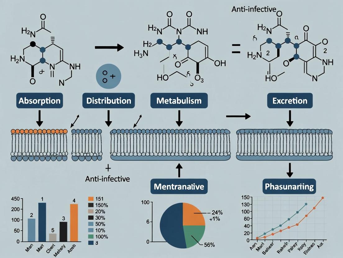
Understanding Lipoglycopeptides: Core Structures and Pharmacokinetic Breakthroughs
This whitepaper defines the long-acting lipoglycopeptide (LALG) antibiotic class, focusing on the core agents dalbavancin, oritavancin, and telavancin. Within the broader thesis on the Absorption, Distribution, Metabolism, and Excretion (ADME) profiles of these agents, this document serves as a technical guide to their chemical, pharmacological, and experimental characterization. Their unique pharmacokinetic properties, primarily driven by extensive tissue binding and prolonged half-lives, differentiate them from traditional glycopeptides and underpin their clinical utility for single- or infrequent-dose regimens.
Core Chemical and Pharmacological Properties
LALGs are semi-synthetic derivatives of natural glycopeptides, modified with lipophilic side chains to enhance antibacterial potency and pharmacokinetic profiles.
Table 1: Core Molecular and In Vitro Pharmacological Properties
| Property | Dalbavancin | Oritavancin | Telavancin |
|---|---|---|---|
| Parent Compound | A40926 (Teicoplanin-like) | Chloroeremomycin (LY264826) | Vancomycin |
| Key Structural Modifications | Lipophilic side chain (C11) on peptide core; amide group on sugar. | 4'-chlorobiphenylmethyl on disaccharide; epi-vancosamine on amino acid 6. | Decylaminoethyl side chain on vancosamine; hydrophilic group (phosphonomethyl) on amino acid 7. |
| Molecular Weight (Da) | ~1816.7 | ~1792.6 | ~1755.6 |
| Primary Mechanism of Action | Inhibits transglycosylation and transpeptidation (cell wall synthesis). | Inhibits transglycosylation; disrupts membrane integrity and potential; inhibits RNA synthesis. | Inhibits transglycosylation and transpeptidation; disrupts membrane potential and permeability. |
| Plasma Protein Binding (%) | 93-98% (concentration-dependent) | ~85% | 90-93% |
| Key In Vitro Activity (MIC90, μg/mL) | S. aureus (MSSA/MRSA): 0.06; S. pyogenes: 0.03; S. agalactiae: 0.06 | S. aureus (MSSA/MRSA): 0.12; Enterococcus spp. (VSE/VRE): 0.25/0.5 | S. aureus (MSSA/MRSA): 0.12-0.5; S. pyogenes: 0.03 |
The defining feature of the LALG class is its extended elimination half-life, enabling long-acting dosing.
Table 2: Comparative Human ADME Profiles
| ADME Parameter | Dalbavancin | Oritavancin | Telavancin |
|---|---|---|---|
| Half-life (t½, days) | ~14.4 (after single 1000 mg dose) | ~13.5 (after single 1200 mg dose) | ~8.0 (after multiple 10 mg/kg doses) |
| Volume of Distribution (Vd, L/kg) | ~0.14 (Low, extensive tissue binding) | ~0.11 (Low, extensive tissue binding) | ~0.13 (Low, extensive tissue binding) |
| Clearance (CL, mL/h/kg) | ~8.7 | ~10.0 | ~15.0 |
| Renal Excretion (% unchanged) | ~42% (over 42 days) | ~5% (over 7 days) | ~76% (over 72 hours) |
| Metabolism | Minimal hepatic; slow hydrolysis of amide bond. | Minimal hepatic; no CYP450. | Minimal hepatic; minor metabolite (hydroxydecyl). |
| Dosing Regimen (ABSSSI) | Single 1500 mg IV dose OR 1000 mg IV, then 500 mg IV at week 1. | Single 1200 mg IV dose. | 10 mg/kg IV every 24 hours for 7-14 days. |
Key Experimental Protocols in LALG Research
Protocol: Determination of Tissue-to-Plasma Partition Coefficients (Kp)
Objective: To quantify drug distribution into tissues, a critical parameter for PK/PD modeling of LALGs. Methodology:
- Animal Dosing & Sacrifice: Administer a single IV bolus of the LALG to rats (n=3-5/time point). At predetermined times post-dose (e.g., 1, 24, 168 h), collect terminal blood (into heparinized tubes) and excise target tissues (e.g., skin, muscle, liver, kidney).
- Sample Processing: Centrifuge blood to obtain plasma. Homogenize weighed tissue samples in buffer (1:3 w/v).
- Bioanalysis: Analyze plasma and tissue homogenate supernatant concentrations using a validated liquid chromatography-tandem mass spectrometry (LC-MS/MS) method. Use matrix-matched calibration standards.
- Calculation: Determine Kp for each tissue at each time point: Kp = [Drug]tissue / [Drug]plasma. Report as mean ± SD. Time-averaged Kp is used for physiologically-based pharmacokinetic (PBPK) modeling.
Protocol:In VitroAssessment of Bacterial Membrane Disruption
Objective: To evaluate the secondary mechanism of action of oritavancin and telavancin. Methodology (Membrane Depolarization using DiSC3(5) dye):
- Bacterial Preparation: Grow S. aureus (e.g., ATCC 29213) to mid-log phase in cation-adjusted Mueller-Hinton broth (CAMHB). Wash and resuspend in buffer with 20 mM glucose.
- Dye Loading: Incubate bacterial suspension with the potentiometric dye DiSC3(5) (final conc. 0.4 μM) until quenched (~1 h).
- Fluorometric Measurement: Aliquot dye-loaded bacteria into a 96-well black plate. Add increasing concentrations of LALG (or vancomycin as control). Immediately monitor fluorescence (excitation 622 nm, emission 670 nm) kinetically for 30-60 min using a plate reader.
- Data Analysis: Calculate maximum depolarization rate or relative fluorescence increase at a fixed time. Plot dose-response to determine effective concentrations.
The Scientist's Toolkit: Key Research Reagent Solutions
Table 3: Essential Materials for LALG ADME and Mechanism Studies
| Research Reagent | Primary Function & Application |
|---|---|
| Synthetic LALG Reference Standard | High-purity compound for preparing calibration curves, quality control samples, and in vitro assays. Critical for bioanalytical method validation. |
| Stable Isotope-Labeled LALG (e.g., ^13C/^15N) | Internal standard for LC-MS/MS quantification. Corrects for matrix effects and recovery variations during sample preparation. |
| Cation-Adjusted Mueller Hinton Broth (CAMHB) | Standardized medium for in vitro susceptibility testing (MIC determination) and time-kill kinetics studies, ensuring reproducible cation concentrations. |
| DiSC3(5) (3,3'-Dipropylthiadicarbocyanine Iodide) | Potentiometric fluorescent dye used to assess bacterial membrane potential disruption, a key mechanism for oritavancin and telavancin. |
| Human Serum Albumin (HSA) Solution | Used in equilibrium dialysis or ultrafiltration experiments to determine plasma protein binding parameters, a major determinant of LALG distribution. |
| Rat Tissue Homogenization Buffer (e.g., Phosphate Buffer, pH 7.4) | Isotonic buffer for preparing homogeneous tissue samples for drug extraction and quantification in tissue distribution studies. |
Visualization of Key Concepts
Diagram 1: LALG Mechanisms of Action
Title: Mechanisms of Action for Long-Acting Lipoglycopeptides
Diagram 2: Experimental PK Workflow for Tissue Distribution
Title: Tissue Distribution Study Workflow for LALGs
Within the broader research on the ADME (Absorption, Distribution, Metabolism, Excretion) profile of long-acting lipoglycopeptide antibiotics, structural modification via lipophilic side chains has emerged as a pivotal strategy. These modifications, often involving the covalent attachment of hydrophobic groups (e.g., alkyl, arylalkyl chains) to the glycopeptide core, are engineered to alter pharmacokinetic properties profoundly. This whitepaper provides an in-depth technical guide on the core structural modifications, their mechanistic impact on ADME parameters, and associated experimental protocols, framed specifically within contemporary lipoglycopeptide research.
Core Structural Modifications & Their Rationale
Lipoglycopeptides, such as telavancin, dalbavancin, and oritavancin, are semisynthetic derivatives of vancomycin. Key modifications include:
- N-Acylated Amino Sugars: Addition of lipophilic chains (e.g., decylaminoethyl in telavancin) to the vancosamine sugar.
- Lipophilic Modifications to the Peptide Core: Addition of chlorobiphenylmethyl (oritavancin) or other hydrophobic groups to different positions on the heptapeptide backbone.
- Dual-Mechanism Designs: Combining lipophilic side chains with other pharmacophores (e.g., a disaccharide in dalbavancin) to enhance both membrane anchoring and target binding.
The primary rationales are:
- Increase Plasma Protein Binding (PPB): Enhancing binding to human serum albumin (HSA) prolongs circulation half-life.
- Promote Tissue Retention: Facilitating partitioning into phospholipid membranes creates a depot effect.
- Alter Renal Clearance: Reducing unbound drug fraction minimizes glomerular filtration.
Quantitative Impact on ADME Properties
The following table summarizes the comparative impact of key lipophilic side chains on the ADME profiles of prominent lipoglycopeptides.
Table 1: Impact of Lipophilic Side Chains on Key ADME Parameters of Select Lipoglycopeptides
| Compound (Core Modification) | Lipophilic Chain (Position) | Plasma Half-life (hrs) | Plasma Protein Binding (%) | Vdss (L/kg) | Primary Clearance Route | Key ADME Impact Reference |
|---|---|---|---|---|---|---|
| Vancomycin (Natural) | None | 4-6 | ~55 | 0.4-0.7 | Renal (GFR) | Baseline comparator |
| Telavancin (Hydrophobic side chain on vancosamine) | Decylaminoethyl | 7-9 | >90 | 0.12-0.14 | Renal (Mixed mechanisms) | Increased PPB, reduced Vd, moderate t1/2 extension |
| Dalbavancin (Lipophilic tail on peptide core + disaccharide) | Dimethylaminopropyl (on Asn) | 346 (≈14 days) | >93 | 0.11-0.15 | Non-renal (catabolism) | Extreme t1/2 due to high PPB & tissue sequestration |
| Oritavancin (Chlorobiphenylmethyl on peptide crosslink) | 4'-Chlorobiphenylmethyl | 393 (≈13 days) | ~85 | 0.10-0.13 | Non-renal (slow hepatic) | Extreme t1/2 due to high tissue binding & slow release |
Vdss: Volume of Distribution at steady state; GFR: Glomerular Filtration Rate.
Experimental Protocols for Characterizing Impact
Protocol: Determination of Plasma Protein Binding (PPB) via Equilibrium Dialysis
Objective: Quantify the fraction of lipoglycopeptide bound to plasma proteins. Materials:
- Test Compound: Lipoglycopeptide stock solution.
- Matrix: Human plasma (heparinized).
- Device: 96-well equilibrium dialyzer (e.g., HTDialysis) with regenerated cellulose membranes (MWCO 12-14 kDa).
- Buffer: Phosphate Buffered Saline (PBS), pH 7.4.
- Analysis: LC-MS/MS system.
Methodology:
- Preparation: Spike the test compound into human plasma to a final therapeutic concentration (e.g., 50 µg/mL). Fill the donor chamber with 150 µL of spiked plasma.
- Dialysis: Fill the receiver chamber with 150 µL of PBS. Seal the plate and incubate at 37°C with gentle agitation for 6-8 hours (ensure equilibrium).
- Sampling: Post-incubation, collect aliquots from both donor (plasma) and receiver (buffer) chambers.
- Sample Processing: Precipitate proteins in the plasma sample using acetonitrile containing an internal standard. Buffer samples may be directly analyzed or diluted with blank plasma for matrix matching.
- Analysis: Quantify drug concentrations in both matrices using a validated LC-MS/MS method.
- Calculation:
- Fraction Unbound (fu) = [Drug]Receiver / [Drug]Donor
- % PPB = (1 - fu) × 100
Protocol: Assessment of Tissue Distribution & Pharmacokinetics in Rodents
Objective: Determine the volume of distribution (Vd) and elimination half-life. Materials:
- Animals: Sprague-Dawley rats or CD-1 mice (n=3-4/time point).
- Formulation: Test lipoglycopeptide in saline (for IV bolus).
- Equipment: Surgical tools for blood collection, LC-MS/MS, pharmacokinetic analysis software (e.g., Phoenix WinNonlin).
Methodology:
- Dosing & Sampling: Administer a single intravenous bolus dose (e.g., 10 mg/kg). Collect serial blood samples (e.g., at 2 min, 30 min, 2, 8, 24, 48, 72, 168 hrs for long-acting agents) via a pre-implanted catheter or terminal cardiac puncture.
- Plasma Processing: Centrifuge blood samples, harvest plasma, and store at -80°C until analysis.
- Bioanalysis: Extract drug from plasma and quantify using LC-MS/MS.
- PK Analysis: Perform non-compartmental analysis (NCA) on mean plasma concentration-time profiles.
- Terminal Half-life (t1/2): Calculated as 0.693/λz, where λz is the terminal slope.
- Volume of Distribution at Steady State (Vdss): Calculated using standard NCA equations: Vdss = Dose * AUMC / (AUC)2, where AUMC is the area under the first moment curve and AUC is the area under the concentration-time curve.
Visualizing the Mechanistic Impact
Diagram 1: Lipophilic Chain Impact on PK Pathways
Diagram 2: Experimental ADME Profiling Workflow
The Scientist's Toolkit: Key Research Reagent Solutions
Table 2: Essential Materials for Lipoglycopeptide ADME Studies
| Item / Reagent | Function in Research | Example / Specification |
|---|---|---|
| Human Serum Albumin (HSA) | Primary binding protein for in vitro PPB and binding constant (Kd) determination via ITC or spectroscopy. | >99% purity, fatty acid-free. |
| Pooled Human Liver Microsomes (HLM) / Hepatocytes | Assessment of metabolic stability and identification of Phase I oxidative metabolites. | Donor-pooled, characterized for major CYP450 activities. |
| Equilibrium Dialysis Devices | Gold-standard method for determining fraction unbound (fu) in plasma. | 96-well format, regenerated cellulose membranes (MWCO 12-14 kDa). |
| Artificial Phospholipid Membranes (e.g., Vesicles, Micelles) | Modeling drug-membrane interactions and tissue partitioning. | DMPC/DMPG liposomes for surface plasmon resonance (SPR) or dialysis studies. |
| Stable Isotope-Labeled Internal Standards (e.g., ¹³C-Vancomycin) | Critical for accurate and precise quantification of glycopeptides in complex biological matrices via LC-MS/MS. | ¹³C6- or ²H4-labeled analogs. |
| LC-MS/MS System with Polar Analytics Column | Bioanalysis of polar, large-molecule antibiotics in plasma, urine, and tissue homogenates. | HILIC or polar-endcapped C18 columns; Triple-quadrupole MS. |
The optimization of Absorption, Distribution, Metabolism, and Excretion (ADME) properties represents a central challenge in modern medicinal chemistry, particularly for antibiotic classes like lipoglycopeptides. The research goal is to engineer molecules that maintain potent antimicrobial activity while achieving a prolonged plasma half-life, enabling less frequent dosing and improving patient adherence. This whitepaper examines the fundamental principles and experimental approaches for extending plasma half-life, framed explicitly within ongoing research on next-generation lipoglycopeptides (e.g., derivatives of dalbavancin and oritavancin). The core thesis is that strategic molecular design, targeting specific ADME parameters, can systematically enhance residence time in the systemic circulation.
Core ADME Principles Governing Plasma Half-Life
Plasma half-life (t₁/₂) is determined by the volume of distribution (Vd) and total clearance (CL): t₁/₂ = (0.693 × Vd) / CL. Designing for extended t₁/₂ therefore involves either increasing Vd (to a point) or, more effectively, reducing CL. Clearance is a sum of metabolic (CLm) and renal/biliary excretion (CLr).
For lipoglycopeptides, key levers include:
- Reducing Renal Clearance: Minimizing glomerular filtration by increasing plasma protein binding (PPB) and molecular size above the renal filtration threshold (~45-50 kDa).
- Altering Distribution: Engineering controlled tissue distribution to create a depot effect, followed by slow redistribution back to plasma.
- Evading Metabolism: Designing molecular motifs resistant to hepatic cytochrome P450 and other metabolic enzymes.
- Modulating Transporters: Avoiding recognition by active efflux transporters in the liver (biliary clearance) and kidney.
Table 1: Comparative ADME Properties of Long-Acting Lipoglycopeptides
| Property | Dalbavancin | Oritavancin | Vancomycin (Reference) | Design Target for Next-Gen |
|---|---|---|---|---|
| Approx. Molecular Weight (Da) | 1,816 | 1,790 | 1,449 | >1,800 |
| Plasma Half-Life (t₁/₂, days) | ~14 | ~10 | 0.5 | >14 |
| Plasma Protein Binding (%) | >93% | ~85% | 30-55% | >90% |
| Volume of Distribution (Vd, L/kg) | 0.11-0.14 | 0.10-0.13 | 0.4-0.9 | Low (0.1-0.3) |
| Primary Route of Elimination | Biliary/Fecal (~50%), Renal | Biliary/Fecal, Renal | Renal (>90%) | Balanced non-renal |
| Key Feature for Long t₁/₂ | High PPB, tissue binding | Self-association, tissue binding | N/A | High PPB, controlled tissue depot |
Detailed Experimental Protocols for Key Assays
Protocol:In VitroPlasma Protein Binding (Ultrafiltration)
Objective: Quantify the fraction of drug bound to plasma proteins. Materials: Human plasma (heparinized), test compound, PBS (pH 7.4), centrifugal ultrafiltration devices (e.g., Amicon Ultra, 10 kDa MWCO), 37°C incubator/shaker, LC-MS/MS.
- Spike test compound into human plasma to a therapeutic concentration (e.g., 50 µg/mL). Incubate at 37°C for 15 min.
- Load 500 µL of spiked plasma into the upper chamber of a pre-rinsed ultrafiltration device.
- Centrifuge at 2000 × g, 37°C, for 30 min to obtain ~100 µL of ultrafiltrate.
- Analyze the total concentration in the initial plasma (Ctotal) and the free concentration in the ultrafiltrate (Cfree) using a validated LC-MS/MS method.
- Calculate % bound = [1 - (Cfree / Ctotal)] × 100.
Protocol:In VivoPharmacokinetic Study in Rodents
Objective: Determine key PK parameters (t₁/₂, Vd, CL, AUC) after a single intravenous dose. Materials: Rats/mice, test compound in sterile saline, catheters, heparinized blood collection tubes, LC-MS/MS.
- Administer test compound intravenously via tail vein (rats) or retro-orbital route at a defined dose (e.g., 5 mg/kg).
- Collect serial blood samples (e.g., 5, 15, 30 min, 1, 2, 4, 8, 24, 48, 72, 96 h) into heparinized tubes.
- Centrifuge samples immediately (3000 × g, 10 min, 4°C). Harvest plasma and store at -80°C.
- Extract analytes from plasma via protein precipitation. Quantify plasma concentrations using LC-MS/MS.
- Analyze concentration-time data using non-compartmental methods (e.g., Phoenix WinNonlin) to calculate PK parameters.
Protocol: Hepatocyte Stability Assay
Objective: Assess metabolic clearance potential in liver cells. Materials: Cryopreserved human hepatocytes, Williams' E medium, test compound, 96-well plates, CO₂ incubator.
- Thaw cryopreserved hepatocytes and suspend in Williams' E medium at 1.0 × 10⁶ viable cells/mL.
- Incubate cells with 1 µM test compound in a 96-well plate at 37°C, 5% CO₂.
- At time points (0, 15, 30, 60, 90, 120 min), remove an aliquot of supernatant and quench with cold acetonitrile.
- Analyze parent compound disappearance by LC-MS/MS.
- Calculate intrinsic clearance (CLint) from the half-life of disappearance.
Design Logic for Extended Plasma Half-Life
The Scientist's Toolkit: Key Research Reagent Solutions
Table 2: Essential Reagents & Materials for ADME/PK Studies
| Item | Function & Rationale | Example Product/Supplier |
|---|---|---|
| Human Plasma (Pooled) | In vitro PPB studies; provides physiologically relevant protein matrix. | BioIVT, Sigma-Aldrich |
| Cryopreserved Hepatocytes | Gold-standard for in vitro metabolic stability assessment; retain Phase I/II enzyme activity. | Thermo Fisher (Gibco), Lonza |
| LC-MS/MS System | Sensitive and specific quantification of drug concentrations in complex biological matrices. | Waters Xevo TQ-XS, Sciex Triple Quad 6500+ |
| Centrifugal Ultrafiltration Units | Rapid separation of protein-bound vs. free drug for PPB calculation. | Millipore Amicon Ultra (10 kDa MWCO) |
| In Vivo Pharmacokinetic Software | Non-compartmental analysis (NCA) of concentration-time data to derive t₁/₂, Vd, CL. | Certara Phoenix WinNonlin |
| Stable Isotope-Labeled Internal Standards | Critical for accurate LC-MS/MS quantification, correcting for matrix effects and recovery. | Custom synthesis (e.g., Alsachim, TLC) |
| Animal Disease Models (e.g., Neutropenic Thigh) | Evaluates PK/PD relationship and efficacy of extended half-life in an infection context. | Charles River, Inotiv |
Molecular Design Strategies: Lessons from Lipoglycopeptides
Recent research on lipoglycopeptides highlights successful strategies:
- Lipidation: Adding a lipid tail (e.g., dalbavancin) dramatically increases affinity for serum albumin and promotes binding to cell membranes, creating a dual-depot effect. The lipid tail is the primary determinant of its >90% PPB and 14-day half-life.
- Controlled Self-Association: Oritavancin exhibits concentration-dependent self-association, which may protect it from renal filtration and enzymatic degradation.
- Glycosylation Optimization: Modifying the sugar moieties can alter hydrogen bonding and polarity, fine-tuning Vd and interactions with transporters without compromising antimicrobial activity.
Table 3: Impact of Specific Modifications on ADME Parameters
| Molecular Modification | Effect on PPB | Effect on Vd | Effect on CL | Net Impact on t₁/₂ |
|---|---|---|---|---|
| Addition of C16 Lipid Chain | ↑↑↑ (Major increase) | ↑ (Slight increase) | ↓↓ (Decrease) | ↑↑↑ |
| Increased Glycosylation | ↑ (Moderate increase) | ↓ (Decrease) | ↓ (Moderate decrease) | ↑ |
| Dimerization | ↑↑ | Variable | ↓↓ (Reduced renal filtration) | ↑↑ |
Long-Acting Lipoglycopeptide PK Model
The fundamental challenge of designing molecules for extended plasma half-life is systematically addressed by targeting the components of clearance and distribution. Research on long-acting lipoglycopeptides provides a proven blueprint: strategic lipidation and modification to achieve ultra-high plasma protein binding and controlled tissue distribution. The integration of robust in vitro assays (PPB, hepatocyte stability) with definitive in vivo PK studies, as outlined in this guide, forms an iterative feedback loop for molecular optimization. Success in this endeavor directly translates to next-generation therapeutics with improved dosing regimens and enhanced therapeutic outcomes.
Within the framework of research on the ADME (Absorption, Distribution, Metabolism, Excretion) profile of long-acting lipoglycopeptides (LGPs), understanding the mechanisms enabling prolonged antimicrobial activity is paramount. The extended half-life and sustained efficacy of agents like dalbavancin and oritavancin are not attributable to a single factor but result from the synergistic interplay of high plasma protein binding, extensive tissue distribution, and slow release from peripheral compartments. This whitepaper provides a technical deconstruction of these core mechanisms, serving as a guide for researchers in antibiotic development.
Core Mechanisms of Prolonged Action
Plasma Protein Binding
High affinity for serum proteins, predominantly albumin, creates a significant circulating reservoir of drug, preventing rapid renal filtration and maintaining a stable, sub-MIC concentration gradient that drives tissue distribution.
Table 1: Protein Binding and Pharmacokinetic Parameters of Key Lipoglycopeptides
| Compound | % Protein Binding (Human) | Terminal Half-life (t1/2) | Primary Binding Protein | Reference |
|---|---|---|---|---|
| Dalbavancin | >93% | ~347 hours | Human Serum Albumin | (Dunne et al., 2016) |
| Oritavancin | ~85% | ~393 hours | Human Serum Albumin | (Bhavnani et al., 2014) |
| Telavancin | ~90% | ~8 hours | Human Serum Albumin | (Higgins et al., 2009) |
Experimental Protocol: Determination of Protein Binding via Equilibrium Dialysis
- Principle: Separation of protein-bound and free drug fractions across a semi-permeable membrane at equilibrium.
- Materials:
- Equilibrium dialysis device (e.g., HTD96b dialysis block).
- Regenerated cellulose membranes (MWCO 12-14 kDa).
- Test compound (LGP) in DMSO stock.
- Human plasma or purified human serum albumin (HSA) solution (e.g., 40 g/L in phosphate buffer).
- Matching buffer (e.g., 67 mM phosphate, pH 7.4).
- LC-MS/MS system for quantification.
- Method:
- Pre-hydrate dialysis membranes in buffer for 30 minutes.
- Spike the plasma/HSA compartment with LGP to a therapeutic concentration (e.g., 100 µg/mL).
- Fill the adjacent compartment with equal volume of buffer.
- Seal plates and incubate at 37°C with gentle agitation for 6-8 hours (time to equilibrium predetermined).
- Post-incubation, aliquot samples from both compartments.
- For the plasma compartment, precipitate proteins with acetonitrile containing internal standard, vortex, and centrifuge. Analyze supernatant (free drug).
- Directly analyze buffer compartment (free drug).
- Calculate % bound = [(Cplasma - Cbuffer) / C_plasma] * 100.
Tissue Distribution and Pharmacokinetic/Pharmacodynamic (PK/PD) Drivers
LGPs exhibit multi-compartmental pharmacokinetics, with slow distribution into and prolonged retention within peripheral tissues, often measured as the volume of distribution at steady state (Vss).
Table 2: Tissue Distribution Metrics for Lipoglycopeptides
| Compound | Vss (L/kg) | Key Target Tissues (High Concentration) | Critical PK/PD Index for Efficacy |
|---|---|---|---|
| Dalbavancin | ~0.14 | Skin, Bone, Renal Cortex | AUC0-24/MIC |
| Oritavancin | ~1.07 | Reticuloendothelial System, Skin | AUC/MIC |
| Telavancin | ~0.14 | Pulmonary Epithelial Lining Fluid | AUC/MIC |
Experimental Protocol: Quantitative Whole-Body Autoradiography (QWBA) for Tissue Distribution
- Principle: Radiolabeled drug is administered in vivo, followed by cryosectioning of the entire animal and imaging to quantify radioactivity in discrete tissues.
- Materials:
- Radiolabeled LGP (e.g., ³H or ¹⁴C).
- Laboratory animals (rats, mice).
- Cryomicrotome.
- Phosphor-imaging plates and scanner.
- Calibrated radioactive standards.
- Image analysis software (e.g., MCID).
- Method:
- Administer a single intravenous dose of radiolabeled LGP to animals (n=3-4 per time point).
- Euthanize animals at predetermined time points (e.g., 1, 24, 168, 336 hours post-dose).
- Embed carcasses in carboxymethyl cellulose and freeze in a hexane/dry ice bath.
- Section sagittally at 30-40 µm thickness in a cryomicrotome at -20°C.
- Thaw-mount sections on adhesive tape and freeze-dry.
- Expose sections against phosphor-imaging plates alongside calibrated standards for 3-7 days.
- Scan plates and use software to convert optical density to radioactivity concentration (nCi/g) for each tissue, correcting for decay.
- Calculate tissue-to-plasma ratios over time.
Slow Release from Peripheral Compartments
The slow terminal half-life is primarily due to the rate-limiting redistribution of drug from deep tissue compartments back into the systemic circulation, not prolonged plasma exposure.
Diagram 1: Multi-Compartment Pharmacokinetic Model of LGP Disposition
The Scientist's Toolkit: Research Reagent Solutions
Table 3: Essential Research Materials for Investigating LGP ADME
| Item | Function/Application | Example Product/Specification |
|---|---|---|
| Human Serum Albumin (HSA) | Key binding partner for in vitro protein binding and displacement studies. | Sigma-Aldrich, Fraction V, ≥96% (A1653), fatty acid-free. |
| Equilibrium Dialysis Kit | Gold-standard method for quantifying free vs. protein-bound drug fraction. | HTDialysis, HTD96b 96-well plate system with 12-14 kDa MWCO membranes. |
| ³H or ¹⁴C Radiolabeled LGP | Critical tracer for mass balance, tissue distribution (QWBA), and metabolite profiling studies. | Custom synthesis from radiochemistry suppliers (e.g., American Radiolabeled Chemicals). |
| Caco-2 Cell Line | Model for assessing passive and active transport across intestinal epithelium (relevant for oral prodrug development). | ATCC, HTB-37, passages 20-40 preferred for consistent monolayer formation. |
| LC-MS/MS System | Quantitative bioanalysis of LGPs and metabolites in complex matrices (plasma, tissue homogenates). | Triple quadrupole MS (e.g., Sciex 6500+, Waters Xevo TQ-S) with reverse-phase UPLC. |
| Phospholipid Vesicles (SUVs/LUVs) | Model membranes to study drug-lipid bilayer interactions influencing tissue distribution. | Prepared from DMPC/DPPC/cholesterol via extrusion (100 nm filters). |
| Cryomicrotome | Essential for preparing whole-body tissue sections for QWBA studies. | Leica CM3600, maintained at -20°C. |
| Pooled Human Liver Microsomes (pHLM) | Initial screening for Phase I oxidative metabolic stability and potential drug-drug interactions. | Corning Life Sciences, 150-donor pool, mixed gender. |
Integrated Experimental Workflow
Diagram 2: Integrated Workflow for Profiling Prolonged Action Mechanisms
The prolonged action of modern lipoglycopeptides is a sophisticated pharmacokinetic phenomenon engineered through molecular design. It is predicated on high plasma protein binding to create a stable reservoir, favorable physicochemical properties driving extensive tissue distribution, and complex multi-compartmental kinetics governed by slow release from deep tissue sites. Deconvoluting these mechanisms requires an integrated experimental approach, combining highly quantitative in vitro and in vivo techniques, as outlined herein. This framework is essential for advancing the next generation of long-acting antimicrobial therapies.
Characterizing ADME: Advanced Methods for Lipoglycopeptide Analysis
Bioanalytical Techniques for Quantifying Ultra-Low Plasma Concentrations Over Weeks
Within the broader thesis on the Absorption, Distribution, Metabolism, and Excretion (ADME) profile of long-acting lipoglycopeptides, accurately quantifying ultra-low plasma concentrations over extended periods (weeks) is paramount. These antibiotics, such as dalbavancin, exhibit terminal half-lives exceeding 300 hours, requiring exceptionally sensitive and stable bioanalytical methods to characterize their complete pharmacokinetic profile. This guide details the advanced techniques enabling this critical measurement.
Core Technical Challenges
The quantification of analytes at low pg/mL concentrations over weeks post-dose presents distinct challenges:
- Ultra-Low Limits of Quantification (LLOQ): Required sensitivity often down to 1-10 pg/mL.
- Extended Sample Stability: Analyte and internal standard stability must be demonstrated over the entire study timeline, including freeze-thaw cycles.
- Matrix Effects: Overcoming ion suppression/enhancement from complex plasma matrices over long chromatographic runs.
- Carryover: Minimizing carryover is critical when samples with million-fold concentration differences are analyzed sequentially.
Key Techniques and Methodologies
Immunocapture-LC-MS/MS
This technique combines the specificity of immunoaffinity enrichment with the sensitivity of mass spectrometry.
Detailed Protocol:
- Reagent Preparation: Coat a 96-well plate with a monoclonal antibody specific to the lipoglycopeptide of interest. Block with 1% BSA in PBS.
- Sample Preparation: Dilute 100 µL of plasma sample with 200 µL of PBS buffer (pH 7.4).
- Immunocapture: Add diluted sample to antibody-coated wells. Incubate for 2 hours at room temperature with gentle shaking.
- Washing: Wash wells 5x with PBS-Tween 20 (0.05% v/v) to remove non-specifically bound matrix components.
- Elution: Elute the captured analyte using 100 µL of a low-pH elution buffer (e.g., 0.1% Formic Acid). Neutralize immediately.
- LC-MS/MS Analysis: Inject eluent onto a nano-flow or micro-flow LC system coupled to a triple quadrupole MS.
Diagram Title: Immunocapture-LC-MS/MS Workflow
Microflow LC-Nanospray MS/MS
Reducing LC flow rates to the µL/min scale increases ionization efficiency, enhancing signal-to-noise ratio.
Detailed Protocol:
- Column: Use a capillary column (e.g., 0.3 mm ID, 50-100 mm length) packed with 1.7 µm C18 particles.
- LC Conditions: Flow rate: 3-10 µL/min. Gradient: 5-95% mobile phase B (0.1% FA in ACN) over 8-15 minutes.
- Ion Source: Nanospray or microspray emitter (1-10 µm tip).
- MS Parameters: Operate in positive MRM mode. Optimize collision energy for 3-5 specific precursor-to-product ion transitions.
Diagram Title: Microflow LC-Nanospray MS/MS Pathway
Stability-Enhanced Sample Preparation
Critical for long-term study integrity.
Detailed Protocol for Stabilizing Lipoglycopeptides in Plasma:
- Immediate Post-Collection: Add stabilizers to K2EDTA tubes (e.g., protease inhibitors, metal chelators like EDTA).
- Acidification: For base-labile compounds, immediately acidify plasma with 10% v/v of 1M phosphate buffer (pH 3.0).
- Storage: Snap-freeze in liquid N₂ within 1 hour. Store at ≤ -70°C.
- Freeze-Thaw: Conduct stability tests for a minimum of 5 cycles. Thaw samples on wet ice.
Table 1: Comparison of Bioanalytical Techniques for Ultra-Low Quantification
| Technique | Typical LLOQ (pg/mL) | Linear Dynamic Range | Key Advantage | Major Challenge for Long-Term Studies |
|---|---|---|---|---|
| Immunocapture-LC-MS/MS | 1 - 5 | 3-4 orders of magnitude | Exceptional specificity; high matrix removal | Antibody reagent stability and cost |
| Microflow LC-MS/MS | 5 - 20 | 3-4 orders of magnitude | Enhanced ionization efficiency; reduced solvent use | System robustness & potential for carryover |
| Solid Phase Extraction (SPE)-LC-MS/MS | 50 - 100 | 2-3 orders of magnitude | Robust, high-throughput | May lack sensitivity for terminal phase |
| 2D-LC (Online SPE)-MS/MS | 10 - 50 | 3 orders of magnitude | Full automation; high reproducibility | Complex instrumentation |
Table 2: Example Method Validation Parameters for a Lipoglycopeptide (e.g., Dalbavancin)
| Validation Parameter | Result | Acceptance Criteria |
|---|---|---|
| LLOQ | 2.0 pg/mL | CV ≤20%, Accuracy 80-120% |
| Intra-day Accuracy | 94.2 - 102.5% | 85-115% |
| Intra-day Precision (CV%) | ≤6.8% | ≤15% |
| Extraction Recovery | 88.5% | Consistent and reproducible |
| Matrix Effect (CV%) | ≤5.2% | ≤15% |
| Processed Sample Stability (4°C) | 72 hours | No significant degradation |
| Freeze-Thaw Stability (5 cycles) | Stable | Accuracy within ±15% of nominal |
| Long-Term Stability (-70°C) | 24 months (tested) | Accuracy within ±15% of nominal |
The Scientist's Toolkit: Research Reagent Solutions
- Stable Isotope-Labeled Internal Standard (SIL-IS): (e.g., ^13C/^15N-labeled lipoglycopeptide). Function: Compensates for variability in sample preparation and ionization efficiency; essential for accurate quantification.
- Anti-Lipoglycopeptide Monoclonal Antibody (Capture Grade): Function: Provides high-affinity, specific immunoenrichment of the target analyte from plasma, removing >95% of matrix interferents.
- Acidified Plasma Collection Tubes: Function: Immediately stabilizes the analyte upon blood draw by inhibiting enzymatic degradation or conformation changes.
- Low-Binding Microcentrifuge Tubes/Plates (e.g., Polypropylene): Function: Minimizes adsorptive loss of ultra-low concentration analytes to container surfaces.
- Nano-LC Column (e.g., 0.3mm ID, C18, 1.7µm): Function: Enables low-flow chromatography for improved ionization efficiency and sensitivity.
- High-Purity MS-Grade Solvents & Additives: Function: Reduces chemical noise, ensuring consistent chromatographic baselines and ion source cleanliness over thousands of injections.
- Quality Control (QC) Materials: Function: Spiked plasma at Low, Mid, and High concentrations within the calibration curve. Used to monitor method performance and batch acceptance throughout the analysis of long-term study samples.
Within a broader thesis investigating the Absorption, Distribution, Metabolism, and Excretion (ADME) profile of long-acting lipoglycopeptides (LGPs), understanding tissue-specific pharmacokinetics is paramount. The therapeutic efficacy of LGPs against multidrug-resistant Gram-positive infections in deep-seated sites like bone, skin/soft tissue, and lung parenchyma is directly contingent on their ability to achieve sufficient unbound concentrations at the target site. This guide details the principal ex vivo and in vivo methodologies for quantifying drug concentrations in these complex matrices, critical for validating the extended tissue penetration hypothesized for novel LGPs and linking PK/PD indices to clinical outcomes.
Table 1: Core Analytical Metrics for Tissue Penetration Studies
| Metric | Typical Target Value (for LC-MS/MS) | Importance for Tissue Analysis |
|---|---|---|
| Lower Limit of Quantification (LLOQ) | 0.5-10 ng/mL (or ng/g) | Defines sensitivity for low-concentration tissue homogenates. |
| Accuracy (% Nominal) | 85-115% | Ensures reliability of concentration data from complex matrices. |
| Precision (%CV) | ≤15% (≤20% at LLOQ) | Critical for reproducible measurement in heterogeneous tissues. |
| Extraction Recovery | >50% (tissue-dependent) | Indicates efficiency of analyte release from tissue matrix. |
| Matrix Effect (%CV) | ≤15% | Assesses ion suppression/enhancement from tissue components. |
Table 2: Common Tissue Penetration Indices
| Index | Formula | Interpretation |
|---|---|---|
| Tissue-to-Plasma Ratio (Kp) | [Tissue] / [Plasma] | Basic measure of distribution. Kp >1 suggests tissue affinity. |
| Unbound Tissue-to-Plasma Ratio (Kp,uu) | [Tissueunbound] / [Plasmaunbound] | Mechanistic measure of active transport/diffusion. |
| Area Under the Curve Ratio (AUCtissue / AUCplasma) | AUC(0-∞)tissue / AUC(0-∞)plasma | Gold standard for steady-state penetration assessment. |
Experimental Protocols for Tissue Concentration Analysis
3.1. Sample Collection & Preparation Protocol
- Animal Dosing & Sacrifice: Administer LGP via relevant route (IV/SC). Euthanize animals at serial time points (n≥3/time). Collect blood (for plasma) and target tissues (e.g., femur, skin full-thickness punch, whole lung lobe).
- Tissue Processing: Weigh tissue wet. Rinse in saline, blot dry. For bone (cortical): grind under liquid N₂. For skin: remove subcutaneous fat, slice finely. For lung: homogenize whole or regional lobes.
- Homogenization: Homogenize tissue in buffer (e.g., phosphate buffer saline, pH 7.4) at 1:4 (w/v) ratio using a bead homogenizer or rotor-stator. Keep samples on ice.
- Bioanalysis: Use protein precipitation, solid-phase extraction (SPE), or liquid-liquid extraction (LLE) for sample clean-up. Quantify using a validated LC-MS/MS method with stable isotope-labeled internal standard.
3.2. Determination of Unbound Tissue Fraction (fu,t)
- Method: Equilibrium Dialysis or Centrifugal Ultrafiltration of tissue homogenate.
- Protocol: Dilute homogenate (e.g., 1:4 in buffer). Load into donor chamber of a semi-permeable membrane device (MWCO 10-30 kDa). Dialyze against isotonic buffer for 4-6 hours at 37°C. Quantify drug concentration in buffer (receiver) and homogenate (donor) chambers via LC-MS/MS.
- Calculation: fu,t = (Creceiver / Cdonor) * Dilution Factor.
3.3. Microdialysis for In Vivo, Unbound Concentration Sampling
- Probe Implantation: Surgically implant linear or concentric microdialysis probes into target tissue (e.g., subcutaneous, bone marrow, lung parenchyma). Allow stabilization (1-2 hrs).
- Perfusion & Sampling: Perfuse probe with sterile isotonic saline or Ringer's solution at low flow rate (0.5-2 µL/min). Collect dialysate at fixed intervals over the PK period.
- Calibration: Perform in vivo retrodialysis or zero-flow method post-sampling to determine relative recovery for each probe.
- Analysis: Correct dialysate concentration using recovery factor to calculate unbound tissue concentration in the extracellular fluid.
Visualizing Experimental Workflows and Concepts
Diagram 1: Workflow for tissue penetration PK studies.
Diagram 2: Drug distribution between plasma and tissue compartments.
The Scientist's Toolkit: Key Research Reagent Solutions
Table 3: Essential Materials for Tissue Penetration Studies
| Item | Function & Rationale |
|---|---|
| Stable Isotope-Labeled Internal Standard (e.g., ^13C- or ^2H-LGP) | Normalizes for variability in sample preparation and ionization efficiency in LC-MS/MS, ensuring accuracy. |
| Phosphate Buffered Saline (PBS), pH 7.4 | Isotonic homogenization buffer that maintains physiological pH, preserving tissue integrity and drug stability. |
| Equilibrium Dialysis Blocks (e.g., HTD96b) | High-throughput devices for determining unbound fraction (fu,t) via semi-permeable membranes at constant temperature. |
| LC-MS/MS Mobile Phase Additives (e.g., Formic Acid, Ammonium Acetate) | Critical for optimal chromatographic separation and ionization efficiency of polar, amphiphilic LGPs. |
| Microdialysis Probes (Linear/Membrane) | Allow continuous, minimally invasive sampling of unbound drug from the extracellular space of specific tissues in vivo. |
| Protein Precipitation Reagents (e.g., Acetonitrile, Methanol) | Rapidly deproteinize plasma and tissue homogenates, precipitating interfering macromolecules prior to LC-MS/MS. |
| Solid-Phase Extraction (SPE) Cartridges (e.g., Mixed-mode Cation Exchange) | Provide selective clean-up of complex tissue lysates, enhancing sensitivity by removing lipids and other matrix interferences. |
| Artificial Interstitial Fluid (Perfusate for Microdialysis) | Isotonic, biocompatible solution that mimics extracellular fluid, minimizing osmotic disruption during in vivo sampling. |
Within the broader thesis on the ADME (Absorption, Distribution, Metabolism, Excretion) profile of long-acting lipoglycopeptides, pharmacokinetic/pharmacodynamic (PK/PD) modeling stands as the pivotal translational bridge. For novel entities like lipoglycopeptides designed for once-weekly or single-dose administration, robust PK/PD models are indispensable. They define the critical parameters governing efficacy, safety, and optimal dosing, transforming complex physiological and microbiological data into actionable regimens. This technical guide elucidates the core principles, key parameters, and simulation strategies for PK/PD modeling specific to these extended-interval therapies.
Core PK/PD Parameters for Long-Acting Lipoglycopeptides
The extended dosing intervals of once-weekly or single-dose regimens necessitate a focus on parameters that predict sustained target engagement. Key parameters are summarized in the table below.
Table 1: Key PK/PD Parameters and Their Significance
| Parameter | Description | Significance for Once-Weekly/Single-Dose Regimens |
|---|---|---|
| AUC/MIC | Area Under the Curve (exposure) to Minimum Inhibitory Concentration ratio. | For concentration-dependent antibiotics (e.g., lipoglycopeptides), a high AUC/MIC is the primary driver of efficacy and bactericidal activity. Predicts cumulative kill. |
| Cmax/MIC | Peak Concentration to MIC ratio. | Correlates with initial bacterial killing rate and prevention of resistance emergence. Critical for front-loaded, single-dose strategies. |
| T > MIC | Time plasma concentration exceeds the MIC. | For time-dependent killers, this is paramount. For lipoglycopeptides, a long T > MIC is inherent but still validated for efficacy. |
| Terminal Half-life (t1/2,β) | Time for plasma concentration to reduce by 50% in the elimination phase. | Must be sufficiently long (often > 7 days) to support the dosing interval. Driven by slow tissue redistribution and renal clearance. |
| Volume of Distribution (Vd) | Apparent volume into which a drug distributes. | A large Vd (e.g., >0.1 L/kg) indicates extensive tissue penetration, crucial for treating deep-seated infections. |
| Protein Binding (%) | Fraction of drug bound to plasma proteins. | High protein binding (>90% for many lipoglycopeptides) can influence free drug concentration, the active moiety for PK/PD targets. |
| Post-Antibiotic Effect (PAE) | Persistent suppression of bacterial growth after drug removal. | A long PAE extends the effective dosing interval beyond measurable T > MIC, a key feature for sparse dosing. |
Critical Experiments and Protocols for Model Development
In VitroHollow-Fiber Infection Model (HFIM) Time-Kill Studies
This system simulates human PK profiles in vitro to establish exposure-response relationships.
Protocol:
- Setup: Inoculate the extracapillary space of hollow-fiber cartridges with a standardized bacterial inoculum (~10⁸ CFU/mL) of target pathogens (e.g., S. aureus, E. faecalis).
- Drug Administration: Program the central reservoir and pump system to simulate human single-dose or once-weekly PK profiles of the lipoglycopeptide in the circulatory (intracapillary) space. Multiple profiles (simulating different doses) are run in parallel.
- Sampling: At predetermined time points (e.g., 0, 1, 2, 4, 8, 24, 48, 72, 168h), sample from the bacterial compartment for quantitative culture.
- Analysis: Plot bacterial density (log₁₀ CFU/mL) over time. Determine the PK/PD index (AUC/MIC, Cmax/MIC) that best correlates with 1-log kill, stasis, and resistance suppression.
Murine Thigh or Lung Infection Model
An in vivo system to validate PK/PD targets in the context of host immunity.
Protocol:
- Infection: Render mice neutropenic via cyclophosphamide. Inoculate thigh muscles or lungs intranasally with ~10⁶ CFU of the target pathogen.
- Dosing: Administer single subcutaneous doses of the lipoglycopeptide at varying levels (simulating a range of human-equivalent exposures) 2h post-infection.
- PK Sampling: Collect serial plasma samples from satellite groups of mice to characterize the drug's PK profile in the model.
- Efficacy Endpoint: Sacrifice mice 24h post-treatment, homogenize thighs/lungs, and perform quantitative culture. Calculate the change in bacterial density relative to untreated controls.
- Modeling: Link the measured PK profiles to the observed PD effect (CFU reduction) using an inhibitory sigmoid Emax model to identify the target exposure (e.g., AUC/MIC for stasis or 1-log kill).
PK/PD Modeling and Simulation Workflow
The logical flow from data generation to clinical regimen simulation is depicted below.
Diagram Title: PK/PD Modeling & Simulation Workflow for Dose Optimization
Key Simulation Outputs:
- Probability of Target Attainment (PTA): The percentage of virtual patients achieving the PK/PD target at a given MIC.
- Cumulative Fraction of Response (CFR): The weighted average of PTA across the MIC distribution of a target bacterial population. A CFR ≥90% is typically desired.
Table 2: Example Monte Carlo Simulation Output for a Hypothetical Lipoglycopeptide
| Dose | Interval | Target (fAUC/MIC=100) | PTA at MIC=0.5 mg/L | CFR vs. S. aureus Population |
|---|---|---|---|---|
| 1000 mg | Single Dose | 1-log kill | 99.5% | 95.2% |
| 1500 mg | Single Dose | 1-log kill | 99.9% | 98.7% |
| 750 mg | Once Weekly | Stasis (fAUC/MIC=50) | 98.1% | 93.5% |
The Scientist's Toolkit: Research Reagent Solutions
Table 3: Essential Materials for Key Experiments
| Item | Function | Example/Supplier Note |
|---|---|---|
| Hollow-Fiber Infection Model (HFIM) System | Provides a dynamic in vitro system to simulate human PK profiles with bacteria. | CellPhire cartridges; AvaCell systems. |
| Cation-Adjusted Mueller Hinton Broth (CAMHB) | Standardized growth medium for antimicrobial susceptibility and time-kill assays. | Must be prepared according to CLSI guidelines. |
| Cyclophosphamide | Immunosuppressant used to induce neutropenia in murine infection models. | Requires careful handling and institutional IACUC protocols. |
| Stable Isotope-Labeled Internal Standard | Essential for accurate and precise quantification of drug concentrations in biological matrices via LC-MS/MS. | e.g., ¹³C/¹⁵N-labeled lipoglycopeptide analog. |
| Population PK Modeling Software | For developing and simulating nonlinear mixed-effects models. | NONMEM, Monolix, Phoenix NLME. |
| Monte Carlo Simulation Software | For performing stochastic simulations to calculate PTA/CFR. | R (with Mrgsolve or PopED), SAS, integrated within PK software. |
In the development of long-acting lipoglycopeptides, sophisticated PK/PD modeling and simulation are non-negotiable for rational regimen design. By rigorously deriving targets from in vitro and in vivo systems, and validating them through population PK models and Monte Carlo simulations, researchers can confidently advance once-weekly or single-dose regimens with a high probability of clinical success. This model-informed drug development approach de-risks later stages and ensures these novel therapies are positioned to maximize therapeutic benefit while minimizing the burden of frequent dosing.
Translating Preclinical ADME Data to Human Dosing Predictions and Clinical Trial Design
Within the broader thesis on the ADME profile of long-acting lipoglycopeptides, this guide details the technical process of translating preclinical Absorption, Distribution, Metabolism, and Excretion (ADME) data into informed human dose predictions and efficient clinical trial designs. These antibiotics, characterized by lipid moieties attached to glycopeptide cores (e.g., dalbavancin, oritavancin), exhibit unique pharmacokinetics (PK) dominated by extensive tissue distribution, prolonged half-life, and negligible renal metabolism, necessitating specialized translation strategies.
Key Preclinical ADME Assays & Data Translation
Core Quantitative ADME Parameters
Preclinical studies for lipoglycopeptides generate data that feed into pharmacokinetic (PK) and pharmacodynamic (PD) models. The table below summarizes the critical parameters and their role in translation.
Table 1: Core Preclinical ADME Parameters for Lipoglycopeptides & Their Translational Purpose
| Parameter | Typical In Vitro / In Vivo Assay | Translational Purpose & Human Prediction Method |
|---|---|---|
| Solubility & Permeability | Thermodynamic solubility assays; Caco-2/PAMPA permeability. | Predicts absorption potential. Low permeability is expected; confirms parenteral route. |
| Plasma Protein Binding | Equilibrium dialysis or ultrafiltration using radiolabeled compound. | Determines free (active) drug fraction. Critical for scaling PK and estimating effective dose. |
| Metabolic Stability | Incubation with human/microsomes/hepatocytes; LC-MS/MS analysis. | Predicts clearance (CL) route. Lipoglycopeptides show minimal hepatic metabolism; primary clearance via renal filtration. |
| Cytochrome P450 Interaction | CYP inhibition (IC50) and induction assays. | Assess DDI risk. Typically low for this class, informing co-medication in trial protocols. |
| Blood-to-Plasma Ratio | Incubation of compound in fresh blood; partition measurement. | Informs volume of distribution (Vd) scaling and PK sampling matrix. |
| In Vivo Clearance | IV PK study in rodents/non-rodents; non-compartmental analysis. | Allometric scaling (e.g., fixed exponent, MLP) to predict human CL. |
| In Vivo Volume of Distribution | IV PK study; non-compartmental analysis. | Allometric scaling to predict human Vd. High Vd (≥1 L/kg) indicates significant tissue distribution. |
| In Vivo Half-life | Derived from CL and Vd. | Indicates dosing frequency. Long half-life (>24h) supports single-dose or weekly regimens. |
| Tissue Distribution | Quantitative Whole-Body Autoradiography (QWBA) or tissue homogenization. | Validates high Vd; identifies reservoir tissues (e.g., skin, bone) for efficacy against SSTIs. |
| Excretion Balance | Mass balance study with radiolabeled compound in bile-duct cannulated animals. | Confirms primary excretion pathways (feces vs. urine). |
Experimental Protocols for Key Assays
Protocol: Plasma Protein Binding via Equilibrium Dialysis
- Materials: Equilibrium dialysis device (e.g., RED plate), dialysis membrane (8-10 kDa MWCO), test compound (radiolabeled or cold), blank plasma (human/preclinical species), phosphate buffer (pH 7.4).
- Procedure: Spike compound into plasma side to a therapeutically relevant concentration. Load plasma into donor chamber and buffer into receiver chamber. Seal and incubate at 37°C with gentle rotation for 4-6 hours (time to equilibrium). Post-incubation, aliquot from both chambers.
- Analysis: Quantify concentrations using LC-MS/MS. Calculate fraction unbound (fu) = [Receiver] / [Donor]. For lipoglycopeptides, fu is typically low (<10%).
Protocol: In Vivo Pharmacokinetic Study in Rats
- Formulation: Prepare a sterile IV solution of the lipoglycopeptide in saline or a suitable vehicle.
- Dosing & Sampling: Administer a single IV bolus dose (e.g., 3 mg/kg) to cannulated rats (n=3-4). Collect serial blood samples (e.g., at 2, 15, 30 min, 1, 2, 4, 8, 24, 48, 72h) into EDTA tubes.
- Sample Processing: Centrifuge to obtain plasma. Store at -80°C.
- Bioanalysis: Analyze plasma samples using a validated protein precipitation followed by LC-MS/MS method.
- PK Analysis: Use non-compartmental analysis (NCA) software (e.g., Phoenix WinNonlin) to estimate CL, Vd, and half-life (t1/2).
Predictive Modeling for Human Dosing
Allometric Scaling for Human PK Parameters
Empirical allometric scaling uses body weight to extrapolate PK parameters from animals to humans.
Table 2: Example Allometric Scaling of a Hypothetical Lipoglycopeptide
| Species | Body Weight (kg) | Clearance (mL/min) | Volume of Distribution (L) |
|---|---|---|---|
| Mouse | 0.025 | 0.25 | 0.03 |
| Rat | 0.25 | 1.8 | 0.25 |
| Dog | 10 | 35 | 8.5 |
| Human (Predicted) | 70 | ~110 | ~90 |
Method: Plot log(CL) vs. log(Body Weight) across species. The slope of the line provides the allometric exponent. A typical exponent for clearance is 0.75. For lipoglycopeptides with minimal metabolism, scaling may incorporate brain weight or a fixed exponent of 0.65-0.8.
Physiologically-Based Pharmacokinetic (PBPK) Modeling
PBPK modeling offers a mechanistic alternative, crucial for molecules with complex distribution like lipoglycopeptides.
Title: PBPK Model Workflow for Lipoglycopeptide Translation
Predicting Human Efficacy Dose: Integrating PK/PD
The target efficacious dose is derived by achieving a human PK profile that meets a validated PD index.
Title: Integrating PK/PD to Predict Human Dose
Informing Clinical Trial Design
First-in-Human (FIH) Trial Design
Preclinical ADME data directly inform the FIH trial structure.
Table 3: From ADME to FIH Trial Design Elements
| Preclinical ADME Insight | FIH Trial Design Implication |
|---|---|
| Long half-life (>24 hours) | Single ascending dose (SAD) phase followed by a long PK sampling period (e.g., 2-4 weeks). Multiple ascending dose (MAD) phase will have widely spaced doses (weekly). |
| High volume of distribution, tissue binding | Potential for long terminal phase; need for sensitive bioanalytical assay to characterize full profile. |
| Low metabolic clearance, renal excretion | Minimal risk for hepatic DDI; focus on renal impairment study planning. PK sampling can be less frequent early on. |
| High plasma protein binding | Efficacy driven by free drug; consider implications for in vitro susceptibility breakpoints. |
| Low oral bioavailability | Trial is IV only; no oral formulation development. |
Designing Proof-of-Concept (PoC) Trials
For lipoglycopeptides in skin infections, tissue distribution data are critical.
Title: Tissue Distribution Drives PoC Trial Design
The Scientist's Toolkit: Research Reagent Solutions
Table 4: Essential Reagents for Lipoglycopeptide ADME Studies
| Reagent / Material | Supplier Examples | Function in ADME Studies |
|---|---|---|
| Pooled Human Liver Microsomes (HLM) | Corning, Xenotech | Assess Phase I metabolic stability and metabolite identification. |
| Cryopreserved Human Hepatocytes | BioIVT, Lonza | Gold standard for assessing intrinsic clearance and metabolic pathways. |
| Caco-2 Cell Line | ATCC, Sigma-Aldrich | Model for intestinal permeability assessment (less critical for IV drugs). |
| Rapid Equilibrium Dialysis (RED) Device | Thermo Fisher Scientific | High-throughput assessment of plasma protein binding. |
| Species-Specific Blank Plasma | BioreclamationIVT, Sera Labs | Matrix for protein binding, stability, and blood partitioning studies. |
| Radiolabeled Compound ([14C]- or [3H]-) | Custom synthesis (e.g., PerkinElmer) | Essential for mass balance, tissue distribution (QWBA), and metabolite profiling. |
| Artificial Lipid Membranes (PAMPA) | pION | High-throughput passive permeability screening. |
| Recombinant CYP Enzymes | Corning, BD Biosciences | Reaction phenotyping to identify specific enzymes involved in metabolism. |
| Specific CYP Inhibitors (e.g., Ketoconazole) | Sigma-Aldrich | Used in HLM studies to confirm enzymatic pathways. |
| LC-MS/MS System with High Sensitivity | Sciex, Waters, Agilent | Quantification of drug and metabolites in biological matrices at low concentrations. |
Navigating ADME Complexities: Challenges and Strategic Solutions
Addressing Non-Linear Pharmacokinetics and High Protein Binding in PK Modeling
Within a broader thesis investigating the Absorption, Distribution, Metabolism, and Excretion (ADME) profile of long-acting lipoglycopeptide antibiotics (e.g., dalbavancin, oritavancin), addressing non-linear pharmacokinetics (PK) and high plasma protein binding (PPB) is paramount. These drugs exhibit complex behavior due to saturable distribution, binding to acute phase proteins, and prolonged half-lives. Accurate PK/PD modeling is essential for dose optimization, predicting drug-drug interactions, and ensuring therapeutic efficacy in complex infections.
Mechanistic Foundations and Challenges
Non-Linear PK in Lipoglycopeptides: Non-linearity often arises from capacity-limited processes. For lipoglycopeptides, this is primarily due to saturable tissue distribution or binding, rather than metabolism.
- Saturable Peripheral Distribution: High-affinity binding to biological targets (e.g., bacterial membrane precursors) in tissues can become saturated at therapeutic doses.
- High Protein Binding (>90%): Primarily to Albumin and, critically, to Acute-Phase Proteins (APPs) like Alpha-1-Acid Glycoprotein (AAG). Changes in APP levels during infection (inflammation) can significantly alter free drug concentration, the pharmacologically active moiety.
Key PK Modeling Challenge: Standard compartmental models assuming linear, instantaneous equilibrium fail. High PPB necessitates modeling the free drug concentration, which drives antimicrobial activity and tissue distribution.
Table 1: Key PK Parameters of Long-Acting Lipoglycopeptides
| Parameter | Dalbavancin | Oritavancin | Notes |
|---|---|---|---|
| Human PPB (%) | ~93% (Albumin) | ~85% (AAG) | Primary binding protein differs. |
| Apparent Vd (L/kg) | ~0.11 | ~1.2 | Oritavancin's larger Vd suggests extensive tissue binding. |
| Terminal t½ (days) | ~14 | ~10.5 | Ultra-long half-life enables single-dose regimens. |
| Clearance (L/h) | ~0.04 | ~0.08 | Very low, primarily non-renal (non-linear components suspected). |
| Reported Non-Linearity | Dose-dependent Vd & CL | Dose-dependent Vd & CL | Evident in single vs. multiple dose studies. |
Table 2: Impact of Protein Binding on PK/PD Drivers
| Scenario | Total Drug Concentration | Free Drug Fraction | Active Free Drug Concentration | PK/PD Driver (e.g., fAUC/MIC) |
|---|---|---|---|---|
| Normal Inflammatory State | Baseline | Baseline | Baseline | Baseline Efficacy |
| High Inflammation (↑AAG) | May increase | Decreases | May decrease significantly | Potentially Subtherapeutic |
| Low Albumin (e.g., critically ill) | May decrease | Increases | May increase | Risk of Toxicity |
Advanced PK Modeling Approaches: Protocols & Methodologies
Protocol for Characterizing Protein Binding (Ultrafiltration)
Objective: Determine the free fraction of drug in plasma across a range of clinically relevant concentrations. Reagents: Human plasma (healthy and spiked with AAG), phosphate buffer (pH 7.4), drug stock solution. Equipment: Multi-plate ultrafiltration device (30 kDa MWCO), centrifuge, LC-MS/MS. Procedure:
- Spike plasma with drug across concentration range (e.g., 1-500 μg/mL).
- Incubate at 37°C for 1 hour.
- Load aliquots into ultrafiltration devices.
- Centrifuge at 2000×g, 37°C, for 30 min.
- Analyze total (pre-centrifugation) and ultrafiltrate (free) drug concentrations via validated LC-MS/MS.
- Calculate free fraction: fu = Cultrafiltrate / C_total.
Protocol forIn VivoPK Study to Identify Non-Linearity
Objective: Assess dose-proportionality and saturable distribution. Design: Single-dose, escalating-dose study in a relevant animal model (e.g., rat or rabbit). Procedure:
- Administer three distinct doses (low, medium, high) intravenously (n=6/group).
- Collect serial blood samples over an extended period (≥3 terminal half-lives).
- Measure total plasma drug concentration via LC-MS/MS.
- Perform non-compartmental analysis (NCA) for each dose.
- Key Analysis: Plot AUC and Vd vs. Dose. A non-proportional increase in AUC or a changing Vd with dose indicates non-linearity.
Developing a Mechanistic PK Model: Incorporating Binding and Non-Linearity
A proposed physiologically-based pharmacokinetic (PBPK) or target-mediated drug disposition (TMDD) model structure is illustrated below.
Diagram 1: Mechanistic PK Model for Lipoglycopeptides
The Scientist's Toolkit: Research Reagent Solutions
Table 3: Essential Reagents and Materials for PK Studies
| Item | Function/Application | Key Consideration |
|---|---|---|
| Human Plasma (Pooled) | In vitro protein binding studies. | Use lots with characterized albumin/AAG levels. |
| Alpha-1-Acid Glycoprotein (AAG) | Spiking studies to model inflammation. | Critical for lipoglycopeptides like oritavancin. |
| Ultrafiltration Devices (30 kDa MWCO) | Separation of free drug from protein-bound drug. | Must maintain pH and temperature (37°C) during centrifugation. |
| Stable Isotope-Labeled Drug (Internal Standard) | LC-MS/MS quantification for high precision. | Essential for accurate bioanalysis in complex matrices. |
| PBPK Modeling Software (e.g., GastroPlus, Simcyp) | In silico integration of in vitro and physiological data. | Requires input of binding constants, tissue partition coefficients. |
| LC-MS/MS System with High Sensitivity | Quantification of total and free drug at low concentrations. | Necessary for the long tail of the concentration-time curve. |
Integrating detailed assessments of non-linear PK and high protein binding into PK models is non-negotiable for the rational development of long-acting lipoglycopeptides. Employing the experimental protocols and mechanistic modeling frameworks outlined herein allows researchers to accurately simulate free drug exposure, predict patient variability due to inflammatory status, and ultimately optimize dosing strategies within the comprehensive ADME thesis of these complex therapeutics.
Within the context of advanced ADME (Absorption, Distribution, Metabolism, Excretion) profile research for long-acting lipoglycopeptide antibiotics, optimizing tissue penetration is paramount for clinical efficacy. Achieving therapeutic concentrations at the site of infection, particularly in hard-to-penetrate tissues like bone, lung parenchyma, and abscesses, is a critical determinant of success. This technical guide analyzes the physicochemical, physiological, and formulation factors governing distribution, focusing on insights relevant to the development of next-generation lipoglycopeptides.
Core Factors Influencing Tissue Distribution
Physicochemical Properties of the Drug Molecule
The inherent properties of a lipoglycopeptide directly dictate its passive and active transport capabilities.
- Lipophilicity: A primary driver of passive diffusion across lipid membranes. Optimized lipophilicity (often expressed as log P or log D) enhances penetration into cellular tissues but must be balanced to avoid excessive plasma protein binding.
- Molecular Weight & Size: Impacts diffusion rates through capillary pores and interstitial spaces. Larger molecules (like glycopeptides) rely more on convective transport.
- Ionization State (pKa): Influences the fraction of unionized drug available for passive diffusion at a given tissue pH (e.g., acidic environment of abscesses).
- Plasma Protein Binding (PPB): High PPB (>90% for many glycopeptides) restricts the free drug concentration available to diffuse into tissues, but can also prolong half-life.
Physiological and Pathophysiological Factors
- Tissue Perfusion: Determines the rate of drug delivery to the tissue capillary bed. Highly perfused tissues (e.g., liver, kidney) achieve equilibrium rapidly.
- Capillary Permeability: Varies by tissue type (continuous, fenestrated, sinusoidal endothelium). Inflammation often increases permeability via cytokines.
- Tissue Composition: Binding to tissue proteins or cellular components creates a depot effect, influencing volume of distribution (Vd).
- Presence of Efflux Transporters (e.g., P-glycoprotein): Can actively pump drugs out of specific cells or tissues, reducing intracellular concentrations.
- The Blood-Brain Barrier (BBB): A specialized, restrictive endothelial barrier requiring specific strategies for penetration.
Drug Formulation & Delivery
For long-acting agents, formulation is critical for sustained release and penetration.
- Prodrug Strategies: Modify the drug to enhance penetration, with enzymatic conversion at the target site.
- Nanoparticle/Liposomal Encapsulation: Alters pharmacokinetics, protects drug, and can enable targeted delivery via enhanced permeability and retention (EPR) effect in inflamed tissues.
Table 1: Comparative Physicochemical and PK Parameters of Select Lipoglycopeptides
| Parameter | Dalbavancin | Oritavancin | Telavancin | Relevance to Tissue Penetration |
|---|---|---|---|---|
| Plasma Protein Binding (%) | ~93-98 | ~85-90 | ~90-95 | High binding limits free fraction; oritavancin may have slightly better availability. |
| Apparent Volume of Distribution (Vd, L/kg) | ~0.11-0.14 | ~0.75-1.0 | ~0.10-0.14 | Oritavancin's large Vd suggests extensive tissue distribution and binding. |
| Terminal Half-life (t1/2, days) | ~14-21 | ~10-15 | ~0.7-1.0 | Extremely long t1/2 of dalbavancin/oritavancin enables single-dose regimens. |
| Primary Elimination Route | Biliary/Fecal | Biliary/Fecal | Renal | Non-renal routes favorable for patients with renal impairment. |
| Log D (at pH 7.4) | ~ -1.0 to -2.0* | More lipophilic* | ~ -0.5 to -1.5* | Lipophilicity influences membrane penetration and intracellular uptake. |
*Estimated ranges from research data; precise values are compound-specific.
Table 2: Tissue-to-Plasma Ratios (T/P) in Key Tissues (Representative Values)
| Tissue Type | Dalbavancin (T/P) | Oritavancin (T/P) | Telavancin (T/P) | Experimental Model |
|---|---|---|---|---|
| Skin Blister Fluid | ~0.5-0.7 | ~0.4-0.6 | ~0.5-0.6 | Human dermal blister |
| Bone | ~0.2-0.4 | ~0.3-0.5 | Data limited | Animal/surgical discard models |
| Lung Epithelial Lining Fluid | Data limited | Data limited | ~0.5-1.0 | Bronchoalveolar lavage (BAL) |
| Abscess/Inflammatory Cells | High intracellular uptake | Very high intracellular uptake | Moderate uptake | In vitro phagocyte assays |
Experimental Protocols for Assessing Tissue Penetration
Protocol 1: Determination of Tissue-to-Plasma Ratio in a Rodent Model
Objective: Quantify drug concentration in a target tissue (e.g., bone, lung) relative to plasma. Methodology:
- Dosing & Sacrifice: Administer a single subcutaneous or intravenous dose of the lipoglycopeptide to groups of rats (n=5-6 per time point). Euthanize groups at pre-defined time points (e.g., 2, 24, 72, 168h post-dose).
- Sample Collection: Collect blood via cardiac puncture into heparinized tubes. Centrifuge to obtain plasma. Immediately harvest target tissues, rinse in saline, blot dry, and weigh.
- Tissue Homogenization: Homogenize tissues in an appropriate buffer (e.g., phosphate buffer saline, pH 7.4) using a bead mill or rotor-stator homogenizer. Maintain samples on ice.
- Bioanalysis: Quantify drug concentrations in both plasma and tissue homogenate using a validated LC-MS/MS method. For tissues, report concentration per gram of wet tissue weight.
- Data Analysis: Calculate the tissue-to-plasma ratio (T/P) as [Tissue] / [Plasma] at each time point. Report mean and standard deviation. AUC-based T/P ratios (AUCtissue / AUCplasma) provide a more integrated measure.
Protocol 2:In VitroAssessment of Intracellular Uptake in Phagocytic Cells
Objective: Measure the capacity of a lipoglycopeptide to accumulate within macrophages, relevant for treating intracellular pathogens and abscesses. Methodology:
- Cell Culture: Differentiate human THP-1 monocytic cells into adherent macrophages using PMA (phorbol 12-myristate 13-acetate) or use primary human monocyte-derived macrophages.
- Drug Exposure: Incubate cells with the test lipoglycopeptide at a clinically relevant concentration (e.g., 10 µg/mL) in culture medium for 1-24 hours. Include controls.
- Wash & Lysis: Terminate uptake by placing plates on ice. Wash cells 3x with ice-cold PBS to remove extracellular drug. Lyse cells with a detergent-based lysis buffer or water.
- Quantification: Analyze lysate drug concentration via LC-MS/MS. Normalize to total cellular protein content (determined by BCA or Bradford assay).
- Calculation: Express uptake as ng of drug per mg of cellular protein. Calculate a Cellular-to-Extracellular Concentration Ratio (C/E).
The Scientist's Toolkit: Key Research Reagent Solutions
Table 3: Essential Materials for Tissue Penetration Studies
| Item | Function in Research |
|---|---|
| LC-MS/MS System | Gold-standard for sensitive and specific quantification of drugs in complex biological matrices (plasma, tissue homogenate). |
| Stable Isotope-Labeled Internal Standard | Critical for accurate LC-MS/MS bioanalysis, correcting for matrix effects and extraction efficiency variability. |
| Physiologically Relevant In Vitro Assay Kits | e.g., MDR1-MDCK cell monolayer kits for P-gp efflux assessment; artificial membrane permeability (PAMPA) kits for initial passive diffusion screening. |
| Reconstituted Tissue Homogenates | Commercially available human/animal tissue homogenates for preliminary binding and metabolism studies. |
| Human Plasma (from various donors) | For determining plasma protein binding via ultrafiltration or equilibrium dialysis. |
| 3D Tissue Culture/Organ-on-a-Chip Models | Advanced systems mimicking tissue barriers (e.g., lung, BBB) for more predictive penetration studies than simple monolayers. |
Visualizations
Title: Key Factors Driving Tissue Penetration
Title: In Vivo Tissue Distribution Study Workflow
Mitigating Potential for Drug Accumulation and Long-Term Exposure Risks
This whitepaper examines the critical challenge of drug accumulation and long-term exposure risks within the ADME (Absorption, Distribution, Metabolism, Excretion) profile framework of long-acting lipoglycopeptide antibiotics. As these agents, such as dalbavancin and oritavancin, exhibit extended terminal half-lives enabling weekly or even single-dose regimens, understanding and mitigating their non-linear pharmacokinetics and tissue sequestration is paramount for clinical safety. This guide details experimental strategies to quantify and model accumulation, outlines key risk factors, and proposes methodologies to optimize the therapeutic index of next-generation candidates.
Long-acting lipoglycopeptides represent a significant advancement in treating serious Gram-positive infections. Their prolonged activity stems from extensive plasma protein binding, metabolic stability, and high tissue distribution volumes. The core ADME characteristics driving long-term exposure include:
- High Protein Binding (>99%): Limits renal clearance, extending plasma half-life.
- Hepatic Stability: Minimal cytochrome P450 metabolism reduces drug-drug interaction potential but contributes to persistence.
- Tissue Distribution & Sequestration: Partitioning into peripheral compartments (e.g., skin, bone, liver) creates a reservoir for slow re-distribution.
- Biliary-Fecal Primary Excretion Route: Slow, saturable process influencing non-linear PK.
The primary risk of accumulation lies in the potential for delayed adverse events (e.g., hepatotoxicity) and selective pressure contributing to antimicrobial resistance.
Quantitative Analysis of Accumulation Parameters
Key pharmacokinetic parameters must be systematically evaluated to assess accumulation risk. The following table summarizes critical metrics and their target thresholds for risk assessment.
Table 1: Key PK/PD Parameters for Accumulation Risk Assessment
| Parameter | Symbol | Target Threshold (Concern Zone) | Experimental Method |
|---|---|---|---|
| Terminal Half-life | t₁/₂ | > 168 hrs (7 days) | Non-compartmental analysis (NCA) of plasma concentration-time data. |
| Accumulation Ratio | Rac | > 2.5 | Calculated from AUC0-τ after multiple doses / AUC0-τ after single dose. |
| Volume of Distribution at Steady State | Vss | > 1.0 L/kg | Determined via IV bolus or infusion study using NCA or compartmental modeling. |
| Total Systemic Clearance | CL | < 0.1 L/hr/kg | Derived from Dose / AUC0-∞ after IV administration. |
| Percent Bound to Plasma Proteins | %PPB | > 99% | Equilibrium dialysis or ultrafiltration. |
| Tissue-to-Plasma Ratio (Liver, Skin) | Kp | > 5 | Quantitative Whole-Body Autoradiography (QWBA) or tissue homogenate analysis. |
Core Experimental Protocols for Risk Mitigation
Protocol:Tissue Distribution & Sequestration using QWBA
Objective: To quantitatively visualize and measure the concentration of a radiolabeled ([14C]- or [3H]-) lipoglycopeptide candidate across tissues over an extended period. Methodology:
- Dosing: Administer a single intravenous dose of the radiolabeled compound to Sprague-Dawley rats (n=3-4/time point).
- Time Points: Euthanize animals at pre-determined points (e.g., 1 hr, 24 hrs, 7 days, 28 days post-dose).
- Specimen Preparation: Immediately after euthanasia, flash-freeze the carcass in a hexane/dry ice bath. Embed in carboxymethylcellulose matrix and section sagittally at 40 μm using a cryomicrotome.
- Imaging: Thaw-mount sections on adhesive tape. Expose alongside calibrated radioactive standards on a phosphor imaging plate for 5-7 days.
- Quantification: Analyze images using imaging analysis software. Convert optical density in tissues to drug concentration (μg Eq./g) using the standard curve.
- Data Analysis: Calculate tissue-to-plasma (Kp) ratios and identify tissues with high, persistent concentrations.
Protocol:In Vitro Lysosomal Trapping Assay
Objective: To assess the potential for ionizable lipoglycopeptides to sequester in acidic lysosomes, a key mechanism of cellular accumulation. Methodology:
- Cell Culture: Seed human hepatoma (HepG2) cells or fibroblasts in 24-well plates.
- Compound Incubation: Incubate cells with the test compound (e.g., 10 μM) in culture medium for 4 hours at 37°C.
- Lysosomal Modulation: Include parallel treatments with:
- Control: Standard medium.
- Ammonium Chloride (NH4Cl, 20 mM): Alkalizes lysosomal pH, inhibiting ion trapping.
- Bafilomycin A1 (100 nM): Inhibits V-ATPase, preventing lysosomal acidification.
- Cell Harvest & Lysis: Wash cells with cold PBS, lyse with 70% methanol/water.
- Quantification: Analyze lysates using LC-MS/MS to determine intracellular concentration.
- Calculation: The difference in cellular uptake between control and NH4Cl/Bafilomycin-treated groups indicates the extent of lysosomal trapping.
Visualization of Key Pathways and Workflows
Diagram Title: Primary Accumulation Pathways for Lipoglycopeptides
Diagram Title: QWBA Tissue Distribution Workflow
The Scientist's Toolkit: Key Research Reagent Solutions
Table 2: Essential Reagents for Accumulation Studies
| Reagent / Material | Vendor Examples | Primary Function in Research |
|---|---|---|
| Radiolabeled Test Compound ([14C], [3H]) | American Radiolabeled Chemicals, PerkinElmer, Moravek | Enables definitive tracking of drug and metabolites for mass balance, tissue distribution (QWBA), and excretion studies. |
| Human Plasma (Pooled) | BioIVT, Sigma-Aldrich | Used to determine plasma protein binding (%) via equilibrium dialysis. |
| Equilibrium Dialysis Cells | HTDialysis, Thermo Fisher Scientific | Gold-standard apparatus for measuring unbound fraction of drug in plasma. |
| Phosphor Imaging Plates & Scanner | Fujifilm, GE Healthcare | Critical for detecting and quantifying radiation in QWBA studies. |
| Cryomicrotome | Leica Biosystems | Instrument for preparing thin, whole-body sagittal sections for QWBA. |
| LC-MS/MS System (e.g., Triple Quadrupole) | Sciex, Waters, Agilent | Quantifies drug concentrations in biological matrices (plasma, tissue homogenate, bile) with high sensitivity and specificity. |
| Lysosomal Modulators (NH4Cl, Bafilomycin A1) | Sigma-Aldrich, Tocris | Pharmacological tools to assess the role of lysosomal trapping in cellular accumulation assays. |
| Physiologically Based Pharmacokinetic (PBPK) Software (GastroPlus, Simcyp) | Simulations Plus, Certara | Platforms to model and simulate long-term PK and accumulation scenarios from in vitro and preclinical data. |
Mitigating accumulation risk requires a multi-faceted approach integrated early in development:
- Structural Optimization: Modifying the lipophilic side chain to fine-tune protein binding and tissue affinity.
- Regimen Simulation: Utilizing PBPK modeling to predict safe and effective dosing intervals that minimize peak-to-trough fluctuations and steady-state accumulation.
- Proactive Monitoring: Defining biomarkers (e.g., serum liver enzymes) and imaging strategies for clinical monitoring based on preclinical tissue distribution data.
In conclusion, the extended pharmacokinetics of long-acting lipoglycopeptides demand rigorous, specialized ADME investigations. By systematically applying the quantitative assessments, experimental protocols, and tools outlined herein, researchers can proactively identify, characterize, and mitigate the risks of drug accumulation, thereby ensuring the development of safer therapeutics within this potent antibiotic class.
The ADME (Absorption, Distribution, Metabolism, Excretion) profiling of long-acting lipoglycopeptide antibiotics, such as dalbavancin, oritavancin, and telavancin, presents unique analytical challenges. These complex semi-synthetic molecules, characterized by a lipophilic side chain attached to a glycopeptide core, exhibit extended half-lives and complex distribution into deep tissue compartments. Reliable bioanalytical data is paramount for elucidating their pharmacokinetic/pharmacodynamic (PK/PD) relationships, yet assays are frequently confounded by in vitro and in vivo stability issues, interference from structurally similar metabolites, and a lack of robust cross-validation. This guide details systematic approaches to troubleshoot these critical analytical hurdles, ensuring data integrity for regulatory submission and scientific insight.
Stability Challenges and Mitigation Strategies
Long-acting lipoglycopeptides are susceptible to degradation via multiple pathways, compromising assay accuracy.
Table 1: Primary Stability Challenges for Lipoglycopeptides in Bioanalytical Assays
| Stress Factor | Potential Degradation Pathway | Impact on Assay | Recommended Mitigation |
|---|---|---|---|
| pH Fluctuations | Hydrolysis of glycosidic bonds, deamidation. | Altered analyte recovery, formation of multiple peaks in LC. | Maintain samples and buffers at pH 5.0-6.0; use stable buffer systems (e.g., phosphate, citrate). |
| Oxidative Stress | Oxidation of methionine, tryptophan, or the lipophilic tail. | Reduced active drug concentration, new interfering species. | Add antioxidants (0.1-0.2% ascorbic acid or methionine) to sample preparation buffers. |
| Enzymatic Degradation | Proteolytic cleavage by plasma enzymes (e.g., peptidases). | Time-dependent loss of analyte in matrix during processing. | Immediate chilling, use of protease inhibitor cocktails, and rapid acidification. |
| Adsorption/Loss | Binding to labware (glass, plastics) via hydrophobic interactions. | Non-linear calibration, poor recovery, especially at low concentrations. | Silanize glassware; use polypropylene tubes; add carrier proteins (e.g., 0.1% BSA) or surfactant (0.01% Tween-80) to buffers. |
| Freeze-Thaw Cycles | Repeated phase changes disrupt molecule integrity. | Progressive decrease in measured concentration. | Aliquot samples into single-use vials; limit cycles to ≤3. Validate stability for each cycle. |
Experimental Protocol: Forced Degradation Study for Stability-Indicating Method Assessment
Objective: To demonstrate method specificity and identify degradation products that may interfere. Procedure:
- Sample Preparation: Prepare a 100 µg/mL solution of the lipoglycopeptide in pooled human plasma.
- Stress Conditions:
- Acidic: Adjust aliquot to pH 3.0 with 1M HCl, incubate at 37°C for 4h.
- Basic: Adjust aliquot to pH 10.0 with 1M NaOH, incubate at 37°C for 4h.
- Oxidative: Add 3% H₂O₂ (v/v) to aliquot, incubate at room temperature for 2h.
- Thermal: Incubate aliquot at 60°C for 24h.
- Photolytic: Expose aliquot to UV light (254 nm) and visible light for 48h.
- Quenching & Analysis: Neutralize pH-stressed samples. Precipitate proteins using cold acetonitrile (2:1 v/v). Analyze supernatant via a validated UHPLC-MS/MS method.
- Data Analysis: Compare chromatograms of stressed samples vs. control. Assess peak purity of the main analyte and identify new peaks (potential degradants/metabolites). The method is stability-indicating if the analyte peak is resolved from all degradation product peaks (resolution >1.5).
Metabolite Interference and Resolution
The metabolism of lipoglycopeptides often involves hydrolysis of the peptide core or modifications to the lipid chain, creating metabolites structurally analogous to the parent drug. These can cross-react in ligand-binding assays or cause ion suppression/enhancement in MS assays.
Table 2: Common Lipoglycopeptide Metabolites and Interference Potential
| Compound | Major Metabolite(s) | Primary Metabolic Pathway | Risk of Assay Interference |
|---|---|---|---|
| Dalbavancin | Hydroxy-dalbavancin, desacetyl-dalbavancin. | Hydroxylation, deacetylation. | High (LC-MS ion suppression; potential antibody cross-reactivity in ELISA). |
| Oritavancin | N-dealkylated oritavancin, hydroxylated derivatives. | N-dealkylation, oxidation. | Very High (Metabolites retain pharmacophore; critical for PK/PD). |
| Telavancin | Telavancin hydroxy metabolite (THM). | Hydroxylation of the lipophilic side chain. | Moderate to High (Requires chromatographic separation from parent). |
Experimental Protocol: Assessing Metabolite Cross-Reactivity in Immunoassays
Objective: Quantitatively determine the cross-reactivity of key metabolites in a competitive ELISA. Procedure:
- Plate Coating: Coat microplate wells with a drug-protein conjugate (e.g., dalbavancin-BSA) overnight.
- Competition: Incubate a fixed, sub-saturating concentration of the anti-drug primary antibody with a serial dilution of either the parent drug (standard curve) or a purified metabolite (test sample) in buffer.
- Detection & Quantification: Add enzyme-conjugated secondary antibody, followed by chromogenic substrate. Measure absorbance.
- Calculation: Plot % inhibition vs. log concentration for parent and metabolite. Calculate cross-reactivity (%) as:
(IC₅₀ of Parent Drug / IC₅₀ of Metabolite) × 100%. A value >5% indicates significant interference, necessitating immunoassay refinement or a switch to LC-MS.
Diagram Title: Metabolite Cross-Reactivity Evaluation Flow
The Imperative of Cross-Validation
Cross-validation between different analytical platforms (e.g., ELISA vs. LC-MS/MS) is non-negotiable for long-acting compounds due to their complex PK. It confirms method robustness and identifies platform-specific biases.
Table 3: Cross-Validation Design for Lipoglycopeptide Assays (LC-MS/MS vs. ELISA)
| Validation Parameter | Acceptance Criteria for Concordance | Action if Failed |
|---|---|---|
| Linear Correlation | Pearson's r > 0.95 across the quantifiable range. | Investigate calibration standard integrity or matrix effects. |
| Slope of Regression | 0.85 - 1.15 (indicating proportional agreement). | Bias suggests differential recovery or metabolite interference. |
| Bland-Altman Analysis | >90% of data points within ±20% limits of agreement. | Systemic bias at certain concentration ranges requires method adjustment. |
| Pharmacokinetic Parameters | AUC₀–t and Cₘₐₓ within 15% of each other. | Major discrepancy invalidates the PK study conclusion. |
Experimental Protocol: Method Cross-Validation Study
Objective: To compare bioanalytical data generated from a ligand-binding assay (LBA) and a chromatographic assay (LC-MS/MS). Procedure:
- Sample Set: Analyze a minimum of 50 incurred study samples (post-dose human or animal plasma) by both methods. Samples should span the entire concentration range.
- Blind Analysis: Analyze samples in a randomized order, with analysts blinded to the results from the other method.
- Statistical Analysis:
- Perform Deming regression (accounts for error in both methods).
- Construct a Bland-Altman plot (difference vs. average of the two methods).
- Calculate key PK parameters (AUC, Cmax) from each dataset for a subset of subjects.
- Acceptance: Results meet criteria outlined in Table 3. The more specific method (typically LC-MS/MS) is considered the "reference," and the LBA may require correction factors or be deemed unsuitable for metabolites.
Diagram Title: Cross-Validation Workflow for Bioanalytical Methods
The Scientist's Toolkit: Research Reagent Solutions
Table 4: Essential Reagents for Lipoglycopeptide ADME Analysis
| Reagent / Material | Function & Rationale | Example / Specification |
|---|---|---|
| Stable Isotope-Labeled Internal Standard (SIL-IS) | Compensates for matrix effects and variability in sample preparation for LC-MS/MS. Essential for accuracy. | ^13C/^15N-labeled dalbavancin; identical retention time, distinct mass. |
| Anti-Drug Monoclonal Antibody (mAb) | Critical capture/detection reagent for specific immunoassays. High specificity reduces metabolite cross-reactivity. | mAb raised against the peptide core (not the variable lipid chain). |
| Phospholipid Removal Plate | Minimizes ion suppression in MS caused by phospholipids from plasma, improving sensitivity and reproducibility. | HybridSPE-PPT or equivalent 96-well plate. |
| PBS with Surfactant & Carrier Protein | Sample diluent for immunoassays; reduces nonspecific binding of hydrophobic drugs to plates and pipette tips. | PBS, pH 7.4, with 0.05% Tween-20 and 0.1% bovine serum albumin (BSA). |
| Acidification Solution | Stabilizes analyte in plasma by inhibiting enzymatic degradation immediately upon blood draw. | 0.5% Formic Acid or 10% Phosphoric Acid solution. |
| UHPLC Column for Polar Compounds | Provides optimal separation of polar metabolites from the parent lipoglycopeptide. | HILIC (Hydrophilic Interaction Liquid Chromatography) column, e.g., BEH Amide, 1.7 µm. |
| Certified Negative Control Matrix | Ensures assay specificity by confirming lack of signal in the target matrix from untreated subjects. | Charcoal-stripped human plasma, certified for low immunoglobulin levels. |
Validating Superiority: Comparative ADME and Clinical Implications
Within the broader thesis on the ADME (Absorption, Distribution, Metabolism, Excretion) profile of long-acting lipoglycopeptides, this whitepaper provides a head-to-head comparison against the foundational glycopeptides, vancomycin and teicoplanin. The strategic incorporation of lipophilic side chains in newer agents (e.g., oritavancin, dalbavancin, telavancin) fundamentally alters pharmacokinetic properties, enabling extended dosing intervals and novel mechanisms. This analysis is critical for researchers optimizing next-generation anti-Gram-positive therapeutics.
Comparative ADME Profiles: Quantitative Data
Table 1: Core Pharmacokinetic Parameters
| Parameter | Vancomycin | Teicoplanin | Telavancin | Dalbavancin | Oritavancin |
|---|---|---|---|---|---|
| Half-life (t₁/₂) | 4-6 h | 70-100 h | 6-9 h | 346 h (~14 days) | 393 h (~16 days) |
| Protein Binding (%) | 30-55 | 90-95 | 90-93 | 93-98 | 85 |
| Volume of Distribution (Vd, L/kg) | 0.4-0.9 | 0.8-1.6 | 0.12-0.14 | 0.11-0.15 | 0.13 |
| Primary Route of Excretion | Renal (>90%) | Renal (70-80%) | Renal (~70%) | Renal (~40%), Biliary | Dual: Biliary/Fecal (~65%), Renal (~20%) |
| Metabolism | Negligible | Negligible | Minor (CYP450 not involved) | Hydrolysis (slow) | Not metabolized |
| Dosing Regimen | Multi-daily (IV) | Once-daily (IV/IM) | Once-daily (IV) | Single dose/weekly (IV) | Single dose (IV) |
Table 2: Key ADME-Driven Properties & Implications
| Property | Vancomycin/Teicoplanin | Lipoglycopeptides (Dalbavancin/Oritavancin) | Research Implication |
|---|---|---|---|
| Tissue Penetration | Moderate (Vd~0.7 L/kg). Teicoplanin has better skin/bone penetration. | Extensive & prolonged due to high protein binding & tissue binding. Creates "depot" effect. | Enables treatment of deep-seated infections (e.g., ABSSSI, osteomyelitis) with infrequent dosing. |
| Mechanism Extension | Inhibition of cell wall synthesis (binding D-Ala-D-Ala). | Dual/Multi-mechanism: Cell wall inhibition + membrane depolarization/disruption (telavancin, oritavancin). | Overcomes some resistance mechanisms (e.g., vanB). Requires specialized assays to distinguish effects. |
| Elimination Pathway | Primarily renal clearance. | Mixed renal and non-renal (hepatobiliary). | Safer profile in renal impairment. Biliary excretion requires study of gut microbiome impact. |
Experimental Protocols for Key ADME Assessments
Protocol 1: Determination of Plasma Protein Binding (Ultrafiltration)
- Preparation: Spike known concentrations of the glycopeptide (e.g., 100 µg/mL) into fresh human plasma (pH 7.4).
- Incubation: Incubate at 37°C for 30 minutes to reach equilibrium.
- Ultrafiltration: Transfer aliquot to a centrifugal ultrafiltration device (e.g., Amicon Ultra, 10 kDa MWCO). Centrifuge at 2000 x g, 37°C, for 30 min.
- Analysis: Quantify drug concentration in the initial plasma (Ctotal) and in the protein-free ultrafiltrate (Cfree) using validated HPLC-MS/MS.
- Calculation: % Protein Binding = [(Ctotal - Cfree) / C_total] * 100.
Protocol 2: Tissue Distribution Study Using Quantitative Whole-Body Autoradiography (QWBA)
- Dosing & Sacrifice: Administer a single radiolabeled (e.g., ¹⁴C) dose of the lipoglycopeptide IV to rodents (e.g., rats). Euthanize animals at predetermined time points (e.g., 1h, 24h, 7d).
- Embedding & Sectioning: Flash-freeze carcass in hexane/dry ice. Embed in carboxymethylcellulose gel. Section sagittally at 40 µm thickness in a cryomicrotome at -20°C.
- Imaging: Mount sections on adhesive tape, freeze-dry. Expose against phosphor imaging plates alongside calibrated radioactive standards for 5-21 days.
- Quantification: Scan plates with a phosphor imager. Use imaging software to convert pixel density to tissue concentrations (µg-equiv/g) via the standard curve.
Protocol 3: In Vitro Assessment of Membrane Depolarization (Dual-Mechanism)
- Cell Preparation: Grow Staphylococcus aureus (e.g., ATCC 29213) to mid-log phase in Mueller-Hinton broth. Wash and resuspend in buffer with 5 mM glucose.
- Dye Loading: Incubate bacteria with the membrane potential-sensitive fluorescent dye 3,3'-diethyloxacarbocyanine iodide [DiOC₂(3)] (final conc. 30 µM) for 30 min at 35°C in the dark.
- Fluorometry: Dispense bacterial suspension into a black-walled microplate. Establish baseline fluorescence (Ex/Em: 485/620 nm). Add test antibiotic (vancomycin, telavancin) and the protonophore CCCP (carbonyl cyanide m-chlorophenyl hydrazone) as a positive control.
- Measurement: Monitor fluorescence continuously for 60 min. A rapid increase in fluorescence indicates membrane depolarization.
Visualization: Pathways and Workflows
Diagram 1: Lipoglycopeptide PK/PD & ADME Overview
Diagram 2: ADME Research Workflow for Lipoglycopeptides
The Scientist's Toolkit: Key Research Reagent Solutions
Table 3: Essential Materials for Lipoglycopeptide ADME Research
| Item | Function/Application | Example & Notes |
|---|---|---|
| Human Plasma (Pooled) | In vitro protein binding and plasma stability studies. Ensures clinically relevant protein interactions. | Commercial pools (e.g., BioIVT, Lee Bio). Use lithium heparin as anticoagulant. |
| Ultrafiltration Devices | Separation of protein-bound from free drug for protein binding calculations. | Amicon Ultra Centrifugal Filters (MWCO 10-30 kDa). Maintain at 37°C during spin. |
| Cryomicrotome | Sectioning frozen animal carcasses for tissue distribution visualization via QWBA. | Leica CM 1950. Critical for obtaining intact, thin (20-40 µm) whole-body sections. |
| Phosphor Imaging Plates & Scanner | Detection and quantification of radiolabeled drug distribution in QWBA. | Fujifilm BAS series plates and FLA-9000 scanner. High sensitivity and linear range. |
| Membrane Potential Dye | Fluorescent probe to assess bacterial membrane depolarization (dual mechanism). | DiOC₂(3) (3,3'-Diethyloxacarbocyanine Iodide). Fluorescence shift indicates depolarization. |
| Cryopreserved Hepatocytes | Assessment of metabolic stability and metabolite profiling in a relevant in vitro system. | Human/rodent hepatocytes (e.g., Thermo Fisher, BioIVT). Use with appropriate incubation media. |
| Stable Isotope-Labeled Standards | Internal standards for precise, accurate quantification of drug and metabolites in biological matrices via LC-MS/MS. | e.g., ¹³C/²H-labeled dalbavancin. Corrects for matrix effects and recovery variability. |
| PBPK Modeling Software | Integration of in vitro and preclinical in vivo data to predict human PK and optimize trial design. | GastroPlus, Simcyp Simulator, PK-Sim. Contain compound libraries for glycopeptides. |
Within the broader thesis on the Absorption, Distribution, Metabolism, and Excretion (ADME) profile of long-acting lipoglycopeptides, this document serves as a technical guide for validating the single-dose treatment paradigm. Lipoglycopeptides, such as dalbavancin and oritavancin, exhibit prolonged half-lives due to their unique physicochemical properties, including lipophilic side chains and high plasma protein binding. This enables sustained antimicrobial activity against Gram-positive pathogens from a single dose. The core challenge lies in rigorously defining the pharmacokinetic/pharmacodynamic (PK/PD) targets that drive efficacy and correlating them with clinical outcomes in infections like acute bacterial skin and skin structure infections (ABSSSI). This whitepaper outlines the key PK/PD indices, methodologies for their determination, and the analytical framework for establishing clinical correlations.
Core PK/PD Targets for Lipoglycopeptides
For concentration-dependent antibacterials like lipoglycopeptides, the primary PK/PD index predictive of efficacy is the ratio of the area under the free drug concentration-time curve to the minimum inhibitory concentration (fAUC/MIC). Secondary indices include the ratio of maximum free drug concentration to MIC (fCmax/MIC). Target magnitudes are derived from preclinical in vitro and in vivo infection models.
Table 1: Preclinical PK/PD Target Values for Lipoglycopeptides
| Lipoglycopeptide | Primary PK/PD Index | Target Magnitude (for Staphylococci) | Key Supporting Model |
|---|---|---|---|
| Dalbavancin | fAUC/MIC | ≥111 | Neutropenic Murine Thigh Infection Model |
| Oritavancin | fAUC/MIC | ≥87 | Neutropenic Murine Thigh Infection Model |
| Telavancin | fAUC/MIC | ≥219 | Neutropenic Murine Thigh Infection Model |
Methodologies for Establishing PK/PD Targets
In VitroPharmacodynamic Models (IVPM)
Protocol: A hollow-fiber infection model (HFIM) is utilized to simulate human pharmacokinetics of a single dose against a standardized bacterial inoculum (~10^6 CFU/mL).
- Setup: Bacterial suspension is loaded into the extracapillary space of a hollow-fiber cartridge. Growth medium is circulated through the intracapillary space.
- Dosing: The medium reservoir is spiked with the lipoglycopeptide to achieve a concentration-time profile mimicking a human single-dose regimen (e.g., dalbavancin 1500 mg).
- Sampling: Samples from the bacterial compartment are collected at predefined intervals (e.g., 0, 4, 8, 24, 72, 168 hours).
- Analysis: Bacterial density (CFU/mL) is determined via serial plating. Drug concentration is measured via validated bioanalytical methods (e.g., LC-MS/MS).
- Modeling: Data is fitted using a Hill-type Emax model to define the exposure-response relationship and the fAUC/MIC associated with stasis and 1-log10 kill.
In VivoMurine Thigh Infection Model
Protocol: This model is critical for validating targets in a living host.
- Animal Preparation: Female neutropenic (cyclophosphamide-induced) ICR mice are used.
- Infection: Thighs are inoculated with a target pathogen (e.g., S. aureus ATCC 33591) suspended in saline.
- Dosing: Two hours post-infection, animals receive a single subcutaneous dose of the lipoglycopeptide. Multiple dose groups are used to achieve a range of exposures.
- Euthanasia & Harvest: Mice are euthanized 24 hours post-treatment. Thighs are homogenized, and bacterial burden is quantified.
- PK/PD Analysis: Individual mouse PK parameters (AUC) are estimated from sparse sampling and population PK models. The relationship between fAUC/MIC and change in bacterial load is analyzed using non-linear regression (e.g., sigmoidal Emax model) to identify the target for stasis.
Correlating PK/PD Targets with Clinical Outcomes
The bridge from preclinical targets to clinical validation involves population pharmacokinetic (PopPK) modeling and clinical trial simulation.
Protocol: Population PK Analysis & Probability of Target Attainment (PTA)
- Data: Concentrate-time data from Phase I and Phase III clinical trials are pooled.
- Model Building: A PopPK model (typically 2- or 3-compartmental) is developed using non-linear mixed-effects modeling (NONMEM). Covariates (e.g., weight, renal function) are tested for significance.
- Monte Carlo Simulation: The final PopPK model is used to simulate the concentration-time profile for 5000-10000 virtual patients receiving the single-dose regimen.
- PTA Calculation: For each virtual patient and a range of MICs (e.g., 0.015 to 0.12 mg/L for S. aureus), the fAUC/MIC is calculated. The PTA is the percentage of patients achieving the predefined PK/PD target (e.g., fAUC/MIC ≥111) at each MIC.
- Clinical Outcome Correlation: Clinical cure rates from Phase III trials are stratified by pathogen MIC. A correlation is established between high PTA (>90%) and sustained clinical cure rates (e.g., >90% at Test-of-Cure visit).
Table 2: Clinical PTA Analysis for Dalbavancin (Single 1500 mg Dose)
| Pathogen MIC (mg/L) | fAUC/MIC (Median) | Probability of Target Attainment (PTA, %) for fAUC/MIC ≥111 | Observed Clinical Cure Rate (%)* |
|---|---|---|---|
| 0.015 | 15,733 | 100 | 98.2 |
| 0.03 | 7,867 | 100 | 96.7 |
| 0.06 | 3,933 | 100 | 95.1 |
| 0.12 | 1,967 | 99.8 | 92.3 |
| 0.25 | 983 | 45.2 | N/A |
*Example data based on published ABSSSI trial results; rates are illustrative.
The Scientist's Toolkit: Essential Research Reagents & Materials
Table 3: Key Research Reagent Solutions for Lipoglycopeptide PK/PD Studies
| Item | Function/Application |
|---|---|
| Lyophilized Lipoglycopeptide Reference Standard | Serves as the primary analytical standard for calibrating HPLC/LC-MS/MS systems to quantify drug concentrations in biological matrices. |
| Cation-Adjusted Mueller Hinton Broth (CAMHB) | Standardized growth medium for in vitro susceptibility testing (MIC determination) and hollow-fiber infection model experiments. |
| Human Serum Albumin (HSA) | Used in protein binding studies via equilibrium dialysis or ultrafiltration to determine the free (pharmacologically active) fraction of drug. |
| Stable Isotope-Labeled Internal Standard (e.g., ^13C-dalbavancin) | Critical for LC-MS/MS bioanalysis to correct for matrix effects and variability in sample preparation, ensuring accurate PK quantification. |
| Specific Pathogen-Free ICR Mice | Standard rodent model for in vivo efficacy studies. The neutropenic thigh infection model is a regulatory benchmark for PK/PD target derivation. |
| Polycarbonate Hollow-Fiber Cartridges | The core component of the in vitro pharmacodynamic model, allowing separation of bacteria from the drug-containing medium to simulate in vivo PK profiles. |
Visualizations
Diagram 1: PK/PD to Clinical Outcome Logic Flow
Diagram 2: Target Validation Workflow
Therapeutic Drug Monitoring (TDM) represents a paradigm shift from standardized dosing towards personalized pharmacotherapy. Its necessity is magnified when considering novel agents with complex Absorption, Distribution, Metabolism, and Excretion (ADME) profiles, such as long-acting lipoglycopeptides (e.g., dalbavancin, oritavancin). Within the broader thesis on the ADME of these agents, TDM emerges not as a universal requirement but as a critical tool for optimizing efficacy and minimizing toxicity in specific clinical and pharmacokinetic scenarios. This guide contrasts the necessity and practicality of TDM for these advanced entities against traditional, small-molecule antimicrobials.
Pharmacokinetic Drivers of TDM: A Comparative Analysis
The core pharmacokinetic (PK) and pharmacodynamic (PD) parameters dictating TDM necessity differ fundamentally between long-acting lipoglycopeptides and traditional agents (e.g., vancomycin, aminoglycosides).
Table 1: Key PK/PD Parameters Influencing TDM Necessity
| Parameter | Traditional Agents (e.g., Vancomycin) | Long-Acting Lipoglycopeptides (e.g., Dalbavancin) | TDM Implication |
|---|---|---|---|
| Half-life (t½) | Short (4-8 hrs). Requires multiple daily doses. | Extremely long (~147-393 hrs). Weekly or single-dose regimens. | Traditional: Essential to maintain troughs. LAGs: Trough monitoring is less critical for efficacy; may be used for toxicity or in special populations. |
| Protein Binding | Moderate (e.g., Vancomycin ~50%). | Very High (>90-95%). | Traditional: Total drug levels correlate with free. LAGs: TDM of total drug may not reflect active, free concentration; complex interpretation. |
| Volume of Distribution | Moderate (~0.7 L/kg). | Low (~0.1-0.2 L/kg). Confined largely to plasma. | Traditional: Levels correlate with renal function. LAGs: Less variable, but tissue penetration may be limited; TDM of serum may not reflect site action. |
| Primary Elimination Route | Renal (glomerular filtration). | Non-renal, complex hepatic metabolism and biliary excretion. | Traditional: TDM mandatory with renal impairment. LAGs: Renal impairment has minimal impact; hepatic function is less predictable driver. |
| Therapeutic Index | Narrow (risk of nephrotoxicity with high troughs). | Appears wide in clinical trials. | Traditional: TDM is standard of care to avoid toxicity. LAGs: Routine TDM for toxicity not justified; may be needed in complex cases. |
| PK/PD Target | AUC/MIC for efficacy; trough monitoring as surrogate. | AUC/MIC is primary driver. | Traditional: Trough-based TDM is practical surrogate. LAGs: Single dose provides prolonged AUC; routine TDM for efficacy is unnecessary. |
Practicality and Methodologies for TDM
When is TDM Necessary for Long-Acting Lipoglycopeptides?
While not routine, TDM may be considered in:
- Extreme obesity (altered volume of distribution).
- Severe renal/hepatic failure in combination.
- Critically ill patients with severe sepsis/shock (altered PK).
- Breakthrough infections or treatment failure.
- Off-label prolonged or repeated dosing.
Experimental Protocol: Quantifying Lipoglycopeptide Plasma Concentrations via HPLC-MS/MS
This protocol is the gold standard for TDM of complex molecules like lipoglycopeptides.
1. Sample Preparation (Protein Precipitation):
- Materials: Patient plasma (100 µL), internal standard (IS; e.g., deuterated dalbavancin), ice-cold acetonitrile (300 µL), vortex mixer, microcentrifuge.
- Procedure: Combine 100 µL of plasma with 20 µL of IS working solution. Add 300 µL of ice-cold acetonitrile. Vortex vigorously for 2 minutes. Centrifuge at 15,000 x g for 10 minutes at 4°C. Transfer the clear supernatant to a clean vial for analysis.
2. Chromatographic Separation (HPLC):
- Column: C18 reversed-phase column (e.g., 2.1 x 50 mm, 1.7 µm particle size).
- Mobile Phase A: 0.1% Formic acid in water.
- Mobile Phase B: 0.1% Formic acid in acetonitrile.
- Gradient: 5% B to 95% B over 5 minutes, hold for 1 minute, re-equilibrate.
- Flow Rate: 0.4 mL/min.
- Column Oven: 40°C.
- Injection Volume: 5 µL.
3. Mass Spectrometric Detection (Triple Quadrupole MS/MS):
- Ionization: Electrospray Ionization (ESI), positive mode.
- Detection: Multiple Reaction Monitoring (MRM).
- Key Transitions (Example for Dalbavancin):
- Precursor ion (m/z) → Product ion (m/z): 909.5 → 1089.5 (quantifier), 909.5 → 629.4 (qualifier).
- Internal Standard: Monitor corresponding transition.
- Data Analysis: Plot peak area ratio (analyte/IS) against concentration of calibrators to generate a linear calibration curve (e.g., 1-100 µg/mL). Use curve to quantify unknown samples.
The Scientist's Toolkit: Key Research Reagent Solutions
Table 2: Essential Materials for Lipoglycopeptide TDM and PK Research
| Item | Function/Description | Example/Supplier |
|---|---|---|
| Analytical Standard | High-purity compound for creating calibration curves and QC samples. | Dalbavancin hydrochloride (Sigma-Aldrich, MedChemExpress) |
| Stable Isotope-Labeled Internal Standard (IS) | Corrects for matrix effects and variability in extraction/ionization; critical for MS/MS accuracy. | Dalbavancin-d5 (or similar deuterated analog) |
| Blank Matrix | Drug-free human plasma for preparing calibration standards. | Commercially sourced from biological suppliers. |
| Solid-Phase Extraction (SPE) Cartridges | Alternative to protein precipitation; provides cleaner extracts, lower matrix effect. | Oasis HLB or mixed-mode cation exchange cartridges (Waters) |
| LC-MS/MS System | Instrumentation for high-sensitivity, specific quantification. | Triple quadrupole systems (Sciex, Agilent, Thermo) |
| PK Modeling Software | To interpret TDM data, estimate AUC, and simulate alternative dosing regimens. | NONMEM, Monolix, WinNonlin (Certara) |
Decision Pathway and Integration with ADME Research
The decision to employ TDM is guided by the drug's ADME profile and patient-specific factors. The following logic pathway visualizes this decision-making process within a research context.
TDM Decision Logic for Lipoglycopeptides
TDM for long-acting lipoglycopeptides is not analogous to its application for traditional agents like vancomycin. Its necessity is exceptional rather than routine, driven by complex patient scenarios and specific research questions about drug exposure. Its practicality is hampered by the need for sophisticated assays (LC-MS/MS) and complex interpretation due to high protein binding. Ultimately, TDM for these advanced agents serves as a precision research and clinical tool, fully integrated with a deep understanding of their unique ADME profile, to answer targeted questions about exposure-response and optimize therapy in the most challenging cases.
This whitepaper delineates the critical link between the ADME (Absorption, Distribution, Metabolism, Excretion) profiles of long-acting lipoglycopeptides (LALGPs) and their downstream economic impact on healthcare systems. Within the broader thesis of LALGP research, we posit that optimizing pharmacokinetic/pharmacodynamic (PK/PD) properties through deliberate ADME engineering is the primary driver for cost-effectiveness, directly influencing key healthcare utilization metrics such as hospital length of stay (LOS), readmission rates, and resource allocation.
The development of novel antimicrobials, particularly against multidrug-resistant Gram-positive pathogens, faces a dual challenge: demonstrating clinical efficacy and proving economic viability. Long-acting lipoglycopeptides (e.g., dalbavancin, oritavancin) represent a paradigm shift, where extended half-lives and tissue distribution profiles—core ADME properties—enable single-dose or infrequent dosing regimens. This technical guide explores how these specific ADME characteristics translate into tangible economic benefits, framing the argument within a drug development context.
Core ADME Properties of LALGPs and Economic Translation
The following table summarizes the primary ADME attributes of leading LALGPs and their direct economic implications.
Table 1: ADME Properties and Economic Impact Correlations
| ADME Property | Typical Value (e.g., Dalbavancin) | Direct Clinical Consequence | Economic & Healthcare Utilization Impact |
|---|---|---|---|
| Half-life (t½) | ~346 hours | Weekly or single-dose therapy | Reduces administration costs, enables outpatient treatment, avoids hospital admission for IV therapy. |
| Protein Binding | >90% (albumin) | Sustained free drug concentration, prolonged antimicrobial effect. | Sustained efficacy may reduce clinical failure and readmission. |
| Volume of Distribution (Vd) | ~12 L | Extensive tissue penetration, including bone and skin structures. | Effective for deep-seated infections (e.g., osteomyelitis), potentially preventing costly complications and extended therapy. |
| Metabolism | Not extensively metabolized. | Low risk of drug-drug interactions. | Simplifies management in complex polypharmacy patients, reduces monitoring costs. |
| Excretion | Biliary/fecal elimination; <5% renal. | Usable in renally impaired patients without dose adjustment. | Broadens eligible patient population, avoids need for therapeutic drug monitoring (TDM) costs. |
| Key PK/PD Index | AUC/MIC | Efficacy linked to total exposure over time. | High, sustained AUC from single dose supports early discharge and outpatient care models. |
Experimental Protocols for Validating ADME-Driven Economics
To substantiate the economic argument, specific in vitro, in vivo, and health economic experiments are required.
Protocol 3.1:In VivoTissue Distribution Pharmacokinetics
Objective: Quantify drug concentrations in target (e.g., skin, bone) and non-target tissues over time to model real-world efficacy and potential for early hospital discharge.
- Animal Model: Use a neutropenic murine thigh or tissue cage infection model with target pathogen (e.g., S. aureus).
- Dosing: Administer a single human-equivalent dose of the LALGP intravenously.
- Sampling: Sacrifice cohorts (n=5-8) at serial timepoints (e.g., 1, 24, 72, 168, 336 hours post-dose). Collect plasma, skeletal muscle, bone, and skin.
- Analysis: Homogenize tissues. Quantify drug concentrations using a validated LC-MS/MS method.
- Output: Generate tissue-to-plasma concentration ratios over time. Model AUC for each compartment to predict efficacy at the site of infection.
Protocol 3.2: Healthcare Resource Utilization (HCRU) Simulation Study
Objective: Model the impact of LALGP dosing regimen vs. standard of care (SOC) on direct medical costs.
- Model Design: Construct a decision-tree or Markov microsimulation model.
- Parameters:
- Clinical Inputs: Derived from Phase III trial data (clinical response, relapse).
- Dosing Inputs: SOC (e.g., twice-daily vancomycin for 7-14 days, potentially with TDM) vs. LALGP (single dose).
- Cost Inputs: Assign real-world costs (2024 USD) to: Drug acquisition, administration (nursing time, supplies), LOS (per diem hospital cost), outpatient infusion clinic visit, readmission for failure, AE management, TDM assays.
- Simulation: Run the model for a hypothetical cohort of 10,000 patients with ABSSSI.
- Outcomes: Calculate total cost per patient, incremental cost-effectiveness ratio (ICER) per quality-adjusted life year (QALY) gained.
Visualizing the ADME-to-Economics Pathway
Title: ADME to Economic Value Logical Pathway
Title: Healthcare Utilization Workflow: LALGP vs. Standard Care
The Scientist's Toolkit: Research Reagent Solutions
Table 2: Essential Reagents for ADME and Efficacy Research on LALGPs
| Research Reagent / Material | Function & Rationale |
|---|---|
| Stable Isotope-Labeled LALGP Internal Standard (e.g., 13C/15N-dalbavancin) | Critical for accurate, sensitive, and reproducible quantification of drug concentrations in complex biological matrices (plasma, tissue homogenates) via LC-MS/MS, enabling precise PK modeling. |
| Human Serum Albumin (HSA) Solutions | Used in in vitro assays to determine the impact of high protein binding on free drug fraction and antimicrobial activity, simulating physiological conditions. |
| Specialized Animal Diet (Low in Fluoroquinolones) | Essential for murine infection models to prevent alteration of gut flora and subsequent confounding of PK/PD results, ensuring data translatability. |
| Simulated Epithelial Lining Fluid (sELF) & Simulated Skeletal Fluid (sSF) | In vitro media mimicking the chemical composition of key infection sites. Used to assess potency (MIC) under physiologically relevant conditions, informing tissue distribution efficacy. |
| Polycarbonate Hollow Fiber Infection Model (HFIM) Cartridges | Enables in vitro modeling of complex, time-varying PK profiles (mimicking human single-dose kinetics) against bacterial populations over 7-14 days, generating robust PK/PD data pre-clinically. |
| Validated Clinical Isolate Panels (MRSA, MRSE, VRE) | Well-characterized strains with known resistance genotypes/phenotypes are required for in vitro and in vivo efficacy studies to establish the compound's spectrum and breakpoints. |
| Microtiter Plates with Cation-Adjusted Mueller-Hinton Broth (CAMHB) | The standard medium for conducting broth microdilution MIC assays as per CLSI/EUCAST guidelines, ensuring reproducibility and regulatory acceptance of potency data. |
Conclusion
The distinct ADME profiles of long-acting lipoglycopeptides, characterized by ultra-long half-lives, extensive tissue distribution, and slow release from peripheral compartments, represent a paradigm shift in antimicrobial therapy. This analysis, spanning from foundational chemistry to comparative clinical validation, confirms that strategic structural modifications directly enable simplified, outpatient-compatible dosing regimens with potent efficacy. Future research must focus on expanding these ADME principles to other antibiotic classes, refining PK/PD models for special populations, and exploring the full potential of long-acting agents in antimicrobial stewardship and resistance mitigation. For drug developers, these compounds serve as a robust blueprint for designing next-generation therapeutics where optimized pharmacokinetics is a primary driver of clinical success.